
Clinical Image | DOI: https://doi.org/10.31579/2690-8816/040
1 Department of Surgery of the State University Hospital of Haiti.
*Corresponding Author: Axler Jean Paul
Citation: Axler J. Paul, Novensky Aurelien, Gerald Vernelus, Harry Jr. Dujour and Harry Jeudy (2021). Giant Serous cystadenoma at HUEH. J. Clinical Research Notes. 2(2). DOI: 10.31579/2690-8816/040
Copyright: © 2021 Axler Jean Paul. This is an open access article distributed under the Creative Commons Attribution License, which permits unrestricted use, distribution, and reproduction in any medium, provided the original work is properly cited.
Received: 24 September 2021 | Accepted: 02 October 2021 | Published: 11 October 2021
Keywords: serous cystadenoma; gynecological tumor; HUEH
Giant cystadenomas are one of rare ovaries’ tumors. Serous cystadenomas are the main cause of giant abdominal masses in women in reproductive age but, they are very rare in postmenopausal women. Diagnosis and surgical management are challenge in both general surgery and gynecology. We followed up and operated this 62-year-old woman at the HUEH surgical department. Anatomopathology study confirmed the serous nature of her giant mass, which had been evolving for about 8 months.
Giant ovarian tumor is defined as any mass larger than 10 cm [1,2]. These Giant ovarian tumors are very rare [3,4]. Of these tumors, cystadenomas are the most common and are often associated with digestive symptoms such as abdominal bloating, pain and nausea. [5,6]. Serous forms occur in 58% of cases and represent 25% of all benign ovarian tumors [1,7]. 50% of cases are diagnosed before of 40 years old, with a greater risk of malignancy in postmenopausal women ranging from 8% to 45% [1]. Diagnosis of these giant tumors is a challenge, whether in general surgery or in gynecology. [3] We present this 62-year-old woman who was received and operated on at the HUEH surgical department.
This is a married, postmenopausal woman aged 62 years, nulliparous (G0P0), known hypertensive for 24 years, with a history of myomectomy and unilateral oophorectomy, was seen at the surgical department of the State University of Haiti (HUEH) under referral, for significant enlargement of the abdomen. Eight (8) months later, she observed a gradual increasing of her abdomen accompanied by nausea and bloating. About four (4) months later, edema of the lower limbs was added to the clinical picture. Worried, she went to a hospital where she received furosemide, which improved the edema. However, the enlargement of the abdomen persisted and she was referred to the Surgical Department for evaluation. On arrival, she had an abdominal echo described a cystic mass with multiple septa and complex cystic pockets alternating with simple pockets. The mass was 46-31.7-26.4 cm in size and was pushing out intra-abdominal organs. There was no ascites or adenopathy. Blood tests were within normal limits, except slightly elevated liver enzymes: SGOT 48 IU/L and SGPT 39 IU/L. After clinical and ultrasonographic evaluation, it was concluded to be a benign giant mucinous cystadenoma given her age and she was prepared for surgery.
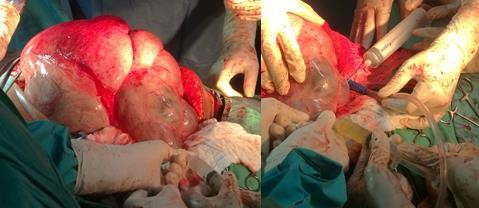
In the OR, the abdomen was approached with a xipho-pubic incision and after dissection of the different abdominal planes and opening of the peritoneum, a large polycystic mass was revealed. Citrine yellow fluid was aspirated from septa to reduce the tension of the mass, and the mass was delivered. (Figure 1)
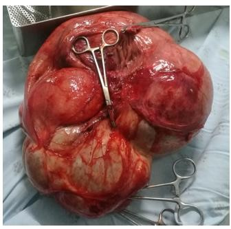
The pedicles were identified, followed by adhesiolysis of the posteroinferior surfaces of the mass and surrounding structures, and finally removal of the mass. Pedicles were ligated, then abdomen was systematically explored for macroscopic metastasis. Washing with saline solution was performed, and then closed after biopsy excision. The exteriorized mass weighed about 5.4 kg. (Figure 2)
Anatomopathological study reveals the presence of a partitioned cavity with serous content, sometimes clear, sometimes lemon-yellow alternating with mucoid content, measuring between 7 and 16 cm in length with a wall between 0.1 and 0.5 cm. The inner surface is smooth greyish congestive with rare nodule attached to the wall. Microscopically, there are sections of cystic ovarian tissue with a wall made of fibrous tissue lined by serous columnar epithelium with serous nuclei. No evidence of malignancy and the diagnosis of serous cystadenoma was retained.
Giant tumors of the ovary are very rare [3,4]. Cystadenomas are the most common cause of these tumors and are often associated with digestive symptoms including abdominal bloating, pain and nausea [5,8]. These symptoms were present in our patient. Serous cystadenomas are usually multilocular, and in some cases they include papillary projections [1]. The most incriminating risk factors are nulliparity, early puberty, and infertility [9]. All these factors are present in our patient who is 62 years old, nulliparous and had puberty at 10 years of age. A few rare cases have been identified in postmenopausal women. The literature presents these masses with a higher incidence in women of reproductive age and 50% in women under 40 years of age [1,10]. Unlike mucinous cystadenomas which are predominant in postmenopausal women [9]. Tumor markers are recommended especially in postmenopausal women, to detect the nature of the mass. The risk of malignancy in these elderly women is estimated to be between 8% and 45% [1], although more than 80% of cases are benign [11]. Sonography is the gold standard to confirm the diagnosis in addition to the anatomopathological study to detect their nature. In the pathological laboratory, the scarcity of papillae, the absence of epithelial stratification and cellular atypia will rule out the diagnosis of a serous borderline tumor, while absence of atypia and invasion of the connective tissue will rule out cancer [7]. On our microscopic specimen, sections of cystic ovarian tissue with a wall made of fibrous tissue lined by serous columnar epithelium with serous nuclei were observed. Hence the benign nature of our mass. Surgical management is recommended especially in cases where digestive symptoms are present and the mass exceeds 8 cm [8]. Many complications have been reported in previous studies during removal of these giant masses. These complications include splanchnic dilatation and venous pooling after sudden removal of these large intra-abdominal masses, and also hypotension due to decreased venous return due to inferior vena cava obstruction and sometimes pulmonary edema due to sudden re-expansion of a collapsed lung, which occurred due to compression by the enlarged abdomen [1]. The procedure was successful with no intraoperative complications and the patient did very well postoperatively. She was followed as an outpatient in the maternity ward, and now she is resuming her daily activities.
Benign serous cystadenomas in postmenopausal women are rare. Surgery as a definitive treatment is a challenge for both the general surgeon and the gynaecologist. It is always important to rule out cystadenosarcoma in the face of these rapidly evolving giant masses.
Dear Editorial Team, Clinical Medical Reviews and Reports. My experience with the journal was highly positive. The peer-review process was rigorous, constructive, and completed in a timely manner. The reviewers provided valuable comments that helped improve the quality and clarity of our manuscript. The editorial office was professional, responsive, and supportive throughout all stages of the publication process. Communication was clear and efficient, and any questions were addressed promptly. Overall, I found the journal to maintain high scientific standards and an excellent publication workflow. I would be pleased to consider submitting future work to this journal. Best wishes from, Elena Popa.

It was my pleasure to submit my testimonial concerning the Reviewer Board of our Scientific Journal “Brain and Neurological Disorders”. The Reviewers focused on some modifications and their contribution was helpful. The ladies of our Editorial Office were also supported my efforts. It was my honor to have such a co-operation and I am looking forward for more collaboration.

Dear Grace Pierce, Editorial Coordinator of Journal of Clinical Research and Reports, Thank you for the speedy and efficient peer review process. I appreciate the fact that your peer reviewers do not take months to respond like with some other journals. I would also like to thank the editorial office for responding quickly to my questions. It is an excellent journal. I plan to submit more manuscripts in the future. Best wishes from, Robert W. McGee

Dear Grace Pierce, Editorial Coordinator of Journal of Clinical Research and Reports, Working with you and your team on our recent publication in JCRR has been a truly wonderful and enjoyable experience. The responses were prompt, and the reviewers were patient, constructive, and highly professional. One reviewer in particular gave me the feeling that a professor was carefully reading and commenting on my coursework, which was deeply touching. The entire process was straightforward and hassle‑free, with no tedious online forms to complete. I highly recommend this journal. Best wishes from, DR Aibing Rao, Head of R&D

I Appreciate the Opportunity to Share my Experience with the Journal of Clinical Research and Reports. The peer review process was timely and constructive, and the feedback provided helped improve the quality of our manuscript. The editorial office was professional, responsive, and supportive throughout the process, ensuring smooth communication and efficient handling of the submission. Overall, it was a positive experience collaborating with your team.

Dear Mercy Grace, Editorial Coordinator of Obstetrics Gynecology and Reproductive Sciences, We would like to express our gratitude for your help at all stages of publishing and editing the article. The editors of the magazine answer all the necessary questions and help at every stage. We will definitely continue to cooperate and publish other works in the Obstetrics Gynecology and Reproductive Sciences! Best wishes from, Alla Konstantinovna Politova,
