
Case report | DOI: https://doi.org/10.31579/2690-4861/602
Department of Neurosurgery, Bethlehem Garden Hospital 3-14-72 Umesono, Kiyose, Tokyo 204-0024, Japan.
*Corresponding Author: Nobuhiko Aoki, Department of Neurosurgery, Bethlehem Garden Hospital 3-14-72 Umesono, Kiyose, Tokyo 204-0024, Japan.
Citation: Nobuhiko Aoki, (2024), Fulminant-type Infantile Acute Subdural Hematoma: Pre- and Post-operative Neuroimaging Analysis, International Journal of Clinical Case Reports and Reviews, 20(1); DOI:10.31579/2690-4861/602
Copyright: © 2024, Nobuhiko Aoki. This is an open-access article distributed under the terms of the Creative Commons Attribution License, which permits unrestricted use, distribution, and reproduction in any medium, provided the original author and source are credited.
Received: 08 November 2024 | Accepted: 18 November 2024 | Published: 26 November 2024
Keywords: infantile acute subdural hematoma; shaken baby syndrome; abusive head trauma; benign enlargement of subarachnoid space; large sylvian fissure; holohemispheric hemorrhage; dural border cell layer; cerebral parenchymal injury
Background:
An acute subdural hematoma (ASDH) in an infant without external signs of head trauma is diagnosed as shaken baby syndrome (SBS) in the United States or as an infantile acute subdural hematoma (IASDH) due to minor head trauma in Japan. The present case report analyzes the pre- and postoperative neuroimaging findings of fulminant-type IASDH and demonstrates that this condition can resolve fully with prompt surgery.
Case description
A 9-month-old, male infant fell at home while trying to stand up by holding on to a sofa. He and struck his occipital region on a mat. and He began crying. Soon thereafter he and displayed signs of altered consciousness status and seizure-like activity. He was taken to an emergency room by ambulance where head computed tomography (CT) revealed a left-sided, holohemispheric, subdural collection with a remarkable midline shift.
The patient’s conscious status deteriorated rapidly, requiring an emergency craniotomy. Intraoperatively, bloody fluid gushed out, and a partially clotted hematoma was removed. The origin of the hemorrhage was unable to be determined. His postoperative course was favorable without any neurological deficits. Fundoscopic examination revealed a left-sided retinal hemorrhage.
Postoperative CT on day 1 visualized a thin line indicative of a subdural hematoma (SDH) with resolution of the mass effect. Magnetic resolution imaging (MRI) on day 16 revealed a thin, tapering SDH. CT on day 34 found a tiny, film-like hemorrhage.
The patient was followed up on an outpatient basis for more than two years but did not present any neurological abnormalities.
Discussion & Conclusion
The present case is invaluable because it demonstrates that the outcome of emergency surgery and the ensuing recovery of fulminant-type IASDH can be favorable in the absence of a cerebral parenchymal injury. Particularly relevant here is the thin, film-like image observed after evacuation of the ASDH, which likely indicated the localization of the hemorrhage in the dural border cell layer (DBCL). Based on the serial neuroimaging analysis, the fulminant-type IASDH was considered secondary to the spread of the hemorrhage in the BDCL into the subdural compartment.
Infantile acute subdural hematomas (IASDH) have beenreported in Japan since the 1960’s [1]. However, because most reports were published in Japanese-language journals, coupled with the frequent criticism that the diagnosis was being used to conceal cases of child abuse, the concept of IASDH was not widely accepted in the English-speaking world [2, 3].
Fulminant-type IASDH is the most severe form of IASDH and differs from shaken baby syndrome (SBS) in that it does not involve a cerebral parenchymal injury [1, 4, 5]. As far as was able to be ascertained, no previous studies have discussed the neuroimaging characteristics of fulminant-type IASDH. The present study therefore aimed to clarify the neuroimaging features and treatment for fulminant-type IASDH.
A 9-month-old, male infant with no remarkable medical history fell and struck his occipital region at home while trying to stand up by holding on to a sofa. He began crying and soon thereafter vomited and displayed signs of altered consciousness status accompanied by seizure-like activity. He was taken to an emergency room by ambulance. On arrival, head computed tomography (CT) revealed a left-sided, mixed-density subdural hematoma with a remarkable midline shift to the right. In addition, an enlarged sylvian fissure (LSF) was noted on the right side (Figure. 1). His conscious status deteriorated rapidly, requiring emergency surgery. Intraoperatively, bloody fluid gushed out, and a partially clotted hematoma was removed.
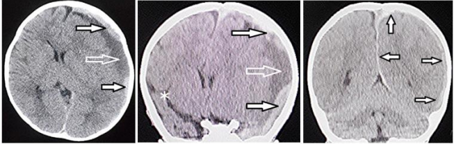
Figure 1: Computed tomography 3 hours after presentation
Left: Axial view, Middle & Right: Coronal view
Left & Middle: High density (black arrows) and low density (white arrow) components of the left-sided subdural hematoma (SDH). Note the remarkable midline shift to the right side and the large sylvian fissure on the right side (asterisk).
Right : SDH covering the entire cerebral hemisphere (i.e, holohemispheric hemorrhage) on the right side (arrows)
The origin of the hemorrhage was unable to be identified within the surgical field. His postoperative course was uneventful, and he achieved recovery without any neurological deficits. Fundoscopic examination revealed a left-sided, retinal hemorrhage.
CT on postoperative day 1 visualized a thin line indicative of a high-density lesion as well as resolution of the mass effect (Figure. 2). Magnetic
resonance imaging (MRI) on day 16 revealed a thin, high-intensity SDH covering the entire left cerebral hemisphere. No parenchymal abnormality indicating a primary brain injury was found (Fig. 3). CT on day 34 found a tiny, film-like hemorrhage at the inner table of the frontal bone (Fig. 4). The patient was followed up on an outpatient basis for more than two years but presented no neurological abnormalities.
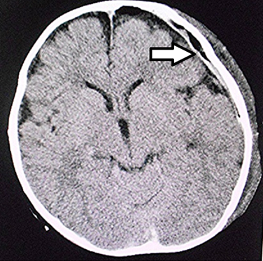
Figure 2. Computed tomography on postoperative day 1
Resolution of the midline shift and the thin, linear, high-density lesion on the left side can be seen (arrow).
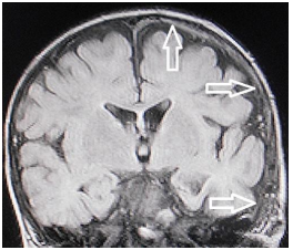
Figure 3. Magnetic resonance imaging (fluid-attenuated inversion recovery, coronal view) on postoperative day 27
The thin, high-intensity subdural hematoma can be seen covering the entire cerebral hemisphere on the left side (arrows). No parenchymal abnormality indicating primary brain injury was observed.
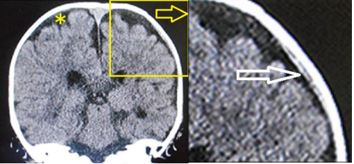
Figure 4: Coronal view on computed tomography on postoperative day 3
Left: Full-sized view
Right: Enlargement of the area in the box on the full-sized view
The thin, film-like, high-density lesion corresponding to the hemorrhage (arrow) can be seen in the dural border cell layer. Note the benign enlargement of the subarachnoid space on the full-sized view (asterisk).
Based on the biophysiological characteristics of infants, IASDH was originally defined in 1984 as an acute, infantile subdural hematoma apparently caused by minor head trauma without loss of consciousness and not associated with a primary brain injury [4]. Most cases of IASDH are also associated with retinal hemorrhage [4].
IASDH is clinically graded as mild (grade I), intermediate (grade II) or fulminant (grade III) in accordance with the findings of a previous study (Table 1) [4]. The pathological features of IASDH, including the fulminant type, as indicated by imaging studies and surgical findings, account for its mostly benign clinical course following conservative management or prompt surgical intervention [5-7]. It is important to emphasize that a favorable outcome depends greatly on the absence of a primary cerebral parenchymal injury. As far as could be ascertained, however, no English-language studies to date have discussed primary cerebral parenchymal injury as a feature differentiating IASDH from SBS (Table 2).
| IASDH | SBS/AHT | |
| Applied force | Minor head trauma | Abuse (high energy impact) |
| Main etiology | Disruption of the bridging vein | Cerebral contusional tears |
| Primary brain injury | None | Common |
| Age distribution | Peak at 6 - 10 months | Widely distributed (including less than 3 months) |
| Sex | Marked preponderance in males | No preponderance |
| Recurrence | Rare | Not rare |
| Prognosis | Depends on hematoma volume (mostly benign clinical courses) | Poor |
| Retinal hemorrhage | Frequent | Common |
(Cited from Ref. 1 with permission of the Society of Japanese Neurosurgery)
Table 1: Comparison of infantile acute subdural hematoma (IASDH) and shaken baby syndrome (SBS) / abusive head trauma (AHT)
| Grade | Type | Clinical features |
| I | Mild | Conscious, no motor disturbance. Vomiting and/or irritability present |
| II | Intermediate | Drowsiness, minimal or mild hemiparesis |
| III | Fulminant | Stupor to coma, moderate to severe hemiparesis. Signs of cerebral herniation present |
Table 2. Clinical grade of infantile acute subdural hematoma on the arrival of patients at the emergency department; proposed in 1984 (Ref. 4)
The dural border cell layer (DBCL), a distinct, soft-tissue layer at the dural-arachnoid interface, is composed of a loose conglomeration of cells having enlarged, extracellular spaces and no extracellular collagen [8]. ASDH occurring in this environment results from the disruption of the DBCL by pooled blood. Furthermore, in the superficial compartment of the DBCL, there is a well-developed dural venous plexus which fills the venous sinuses. Because the DBCL is easily disrupted, a hemorrhage originating in the inner dural plexus may be chiefly responsible for the non-traumatic symptoms of this condition [9]. Particularly relevant here is the thin, film-like image on CT which is observable after evacuation of the ASDH and which may indicate the localization of the hemorrhage in the DBCL [10, 11].
Based on the serial neuroimaging analysis in the present patient, the fulminant- type IASDH was considered secondary to the spread of the hemorrhage from the DBCL into the subdural compartment; the tiny, film-like hemorrhage at the inner table of the frontal bone on CT on postoperative day 34 coincided to be trapped in the DBCL [12] (Figure. 4).
The present case is invaluable because it demonstrates that emergency surgery can produce a favorable outcome in fulminant-type IASDH in the absence of a cerebral parenchymal injury, the fundamental feature distinguishing IASDH from SBS. Particularly relevant here is the thin, film-like appearance observed after evacuation of the ASDH, which likely indicated the localization of the hemorrhage in the DBCL. Based on serial neuroimaging analysis results, fulminant-type IASDH was considered secondary to the spread of the hemorrhage from the DBCL into the subdural compartment.
Dear Editorial Team, Clinical Medical Reviews and Reports. My experience with the journal was highly positive. The peer-review process was rigorous, constructive, and completed in a timely manner. The reviewers provided valuable comments that helped improve the quality and clarity of our manuscript. The editorial office was professional, responsive, and supportive throughout all stages of the publication process. Communication was clear and efficient, and any questions were addressed promptly. Overall, I found the journal to maintain high scientific standards and an excellent publication workflow. I would be pleased to consider submitting future work to this journal. Best wishes from, Elena Popa.

It was my pleasure to submit my testimonial concerning the Reviewer Board of our Scientific Journal “Brain and Neurological Disorders”. The Reviewers focused on some modifications and their contribution was helpful. The ladies of our Editorial Office were also supported my efforts. It was my honor to have such a co-operation and I am looking forward for more collaboration.

Dear Grace Pierce, Editorial Coordinator of Journal of Clinical Research and Reports, Thank you for the speedy and efficient peer review process. I appreciate the fact that your peer reviewers do not take months to respond like with some other journals. I would also like to thank the editorial office for responding quickly to my questions. It is an excellent journal. I plan to submit more manuscripts in the future. Best wishes from, Robert W. McGee

Dear Grace Pierce, Editorial Coordinator of Journal of Clinical Research and Reports, Working with you and your team on our recent publication in JCRR has been a truly wonderful and enjoyable experience. The responses were prompt, and the reviewers were patient, constructive, and highly professional. One reviewer in particular gave me the feeling that a professor was carefully reading and commenting on my coursework, which was deeply touching. The entire process was straightforward and hassle‑free, with no tedious online forms to complete. I highly recommend this journal. Best wishes from, DR Aibing Rao, Head of R&D

I Appreciate the Opportunity to Share my Experience with the Journal of Clinical Research and Reports. The peer review process was timely and constructive, and the feedback provided helped improve the quality of our manuscript. The editorial office was professional, responsive, and supportive throughout the process, ensuring smooth communication and efficient handling of the submission. Overall, it was a positive experience collaborating with your team.

Dear Mercy Grace, Editorial Coordinator of Obstetrics Gynecology and Reproductive Sciences, We would like to express our gratitude for your help at all stages of publishing and editing the article. The editors of the magazine answer all the necessary questions and help at every stage. We will definitely continue to cooperate and publish other works in the Obstetrics Gynecology and Reproductive Sciences! Best wishes from, Alla Konstantinovna Politova,
