
Review Article | DOI: https://doi.org/10.31579/2692-9406/037
Retired Consultant Urologist Surgeon & Independent Investigator, Mansoura University, Faculty of Medicine, Egypt.
*Corresponding Author: Ahmed N. Ghanem, Retired Consultant Urologist Surgeon & Independent Investigator Mansoura University, Faculty of Medicine, No1 President Mubarak Street, Mansoura 35511, Egypt.
Citation: Shoichiro Ozaki. (2020) in clear water no fish can live. Water purification promote global warming, decline of countries. Biomedical Research and Clinical Reviews. 3(2); DOI: 10.31579/2692-9406/037
Copyright: © 2021 Ahmed N. Ghanem, This is an open-access article distributed under the terms of the Creative Commons Attribution License, which permits unrestricted use, distribution, and reproduction in any medium, provided the original author and source are credited.
Received: 18 December 2020 | Accepted: 21 January 2021 | Published: 25 January 2021
Keywords: fluid therapy; ards; fluid overload; volume kinetic; capillary physiology; starling’s law; hydrodynamics, hemodynamics
Fluid therapy (FT) was introduced during WW2. Ever since its complications have been frequently reported but notably some serious complications have been overlooked. The role of volumetric overload (VO) in inducing VU shocks (VOS) and causing the acute respiratory distress syndrome (ARDS) has remained overlooked, unrecognized, and underestimated till recently.
The role of FT complications in inducing VOS and causing ARDS is hard to detect because VOS is a shock that complicates another existing shock seamlessly and un-noticed. The author attributes this to the faulty rules on FT dictated by the wrong Starling’s law that causes many errors and misconceptions of FT which mislead physicians into giving too much fluid during shock resuscitation. The research findings on the wrong Starling’s law and how it has been corrected based on the hydrodynamic of the porous orifice (G) tube and the newly recognized VOS and the new patho-aetiology and therapy of ARDS are summarized here. Other authors in support of this contention are quoted. The errors on current FT and its corrections are given.
Finally, a new prospective cohort study on FT and ARDS is recommended and a call for new guidelines on FT is urgently needed.
Abbreviations
FT fluid therapy
VOS Volumetric Overload Shocks
ARDS Acute Respiratory Distress Syndrome
WW2 World War 2
Introduction
The great benefit that fluid therapy (FT) brought to humanity is undoubted since its introduction during World War 2 (WW2) and has continued in civilian hospital medical practice ever since. However, it has complications and the most serious remain overlooked and underestimated. Fluid therapy is used for fluid maintenance and in the management of shock resuscitation, acutely ill patients and during prolonged major surgeries. The last two are the situation during which FT complications occur.
Despite consistent reporting of FT complications since WW2, the most important and serious complications remain unrecognized and underestimated: This is precisely the current state of FT recommendations that induce Volumetric Overload Shocks (VOS) [1] and cause the Acute Respiratory Distress Syndrome (ARDS) [2,3]. This article addresses this issue by identifying the errors and misconceptions and providing the corrections and the new scientific basis for the future recommendations on FT guidelines. It starts by documenting the evidence that FT induces VOS and causes ARDS.
Evidence provided by the Author
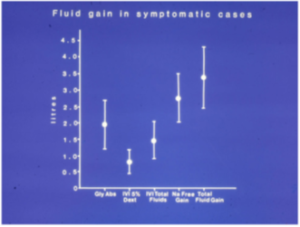
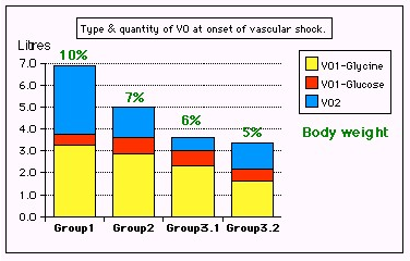
The author reported on VOS or Volume Kinetic (VK) shocks in clinical practice affecting mostly surgical patients [1, 4]. The clinical evidence is summarized in (Figures 1 and 2 and Table 1). It is reported that VOS cause ARDS [2, 3]. The identified underlying culprit is Starling’s law that dictates the faulty rules on FT, causing many errors and misconceptions [4] that mislead physicians into giving too much fluid during the resuscitation of shock [5] that induce VOS [1] and cause ARDS later [2,3]. The correction as based on the hydrodynamic of the porous orifice (G) tube is shown in (Figure 3) that is a diagrammatic representation based on many photographs. It demonstrates the magnetic field-like fluid circulation between fluid in the lumen of the G tube and that around it in surrounding chamber C.
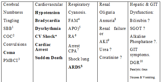
Abbreviations for Table 1
SBB1 Sudden bilateral blindness
COC2 Clouding of consciousness
PMBCI3 Paralysis mimicking bizarre cerebral infarctions, but is recoverable on instant use of HST of 5%NaCl and/or NaCo3, and so is coma and AKI
FAM4 Frothing around the mouth
APO5 Acute pulmonary oedema
RA6 Respiratory arrest
CPA7 Cardiopulmonary arrest
ARDS $ Manifests later on ICU
AKI9 Acute kidney injury
DGR10 Delayed gut recovery
CV Shock* Cardiovascular shock of VOS reported here as VOS 1 and VOS2.
Annuria8 That is unresponsive to diuretics but responds to HST of 5%Ncl and/or 8.4%NaCo3
AKI8 Acute kidney injury
Also occurs the excessive bleeding at the surgical site and
Leukocytosis occurred in the absence of sepsis and septic shock
Evidence provided by other eminent researchers
Other researchers have recently reported that too much fluid infusions causes clinical problems identified and gathered in (Table 1) which are manifestations of the multiple organs dysfunction syndromes (MODS) that include ARDS, Coma, AKI, hepatic and hematological dysfunctions.. However, they have stopped short of recognizing that VOS and ARDS are caused by excessive infusion of FT.
Professor Hahn has reported massively on VK in healthy volunteers and patients [7,8]. He reported in conclusion that: "Guidelines for fluid therapy rarely take into account that adverse effects occur in a dose-dependent fashion. Adverse effects of crystalloid fluids are related to their preferential distribution to the interstitial of the subcutis, the gut, and the lungs. The gastrointestinal recovery time is prolonged by 2 days when more than 2 liters is administered. Infusion of 6-7 liters during open abdominal surgery results in poor wound healing, pulmonary oedema, and pneumonia. There is also a risk of fatal postoperative pulmonary oedema that might develop several days after the surgery. Even larger amounts cause organ dysfunction by breaking up the interstitial matrix and allowing the formation of lacunae of fluid in the skin and central organs, such as the heart. For both crystalloid and colloid fluids, coagulation becomes impaired when the induced hemodilution has reached 40%. Coagulopathy is aggravated by co-existing hypothermia. Although oedema can occur from both crystalloid and colloid fluids, these differ in pathophysiology."
Other authors also found a significant effect of crystalloids overload on morbidity and mortality of ARDS as they did the research during the first 24-48 hours from hospital admission. I have found only one study on adults' trauma patients by Jones et al (2016) [9], and one paediatrics study by Coons et al (2018) [10] and a remarkable review article by Schrier reported in 2010 [11] that incriminate saline overload and recommend judicious use of fluid infusion during resuscitation. In patients of these adult and paediatric trauma trials there is no sepsis involved and both studies were done over a period of 24 and 48 hours, respectively. Both articles detected a significant relationship of VO with morbidity and mortality of ARDS.
Jones et al [9] reported: "Large-volume crystalloid resuscitation is associated with increased mortality and longer time ventilated. Based on this data, we recommend judicious use of crystalloids in the resuscitation of trauma patients.”
The conclusion by Coons et al [10] was: "Early administration of high volumes of crystalloid fluid greater than 60 ml/kg/day significantly correlates with pulmonary complications, days NPO, and hospital length of stay. These results span the first 48 h of a patient's hospital stay and should encourage surgical care providers to exercise judicious use of crystalloid fluid administration in the trauma bay, ICU, and floor"
The huge prospective multicenter trials [12, 13] also documented massive volumetric overload (VO) retained in surviving ARDS patients of 3-10 liters but have neither recognized VOS nor incriminated VO in the patho-aetiology of ARDS. They also did not recognize the high association of VO with the mortality which was estimated at 60 or 90 days not at the immediate period of 24-48 hours after admission as demonstrated by the above reports [9, 10]. Excellent example of these huge multicenter trials is that study reported by Rowan et al in 2017 [12].
In the results section, Rowan et al reported: “Each study day the liberal-strategy group received more fluid than the conservative-strategy group and on days 1 through 4 had a lower urinary output, resulting in a higher cumulative fluid balance (Table 2). During the study, the seven-day cumulative fluid balance was -136±491 ml in the conservative-strategy group, as compared with 6992±502 ml in the liberal-strategy group (P<0.001) (Figure 1 of the Supplementary Material). For patients who were in shock at baseline, the cumulative seven-day fluid balance was 2904±1008 ml in the conservative-strategy group and 10,138±922 ml in the liberal-strategy group (P<0.001). For patients who were not in shock at baseline, the cumulative fluid balance was −1576±519 ml in the conservative-strategy group and 5287±576 ml in the liberal-strategy group (P<0.001)”
Errors and misconceptions on current FT practice
The errors and misconceptions in current clinical practice and its corrections are documented here. For references on this section please see [5].
Error I
Every arterial hypotension is considered synonymous with hypovolemia or at least treated as such with volume expansion in every clinical case of shock, anesthetic induction, or operative period!
Correction I
Hypotension is not synonymous with hypovolemia. The cause of the primary recognized shock and hypotension must be differentiated. The difference between the therapeutic/physiological VO regarding (quantity versus response) in contrast with the paradoxes of pathological VO on arterial pressure and renal response must be precisely identified. Two paradoxical responses of pathological VO require recognition: one is inducing hypotension shock and the second is causing AKI. The transition from the hypovolemic hypotension shock into the VO hypotension shock during overzealous volume expansion occurs seamlessly unnoticed and undetected by any monitoring until it manifests, later on ICU, with torso edema and increased body weight (BW) of ARDS or MODS.
Error II
The volume-pressure relationship of the vascular system is perceived as infinite strait line!?
Correction II
The volume-pressure relationship particularly that of vascular volume and arterial pressure is a limited line segment, beyond which the relation collapses. Within limits, increasing vascular volume (physiological or therapeutic VO) increases arterial pressure but when such limit is exceeded (pathological VO) a paradoxical hypotension occurs. A similar VO paradox exists on the renal function while physiological VO induces diuresis a pathological VO causes AKI as part of the features of MODS. These two paradoxes are not new but hardly recognized.
Error III
The central venous pressure (CVP) and pulmonary capillary wedge pressure (PCWP) as monitoring parameters guiding fluid therapy are given a value of 18 to 22cm water as currently practiced on many ICUs. Although current recommendations indicate that CVP and PCWP are unreliable and no longer being used, evidence from prevalence of ARDS and MODS on ICU testify differently, and it remain part of its definition. The confounded error underlying the misconception of high positive CVP is related to a deeply rooted physiological error.
Correction III
The given figures of CVP and PCWP are erroneously too high yet remain widely practiced. Persistence to achieve such high CVP using massive volume expansion is among the misleading reasons for inducing pathological VO causing ARDS. The infused fluid rapidly shifts out of the vascular system and CVP may drop back to below 10cm water, then another bolus VO is given before the gross torso edema and increase of BW becomes obvious. The correct CVP figures are given in all physiology textbooks that swing around 0 (at mid-axillary line) with a range of +7 to -7cm water. If we do not understand how Nature works, we must faithfully imitate until reliable methods of monitoring fluid therapy are found.
Error IV
The capillary forces responsible for irrigating and oxygenating the ISF space and cells are mixed up with that causing edema, flooding, and drowning.
Correction IV
It is strongly recommended that every physician involved in fluid therapy, ARDS and MODS management should reconsider what is the physiological function of the arterial and venous pressures and which pressure is responsible for what? In particular relating the pathological ISF accumulation or subcutaneous edema with the forces on which the hypothesis that dictates capillary-ISF transfer on the causation of dropsy, proposed by Starling at the Lancet in 1886, reveals the error. The reason is that the forces on which this hypothesis is based govern the volume and pressure regulation of the vascular and ISF compartments, and subsequently cell viability are incorrect. Being false, this hypothesis underlies most mentioned erroneous concepts on fluid therapy. Starling’s hypothesis was wrongly made later into physiological law. It may be realized that this is the major error responsible for the current dilemma on ARDS and MODS concealing its real patho-aetiology of VO.
Error V
The major misconception, and unfortunately the most prevailing, is wrongly assuming that the vascular system is an all positive pressure system, in which not only the mentioned arterial volume-pressure relationship is misconceived as infinite strait line but also keeping high venous pressure and ISF tissue over-hydrated are erroneously believed to enhance cell nourishment and oxygen delivery. This underlies the liberal volume expansion pumping in too much fluid that creates edema, flooding and drowning of the ISF tissue as well as vital organs and cells! This is precisely the error underlying the pathological VO inducing ARDS and MODS.
In the circulatory system is the arterial pressure seems to be so for very good reason: it is the driving force for ejecting fluid through the capillary orifice creating the side negative energy pressure that drives the dynamic autonomous magnetic field-like fluid circulation between capillary lumen and surrounding tissues - keeping the ISF tissue pressure negative, appearing almost dry, while efficiently irrigated and oxygenated!
Correction V
To assume that CVS to be an all positive pressure system is quite simply wrong. In fact, there is a lot of negative physiological pressure under the skin. It is well known that the pleural spaces have negative pressure and the pressure in alveoli alternates. The CVP of normal subjects may swing around Zero, between positive +7 and negative -7 mmhg [14-16]. The intracranial pressure is also negative. Thus, the ISF space of subcutaneous tissues, most organs and parts of the body have negative pressure of -7 cm water that has been demonstrated [20] and re-affirmed [21] but neither considered nor satisfactorily explained.
Identifying Starling’s law as the culprit dictating the faulty rules causing the errors and misconceptions on FT
The wrong Starling’s law is the culprit dictating the above-mentioned errors and misconceptions that mislead physicians into giving too much fluid during the resuscitation of shock, acutely ill patients, and prolonged major surgery [5]. It was investigated at both physics and physiological fronts. As Starling based his hypothesis on the hydrodynamic of Poiseuille’s tube which is large brass tube of uniform diameter, I constructed the porous orifice G tube us based on the capillary ultra-structure of having a narrow orifice that is the precapillary sphincter and intercellular slit wide pores that allow the passage of plasma proteins thus nullifying the oncotic pressure in vivo. The hydrodynamic of G tube proved totally different from Poiseuille’s tube. The G tube has a side pressure that exert negative pressure on the wall near the inlet and positive pressure near the exit. This creates the unique, rapid magnetic fluid-like circulation between fluid in lumen and fluid around it (Figure 3). Thus, Starling’s law is proved wrong on both of its forces. The G tube phenomenon replaces Starling’s law for the capillary-interstitial (ISF) transfer.
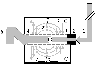
The numbers should read as follows:
1. The inflow pressure pushes fluid through the orifice
2. Creating fluid jet in the lumen of the G tube**.
3. The fluid jet creates negative side pressure gradient causing suction maximal over the
proximal part of the G tube near the inlet that sucks fluid into lumen.
4. The side pressure gradient turns positive pushing fluid out of lumen over the distal
part maximally near the outlet.
5. Thus, the fluid around G tube inside C moves in magnetic field-like circulation (5)
taking an opposite direction to lumen flow of G tube.
6. The inflow pressure 1 and orifice 2 induce the negative side pressure creating the dynamic G-C circulation phenomenon that is rapid, autonomous, and efficient in moving fluid and particles out from the G tube lumen at 4, irrigating C at 5, then sucking it back again at 3,
7. Maintaining net negative energy pressure inside chamber C.
**Note the shape of the fluid jet inside the G tube (Cone shaped), having a diameter of the inlet on right hand side and the diameter of the exit at left hand side (G tube diameter). I lost the photo on which the fluid jet was drawn, using tea leaves of fine and coarse sizes that runs in the centre of G tube leaving the outer zone near the wall of G tube clear. This may explain the finding in real capillary of the protein-free (and erythrocyte-free) sub-endothelial zone in the Glycocalyx paradigm. It was also noted that fine tea leaves exit the distal pores in small amount maintaining a higher concentration in the circulatory system than that in the C chamber- akin to plasma proteins.
The new scientific basis for future guideline recommendations on FT
The G tube hydrodynamic as the correct replacement for the wrong Starling’s law form the scientific bases for future guidelines on the practice of FT. Two other misconceptions on capillary physiology were corrected by the newly discovered tree branching law (under consideration) [13,14]. The two other misconceptions on capillary physiology add to understand the correct model for the capillary-ISF transfer that adequately provide for the cells’ viability at rest and during strenuous exercise.
Therapy of FT complications of VOS and ARDS
Prevention
Being iatrogenic complications of fluid therapy, both VOS and ARDS are preventable.
To prevent VOS and ARDS a limit to the maximum amount of fluid used during shock resuscitation or major surgery must be agreed upon (New guidelines are required).
Surgical care providers must exercise judicious use of crystalloid fluid administration in the trauma bay, ICU, and floor.
Replace the loss in haemorrhagic hypovolemic shocks but do not overdo it.
If hypotension develops despite volume replacement later during ICU stay, inotropic drugs, hydrocortisone 200 mg and hypertonic sodium therapy (HST) should be used-please, see later. The latter restores the pre-capillary sphincter tone (peripheral resistance) so that the capillary works as normal G tube again, but NO isotonic crystalloids or colloids over-infusions is required.
To learn the new correct science, one must unlearn the old incorrect habits.
The following practices should be abandoned:
A. Bolus fluid therapy in surgical patients
B. Abandon the aggressive current liberal regimen of Early Goal-Directed Therapy (EGDT) in treating shocked and septic patients [26]. Multiple huge multicenter trials have proved it to be the wrong practice.
C. Please refrain from persisting to elevate CVP to levels above 12 and up to 18-22 cm saline in shock management. This is a major cause for inducing VOS and ARDS during shock resuscitation, particularly septic shock.
Therapeutic
Hypertonic sodium therapy (HST) of 5%NaCl and/or 8.4%NaCo3 has truly proved lifesaving therapy for the TURP syndrome and acute dilution HN as well as secondary VOS 2 that complicates fluid therapy of VOS 1 causing ARDS. It works by inducing massive diuresis being a potent suppressor of antidiuretic hormone. It may also work on the pre-sphincter capillary restoring its tone.
My experience in using it for treating established ARDS with sepsis and primary VOS 2 that causes ARDS is not tested. However, evidence on HST suggests it will prove successful if given early, promptly and adequately to ARDS patients while refraining from any further isotonic crystalloid or colloid fluid infusions using saline, HES and/or plasma therapy- just give the normal daily fluid requirement and no more. After giving HST over one hour using the CVP catheter already inserted, the patient recovers from AKI and produces through a urinary catheter massive amount of urine of 4-5 liters as you watch. This urine output should not be replaced. Just observe the patient recovering from his AKI, coma and ARDS and asks for a drink. This is done in addition to the cardiovascular, respiratory, and renal support on ICU. Patients with AKI on dialysis, the treating nephrologist should aim at and set the machine for inducing negative fluid balance.
The HST of 5%NaCl and/or 8.4%NaCo3 is given in 200 ml doses over 10 minutes and repeated. I did not have to use more than 1000 ml during the successful treatment of 16 ARDS patients. Any other hypertonic sodium concentration is not recommended. A dose of intravenous diuretic may be given but it does not work in a double or triple the normal dose. A dose of 200 mg of hydrocortisone is most useful. Antibiotic prophylactic therapy is given in appropriate and adequate doses to prevent sepsis and septic shock. No further fluid infusions of any kind crystalloids, colloids and blood is given. The urinary loss should not be replaced as this defeats the objective of treatment.
A suggested recommended future trial urgently needed
I would recommend a small pilot prospective controlled cohort study on 100 patients as a start to try HST in established ARDS cases that would be something to look forward to reading a report on it, hopefully soon. No multicenter trial or high expenses is needed for that. Not much time is required either. If you cannot do it on a hundred patients, you probably cannot (as Mr. JP Ward put it to me before the start of our prospective study [19]. I can assure the investigators that no harm will come to patients. It is a guaranteed win bit; you may win but you do not lose anything. In the worst-case scenario, the patient may not respond because of chronicity of ARDS or after sepsis complicates ARDS and gets the capillary damage established. As the author of all self-referenced articles here, published in open access journals, and as copyright holder I give open permission to any interested investigator to use any of my articles as template, particularly recommended article [19]- the appropriate permission from the editors of BJUI and authors are given. I strongly recommend that hypertonic sodium therapy should be given a trial in the management of ARDS of both sepsis and Covid-19 as it may prove to be my successful positive contribution to the war against the Covid-019 pandemic.
Conclusion
Both VOS and ARDS are iatrogenic complications that remain overlooked and underestimated. The reason for that is the new VOS complicates the shock being treated with fluid therapy and difficult to detect and recognize. The massive use of FT is not the treating physician’s fault. Hence all physician find it offensive to label as fluid overload. Physicians are being mislead by the faulty rules on FT causing many errors and misconceptions that lead to massive fluid retention during shock resuscitation. This VO induces VOS and cause ARDS.
Dear Editorial Team, Clinical Medical Reviews and Reports. My experience with the journal was highly positive. The peer-review process was rigorous, constructive, and completed in a timely manner. The reviewers provided valuable comments that helped improve the quality and clarity of our manuscript. The editorial office was professional, responsive, and supportive throughout all stages of the publication process. Communication was clear and efficient, and any questions were addressed promptly. Overall, I found the journal to maintain high scientific standards and an excellent publication workflow. I would be pleased to consider submitting future work to this journal. Best wishes from, Elena Popa.

It was my pleasure to submit my testimonial concerning the Reviewer Board of our Scientific Journal “Brain and Neurological Disorders”. The Reviewers focused on some modifications and their contribution was helpful. The ladies of our Editorial Office were also supported my efforts. It was my honor to have such a co-operation and I am looking forward for more collaboration.

Dear Grace Pierce, Editorial Coordinator of Journal of Clinical Research and Reports, Thank you for the speedy and efficient peer review process. I appreciate the fact that your peer reviewers do not take months to respond like with some other journals. I would also like to thank the editorial office for responding quickly to my questions. It is an excellent journal. I plan to submit more manuscripts in the future. Best wishes from, Robert W. McGee

Dear Grace Pierce, Editorial Coordinator of Journal of Clinical Research and Reports, Working with you and your team on our recent publication in JCRR has been a truly wonderful and enjoyable experience. The responses were prompt, and the reviewers were patient, constructive, and highly professional. One reviewer in particular gave me the feeling that a professor was carefully reading and commenting on my coursework, which was deeply touching. The entire process was straightforward and hassle‑free, with no tedious online forms to complete. I highly recommend this journal. Best wishes from, DR Aibing Rao, Head of R&D

I Appreciate the Opportunity to Share my Experience with the Journal of Clinical Research and Reports. The peer review process was timely and constructive, and the feedback provided helped improve the quality of our manuscript. The editorial office was professional, responsive, and supportive throughout the process, ensuring smooth communication and efficient handling of the submission. Overall, it was a positive experience collaborating with your team.

Dear Mercy Grace, Editorial Coordinator of Obstetrics Gynecology and Reproductive Sciences, We would like to express our gratitude for your help at all stages of publishing and editing the article. The editors of the magazine answer all the necessary questions and help at every stage. We will definitely continue to cooperate and publish other works in the Obstetrics Gynecology and Reproductive Sciences! Best wishes from, Alla Konstantinovna Politova,
