
Review Article | DOI: https://doi.org/10.31579/2690-1897/174
Candidate of biological science, Assistant professor of pathophysiology department named D. A. Maslakov, Grodno State Medical University; Grodno State Medical University, 80 Gorky.
*Corresponding Author: Bon E.I, Candidate of biological science, Assistant professor of pathophysiology department named D. A. Maslakov, Grodno State Medical University; Grodno State Medical University, 80 Gorky.
Citation: Maksimovich N.E, Bon E.I, Vishnevskaya E.I, Bon E.I. (2024), Features of Histological Changes in Neurons of the Parietal Cortex and Hippocampus in Rats with Cerebral Ischemia and Introduction, J, Surgical Case Reports and Images 7(1); DOI:10.31579/2690-1897/174
Copyright: © 2024, Bon E.I. This is an open access article distributed under the Creative Commons Attribution License, which permits unrestricted use, distribution, and reproduction in any medium, provided the original work is properly cited.
Received: 05 January 2024 | Accepted: 15 January 2024 | Published: 29 January 2024
Keywords: histological changes; neurons; parietal cortex; hippocampus; cerebral ischemia; omega-3 polyunsaturated fatty acids
Acute cerebrovascular accidents are one of the most pressing problems in modern medicine. The incidence of strokes varies in different regions of the world from 1 to 4 cases per 1000 population per year, increasing significantly with age. Cerebrovascular diseases of ischemic origin tend to grow, rejuvenate, and are associated with a severe clinical course, high rates of disability and mortality. Modeling ischemic damage leads to pronounced morphological changes in neurons of the parietal cortex and hippocampus of the brain - a decrease in their size, deformation of perikarya, an increase in the degree of chromatophily of neurons with simultaneous shrinkage and subsequent death, as well as the development of pericellular edema. These disorders were most pronounced in the 3rd subgroup of SCI with the shortest interval between dressings, amounting to 1 day and TCI. Changes in the parietal cortex and hippocampus during CI were unidirectional, but in the parietal cortex, which is most sensitive to oxygen deficiency, they were more pronounced and appeared at an earlier time.
Acute cerebrovascular accidents are one of the most pressing problems in modern medicine. The incidence of strokes varies in different regions of the world from 1 to 4 cases per 1000 population per year, increasing significantly with age. Cerebrovascular diseases of ischemic origin tend to grow, rejuvenate, and are associated with a severe clinical course, high rates of disability and mortality [1-4].
The goal is to study the characteristics of histological changes in neurons of the parietal cortex and hippocampus in rats with cerebral ischemia and administration of omega-3 polyunsaturated fatty acids.
The experiments were performed on 174 male outbred white rats weighing 260±20 g in compliance with the requirements of Directive of the European Parliament and Council No. 2010/63/EU of 22.09.2010 on the protection of animals used for scientific purposes.
The choice of experimental animals is due to the similarity of the angioarchitecture of the brain of rats and humans. Simulation of cerebral ischemia (CI) was carried out under conditions of intravenous thiopental anesthesia (40-50 mg/kg).
The studies used models of total (TCI), subtotal (SCI), partial (PCI) and stepwise subtotal (SSCI) cerebral ischemia. The table shows the experimental groups and the number of animals in them (Table 1).
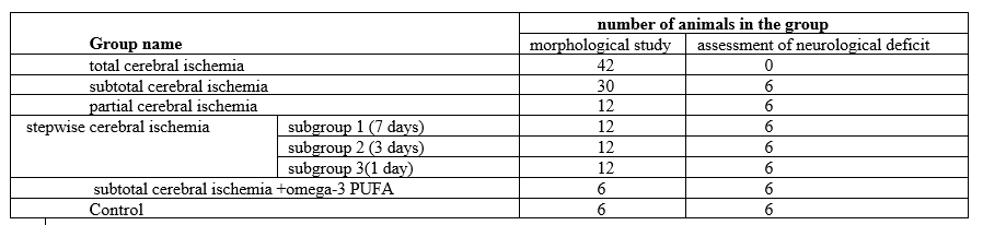
Table 1 – Experimental groups
Note - omega-3 PUFAs - omega-3 polyunsaturated fatty acids
Total cerebral ischemia was modeled by decapitation of animals. Material was collected at the 1st, 5th, 15th, 30th and 60th minutes, as well as 5 hours and 24 hours after decapitation [6].
Subtotal cerebral ischemia was modeled by simultaneous ligation of both common carotid arteries (CCA). The material was collected 1 hour, 2 hours, 3 hours, 6 hours and 24 hours after surgery.
Partial cerebral ischemia was modeled by ligating one CCA on the right. The material was taken 1 hour and 1 day after surgery.
Stepped subtotal was carried out by sequential ligation of both CCAs with an interval of 7 days (subgroup 1), 3 days (subgroup 2) or 1 day (subgroup 3). The material was taken 1 hour and 1 day after ligation of the second CCA in each of the subgroups [1-4].
To study the effects of omega-3 polyunsaturated fatty acids (ω-3 PUFA), animals with IGM were intragastrically administered the drug "Omegamed" at a dose of 5 g/kg body weight for a week. The control group consisted of sham-operated rats of similar sex and weight.
For morphological research, after decapitation of the animal, the brain was quickly removed, and pieces of the anterior cerebral cortex were fixed in Carnoy's fluid. The localization of the parietal cortex and hippocampal cortex in histological preparations of the rat brain was determined using a stereotaxic atlas.
Serial paraffin sections were stained with 0.1% toluidine blue using the Nissl method and for the detection of ribonucleoproteins (RNPs) according to Einarson [4].
The study of histological preparations, their microphotography, morphometry and densitometry of the chromogen sediment was carried out using an Axioscop 2 plus microscope (Zeiss, Germany), a digital video camera (LeicaDFC 320, Germany) and the ImageWarp image analysis program (Bitflow, USA).
In each animal, at least 30 neurons of the fifth layer of the parietal cortex and the pyramidal layer of the CA1 field of the hippocampus were assessed.
In histological studies, the size and shape of perikarya of neurons in the parietal cortex and hippocampus of the rat brain were determined, and changes in cytoplasmic chromatophily were studied [4].
The study of changes in the size and shape of neuronal perikarya was carried out by assessing their area, form factor (4πS/P2 - an indicator of sphericity and folding), elongation factor - an indicator of sphericity (Dmax/Dmin) using the ImageWarp image analysis program (Bitflow, USA).
Among the total number of neurons, cells were distinguished according to the intensity of cytoplasmic staining (chromatophilia): normochromic - moderately stained; hyperchromic – dark; hyperchromic wrinkled – very dark, with deformed perikarya; hypochromic – pale colored; shadow cells – unstained, with vacuolated nuclei; cells with pericellular edema - wrinkled neurons with signs of edema around the perikarya. The number of each cell type was counted [6,8,9,11,16].
To prevent systematic measurement error, brain samples from the compared control and experimental groups of animals were processed under the same conditions.
In this work, we studied the characteristics of histological changes in neurons of the parietal cortex and hippocampus and assessed the neurological deficit in rats with cerebral ischemia and administration of omega-3 polyunsaturated fatty acids.
When modeling PCI, there were no pronounced morphological changes in the perikarya of neurons in the parietal cortex and hippocampus of the brain, which is explained by compensation of blood circulation in the circle of Willis. However, motor tests indicated the development of a minor neurological deficit.
In the dynamics of subtotal cerebral ischemia, a decrease in the size of the perikarya of neurons was observed, and their elongation worsened. The area of neurons and the elongation of their bodies indicate the state of the cytoskeleton and changes in the water-electrolyte balance, which can be disturbed during cerebral ischemia due to the resulting energy deficiency. The degree of cytoplasmic chromatophily can be determined by the state of the cell’s water balance, as well as the activity of synthetic processes.
The number of normochromic and hyperchromic neurons decreased; by the 2-3rd hour of ischemia, the proportion of hyperchromic wrinkled neurons increased, some of which by the 6th hour were transformed into cells with pericellular edema.
Stepwise subtotal, modeled by ligation of the CCA with an interval of 1 and 3 days, led to irreversible damage to neurons in the parietal cortex and hippocampus of rats, which manifested itself in a decrease in their size, deformation of the perikarya, and an increase in the number of wrinkled neurons and shadow cells. These disorders were most pronounced in the 3rd subgroup (the interval between dressings of the CCA was 1 day). When both CCAs were ligated with an interval of 7 days (1st subgroup), the changes were less pronounced, especially in the hippocampus: the size of the perikarya of neurons and the ratio of neurons in terms of the degree of cytoplasmic chromatophily differed slightly from the indicators in the PCI group and in the control group. In the 1st subgroup, neurological deficit was also expressed to a lesser extent than in the 2nd and 3rd subgroups.
Increasing the interval between dressings promotes the adaptation of neurons to a lack of oxygen, which reduces the degree of development of neurological deficits.
The most pronounced morphological changes were observed during total cerebral ischemia, with a maximum increase in the number of hyperchromic neurons already by the 15th minute and their subsequent progressive decrease due to transformation into hyperchromic wrinkled neurons, the maximum of which was noted at the 30-60th minute of TCI. After 5 hours and 24 hours of the ischemic period, cells with pericellular edema predominated in the population of neurons (p<0>
The smallest morphological changes in the neurons of the cortex of both studied sections were noted in the “PCI” groups and in the 1st subgroup “SSCI” with an interval between ligations of the CCA of 7 days.
It is obvious that with these methods of modeling CI, adaptation processes occur that prevent the development of pronounced morphological changes and allow neurons to adapt to conditions of moderate hypoxia.
Rats with experimental CI had less muscle strength, showed less motor activity, and had behavioral disorders. In animals with SCI and in the 3rd subgroup “SSCI”, more pronounced disorders were observed compared to the 1st subgroup “SSCI” and the “PCI” group. The morphological basis of the identified changes in CI is damage to brain neurons as a result of destabilization of nervous processes (the ratio of excitation and inhibition reactions), which affects the performance of brain functions.
In accordance with the literature, 7 days after hypoxia caused by ligation of the CCA, due to the development of compensatory mechanisms, there is a tendency to improve microcirculation: the patency of capillaries is restored, their number and diameter increase, which leads to an improvement in cerebral blood flow, which is one of the important effects of compensation. It is based on an increase in vascular density.
In neurons, the activity of the key enzyme of the respiratory chain, NADPH-cytochrome C oxidoreductase, increases. Its affinity for NADPH decreases, which increases the resistance of mitochondria to oxygen. With a decrease in the intensity of oxidative processes, a more efficient functioning of the respiratory chain was noted - a “paradoxical effect” of adaptation to hypoxia. There is an inverse relationship between the phylogenetic age of the cerebral cortex and the severity of its adaptive variability, which explains the better state of hippocampal neurons in CI compared with the parietal cortex [3].
The administration of the drug ω-3 polyunsaturated fatty acids has a corrective effect on the hippocampus under conditions of subtotal ischemia, reducing the number of shadow cells and hyperchromic wrinkled neurons, without affecting the size and shape of neurons in the parietal cortex of the brain and contributing to a less severe manifestation of neurological deficit [5,7]. The beneficial effect of polyunsaturated fatty acids on the condition of hippocampal neurons in conditions of subtotal cerebral ischemia may be due to an improvement in the rheological properties of blood due to a decrease in the production of thromboxane A by platelets and an increase in the level of tissue plasminogen activator, as well as an improvement in the fluidity of the neuronal membrane and a decrease in blood viscosity [10,12]. Omega-3 PUFAs also have an anti-inflammatory effect due to their integration into the phospholipid layer of cell membranes of monocytes, leukocytes, and endothelial cells, which is accompanied by a decrease in the production of inflammatory mediators and a decrease in the adhesion of leukocytes to the endothelial wall. In addition, polyunsaturated fatty acids, influencing the synthesis of prostaglandins, regulate vascular tone and prevent vascular vasoconstriction under the influence of catecholamines, which causes a moderate hypotensive effect. Neurons of the hippocampus, a phylogenetically older part of the cerebral cortex, are less sensitive to hypoxia, which is the reason for the therapeutic effect of Omega-3 PUFAs in the form of a decrease in the number of hyperchromatic wrinkled neurons and neurological deficits [14,15].
Thus, modeling ischemic damage leads to pronounced morphological changes in neurons of the parietal cortex and hippocampus of the brain - a decrease in their size, deformation of perikarya, an increase in the degree of chromatophily of neurons with simultaneous shrinkage and subsequent death, as well as the development of pericellular edema. These disorders were most pronounced in the 3rd subgroup of SCI with the shortest interval between dressings, amounting to 1 day and TCI. Changes in the parietal cortex and hippocampus during CI were unidirectional, but in the parietal cortex, which is most sensitive to oxygen deficiency, they were more pronounced and appeared at an earlier time.
Dear Editorial Team, Clinical Medical Reviews and Reports. My experience with the journal was highly positive. The peer-review process was rigorous, constructive, and completed in a timely manner. The reviewers provided valuable comments that helped improve the quality and clarity of our manuscript. The editorial office was professional, responsive, and supportive throughout all stages of the publication process. Communication was clear and efficient, and any questions were addressed promptly. Overall, I found the journal to maintain high scientific standards and an excellent publication workflow. I would be pleased to consider submitting future work to this journal. Best wishes from, Elena Popa.

It was my pleasure to submit my testimonial concerning the Reviewer Board of our Scientific Journal “Brain and Neurological Disorders”. The Reviewers focused on some modifications and their contribution was helpful. The ladies of our Editorial Office were also supported my efforts. It was my honor to have such a co-operation and I am looking forward for more collaboration.

Dear Grace Pierce, Editorial Coordinator of Journal of Clinical Research and Reports, Thank you for the speedy and efficient peer review process. I appreciate the fact that your peer reviewers do not take months to respond like with some other journals. I would also like to thank the editorial office for responding quickly to my questions. It is an excellent journal. I plan to submit more manuscripts in the future. Best wishes from, Robert W. McGee

Dear Grace Pierce, Editorial Coordinator of Journal of Clinical Research and Reports, Working with you and your team on our recent publication in JCRR has been a truly wonderful and enjoyable experience. The responses were prompt, and the reviewers were patient, constructive, and highly professional. One reviewer in particular gave me the feeling that a professor was carefully reading and commenting on my coursework, which was deeply touching. The entire process was straightforward and hassle‑free, with no tedious online forms to complete. I highly recommend this journal. Best wishes from, DR Aibing Rao, Head of R&D

I Appreciate the Opportunity to Share my Experience with the Journal of Clinical Research and Reports. The peer review process was timely and constructive, and the feedback provided helped improve the quality of our manuscript. The editorial office was professional, responsive, and supportive throughout the process, ensuring smooth communication and efficient handling of the submission. Overall, it was a positive experience collaborating with your team.

Dear Mercy Grace, Editorial Coordinator of Obstetrics Gynecology and Reproductive Sciences, We would like to express our gratitude for your help at all stages of publishing and editing the article. The editors of the magazine answer all the necessary questions and help at every stage. We will definitely continue to cooperate and publish other works in the Obstetrics Gynecology and Reproductive Sciences! Best wishes from, Alla Konstantinovna Politova,
