
Research Article | DOI: https://doi.org/10.31579/2640-1053/023
1 Department of Orthopaedic Surgery, India
*Corresponding Author: Sakthivel Rajan Rajaram Manoharan, Department of Orthopaedic Surgery, India
Citation: Sakthivel Rajan Rajaram Manoharan, Feasibility of cognitive sparing approaches in children with intracranial tumors requiring partial brain radiotherapy: A dosimetric study using tomotherapy .J Cancer Research and Cellular Therapeutics, Doi: 10.31579/2640-1053/023
Copyright: © 2018 Sakthivel Rajan Rajaram Manoharan . This is an open-access article distributed under the terms of The Creative Commons Attribution License, which permits unrestricted use, distribution, and reproduction in any medium, provided the original author and source are credited.
Received: 05 January 2018 | Accepted: 20 February 2018 | Published: 26 March 2018
Keywords: cognitive sparing; intracranial tumors; brain radiotherapy
Background: To assess feasibility of sparing the neural stem cell compartment (NSC), hippocampus, and limbic circuit during partial brain radiotherapy (PBRT) for pediatric intracranial tumors.
Methods : Treatment plans were generated for the following pediatric intracranial tumors: low and high grade gliomas, low grade brainstem glioma, optic nerve glioma, hypothalamic glioma, localized ependymoma, skull base sarcoma, central nervous system (CNS) germinoma (involved field radiotherapy [IFRT] and whole ventricular radiotherapy [WVRT] ), and craniopharyngioma. For each pathology, standard intensity-modulated radiotherapy (IMRT) plans were generated using helical tomotherapy, as well as IMRT plans which spared limbic circuit, hippocampus, and NSC. Biologically equivalent dose for late effects (BEDlate effects) was generated for limbic circuit, hippocampus, and NSC. Percent reduction in mean, maximum, and minimum physical dose and BED was calculated between plans.
Results: We reduced mean physical dose and BEDlate effects to these critical structures by 44% and 47.9% respectively (range 5.4-78.8% and 7-80.3%). Greatest benefits in relative dose reduction were seen in high grade hemispheric glioma cases; least relative dose reduction was seen in WVRT cases. Dosimetric coverage of treatment target (PTV) was equivalent in all cases as assessed by D95 and V100 metrics. Integral dose to uninvolved brain was reduced by mean of 7.6% (range -19.3% to +0.3%) in sparing plans.
Conclusions: It is possible to spare limbic circuit, NSC, and hippocampus during PBRT for primary pediatric intracranial tumors using helical tomotherapy. This approach reduces integral dose delivered to uninvolved normal brain and may reduce late cognitive sequelae of cranial radiotherapy.
Cranial irradiation plays a role in the treatment of many different primary pediatric intracranial tumors. [1-10] However, the role of radiotherapy in this setting has been gradually diminishing based largely on concerns over the late adverse consequences of cranial irradiation.[11-15] These late effects include cognitive dysfunction, endocrinologic dysfunction, and cerebrovascular morbidity. [13-15] Many of the late adverse cognitive consequences of cranial irradiation may relate to damage to the neural stem cell compartment (NSC), limbic circuit (LC), and hippocampus. [16-18 Sparing of these critical structures dosimetrically may reduce the incidence and/or severity of late adverse cognitive sequelae in treated patients . [17-18] Our group has shown that it is dosimetrically feasible to spare these regions in the setting of whole brain radiotherapy (WBRT), prophylactic cranial irradiation(PCI) and partial brain radiotherapy for adult low and high grade gliomas. [19-21] In this study we demonstrate the feasibility of sparing these structures in the setting of PBRT using common treatment fields and dosing schedules for a number of different primary pediatric intracranial tumors This strategy should reduce the late adverse effects of cranial irradiation for this group of patients.
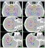
We selected one representative pediatric patient treated in our department within the past 4 years (2007-2010) with each of the following diagnoses: low grade supratentorial hemispheric glioma, high grade supratentorial hemispheric glioma, low grade brainstem glioma (biopsy-proven WHO grade 1 astrocytoma of the midbrain), right optic nerve glioma, suprasellar CNS germ cell tumor, high grade chondrosarcoma of the right sphenoid bone, suprasellar craniopharyngioma, infratentorial ependymoma (without leptomeningeal dissemination), and low grade glioma (WHO grade 1) of the infindibular stalk. Two intensity modulated radiotherapy (IMRT) treatment plans were prepared for each patient using helical tomotherapy (TomoTherapy@, Madison, Wisconsin): one plan (STD: standard) which did not apply optimization criteria to the limbic circuit (LC), hippocampus (HIP), or neural stem cell compartment (NSC), and another plan (SPA: sparing) which attempted to minimize the maximum and mean doses to these same structures For each patient, an appropriate treatment target (PTV: planning target volume) was contoured, and this PTV was applied both the STD and SPA plans. The PTV varied by diagnosis, but generally consisted of the gross tumor as identified on imaging, areas of edema or areas otherwise felt to be at risk for containing microscopic tumor (for example, the ventricular system plus a 1cm margin for CNS germinoma whole ventricular radiotherapy plans), and an additional margin for setup uncertainity on the treatment table.
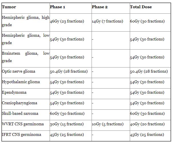
Adequate target coverage, as defined by the D95 (isodose line covering 95% of the PTV) and V100 (percent volume of the PTV receiving at least full dose/100% of the planned treatment dose), was required as the primary treatment objective in all plans (STD and SPA). The dose prescriptions/ treatment schedules for each plan type are shown in (Table 1). Also, standard constraints were applied to the following critical normal structures (OAR: organs at risk) in all plans (STD and SPA): right and left lenses, right and left eyes, right and left optic nerves, optic chiasm, pituitary/infindibulum/ hypothalamus, right and left cochleae, brainstem, and spinal cord. These standard OAR dose constraints are shown in (Table 2)

For the SPA plans, we provided additional optimization criteria to maximally spare the study OAR (LC, HIP, and NSC) by placing restrictions on the mean and maximum doses to these structures (third priority). These study OAR were spared contralaterally for the supratentorial hemispheric low and high grade glioma and skull base sarcoma plans, and bilaterally for the other plans.
For each plan the physical doses and biological equivalent doses (BED) delivered to the following structures were calculated: PTV (D95, V100, minimum dose, and maximum dose) and study OAR (LC, HIP, and NSC: meandose, maximum dose, and minimum dose). Within each tumor subgroup, delivered physical dose and BED to the PTV and study OAR were compared between the STD and SPA plans, and percent relative differences were calculated. The physical doses delivered to the standard OAR (right and left lenses, right and left eyes, right and left optic nerves, optic chiasm, pituitary/infindibulum/hypothalamus, right and left cochleae, brainstem, and spinal cord) were evaluated for each plan (STD and SPA) to ensure that they did not exceed our acceptance criteria (Table 2), but BED were not calculated and the dose delivered to these structures were not compared between the STD and SPA plans. The BED, which represents a measure of the biologic likelihood of a given dose of radiation delivered on a given treatment schedule causing a given effect on a given tissue type (tumor or normal structure) for each of these structures was calculated using the following equation, where n is the number of fractions and d is the dose per fraction in Gy:
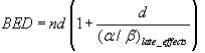
We assumed an alpha/beta (α/β) ratio of 2 for late effects involving LC and HIP. For PTV and NSC we conservatively assumed an α/β ratio of 10 because it is a value previously demonstrated for other tumors and stem cell populations [22]. The α/β ratio represents the ability of a given cellular type to repair sublethal damage to its DNA generated by radiation exposure, and is generally low (around 2-3) for tissues with little or no cellular turnover (and thus plenty of time available to repair damage before the next mitosis) such as muscle cells, fibroblasts, and neurons. The α/β ratio is high (around 10) for cells which are proliferating quickly and thus have little time available for DNA repair between mitoses, such as skin, gut epithelial cells, stem cell populations, and most tumors. No such studies have been completed for human NSC in vivo, and therefore our choice of an α/β ratio of 10 for this cellular population remains speculative.
Since this is a dosimetric comparison study we investigated whether the SPA plans increase the integral dose to the normal uninvolved brain versus the STD plans. Integral dose, expressed in joules (J), represents the total energy deposited in a given mass of tissue, and is generally represented by multiplying the delivered dose (in Gray, or joules/kg of tissue) by the mass of tissue exposed (in kg). For each plan, OAR's designated as "uninvolved brain" which contained all brain parenchyma not otherwise included in standard OAR, study OAR, or treatment targets (PTV) were generated, The integral dose, ID, was computed from differential dose volume histograms using the following equation:
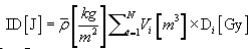
is the average physical density of the uninvolved brain, Vi is the volume in m3 of each dose voxel and Di is the dose, in Gy, in each voxel. All is the average physical density of the uninvolved brain, Vi is the volume in m3 of each dose voxel and Di is the dose, in Gy, in each voxel. All these values are easily extracted from dose volume histograms. Using an average density instead of a voxel specific density in Equation 2 is warranted since the brain density is rather uniform, which is not the case in highly heterogeneous regions such as lung. The integral dose can be expressed as a single value or as a dose-ID histogram d-IDh.
Dosimetric coverage of the treatment target (PTV) was excellent in all STD and SPA plans, with 94.8-96% of PTV receiving full dose in STD plans and 4.9-95% receiving full dose in SPA plans. However, there was greater dose inhomogeniety noted in the SPA plans, with minimum doses 56 to 99% (mean 90%) and maximum doses 101 to 128% (mean 109%) of prescription dose. The corresponding ranges for the STD plans were to 81 to 99% (mean 92%) minimum doses and 101 to 120% (mean 105%) maximum doses relative to the prescription dose. All plans (STD and SPA) were able to meet the dose constraints for all standards OAR as described in (Table 2) (individual plan data not shown).
SPA plans were able to significantly reduce mean physical dose and BED delivered to the study OAR (LC, HIP, and NSC) in all cases: percent reduction in mean physical dose 5.4 to 78.8 (mean 44) and percent reduction in mean BED 7 to 81.5 (mean 47.9). The corresponding percent reduction in mean physical dose and BED for the limbic circuit, hippocampus, and neural stem cell compartment were 5.4 to 77.8 (mean 43.3) and 7 to 80.3 (mean 47.2), 18.2 to 67.4 (mean 46.5) and 25.4 to 81.5 (mean 52.4), and 6.8 to 60 (mean 42.1) and 7.8 to 66.1 (mean 44.1), respectively. In most cases the minimum and maximum physical doses and BED delivered to the study OAR were also reduced in the SPA, although in a some cases the minimum physical dose and BED were higher (craniopharygioma and optic nerve glioma plans: LC absolute minimum physical dose increased by .05 to .1 Gy, mean 0.8 Gy) while in others the maximum physical dose and BED were higher (IFRT, WVRT, high grade glioma, low grade glioma, and craniopharyngioma plans: absolute maximum physical dose increased by .63 to 8.6 Gy, mean 2.5 Gy) for the SPA plan despite a lower mean physical dose and BED, evidence of greater dose inhomogeneity within the study OAR for the SPA plans.
Integral dose (J) delivered to the uninvolved brain was reduced in the SPA plans as compared to the STD plans by a mean of 7.6% (range -19.3% to +0.3%). The greatest reduction in integral dose was noted in the high grade glioma SPA plans (19.3% reduction), The only treatment plan type in which integral dose was increased with sparing techniques was WVRT (0.3% increase in SPA plan versus STD plan) only treatment plan type in which integral dose was increased with sparing techniques was WVRT (0.3% ncrease in SPA plan versus STD plan).
Cranial radiotherapy plays an important role in the treatment of a number of primary pediatric intracranial tumors [1-5]. In the case of CNS germinoma and brainstem glioma, cranial radiotherapy is a standard primary treatment modality, and studies in the setting of CNS germinoma which have attempted to exclude radiotherapy as a component of treatment have shown significantly inferior results [6-8,10].
Unfortunately, the use of cranial radiotherapy in children results in a number of adverse late sequelae include cognitive dysfunction, endocrinologic dysfunction, and vascular damage [13-15]. The cognitive dysfunction can be profound, with St. Jude Children's Hospital and others finding a direct correlation between the dose administered and a decline in overall IQ [14,23-26]. In the St. Jude study, the factors that seem to correlate most strongly were younger age at time of treatment, longer time interval since treatment, female sex, presence of hydrocephalus, higher volume of supratentorial brain irradiated, and higher radiation dose to the supratentorial brain [26]. They also found that irradiation of the supratentorial compartment and temporal lobes resulted in significant declines in IQ regardless of the dose exposure, with each Gy of exposure having a similar impact on declines in IQ [23]. The cognitive deficits seen after treatment are predominantly the inability to develop new skills and process new information, rather than loss of previously acquired function and memories [14].
Changes in fractional anisotropy (FA) on diffusion tensor imaging (DTI) MRI provide evidence of damage to white matter pathways, and these changes can be seen in pediatric patients who have been treated with radiotherapy for medulloblastoma and surgical resection for cerebellar astrocytomas, with one recent study showing a mean reduction in FA of 16.5%in treated patients versus controls [27-29]. These reductions in FA were found to correlate with a younger age at the time of treatment and declines in school performance [28]. Rueckriegel et al. found that supratentorial changes in FA were more prominent in patients treated with radiotherapy and surgical resection than with surgery alone, although the distribution of deficits was similar. Interestingly, the location of most of the changes as identified in (Figure:1 & 2) of their paper lie within the hippocampus, limbic circuit, or neural stem cell compartment [29].
Johannesen and colleagues have shown in a retrospective review of MRI studies from a group of adult patients previously treated with cranial radiotherapy (median dose 54 Gy) that doses of 29.2 Gy or above are associated with grade 3 white matter changes on MRI T2 and FLAIR sequences and worse neurocognitive outcomes and patient-reported quality of life, while doses in the range of 12.5-27.5 Gy delivered to the contralateral hemisphere were not associated with such changes [30]. This study, although performed in adult patients, is consistent with the findings from the group at St. Jude's which found that the percent volume of pediatric supratentorial brain irradiated to varying dose levels (0-20Gy, 20-40Gy, 40-65Gy) correlated with IQ level after cranial irradiation [24].
Since the total dose delivered to the brain in the treatment of primary pediatric brain tumors exceeds this threshold of 20-27.5Gy (Table 1:), it would follow that reduction of dose to non-target regions of the brain in children should improve imaging and clinical outcomes [29-30].
Several investigators have demonstrated the feasibility of sparing NSC, limbic circuit, and/or hippocampus in adults during the administration of partial brain radiotherapy (PBRT) for glioma and whole brain radiotherapy (WBRT) [19,21,32-34]. The Radiation Therapy Oncology Group (RTOG) is currently accruing patients to a phase II study (RTOG 0933) which aims to demonstrate the feasibility of sparing the hippocampus during the administration of whole brain radiotherapy. This study will incorporate baseline and follow up neurocognitive testing to assess the impact of hippocampal sparing on memory and other cognitive domains after treatment [RTOG.org].
Cranial irradiation also produces damage to the hypothalamicpituitary axis, particularly in children at doses as low as 18Gy [13,35-38]. This study was not designed to specifically evaluate dosimetric sparing of the pituitary-hypothalamic axis, but we are able in all plans (STA and SPA) to meet our planning objectives for the hypothalamic-pituitary axis.
Thus, efforts directed toward dosimetrically sparing the study OAR did not compromise dosage to the pituitary-hypothalamic axis.
In the current study, we have demonstrated the feasibility of sparing the limbic circuit, hippocampus, and neural stem cell compartment, with mean physical dose and BED to each structure reduced 44% and 47.9%, respectively. In most cases we selected these structures bilaterally for sparing, but in the hemispheric glioma and skull base sarcoma plans we elected to spare these structures contralaterally as they could not be spared ipsilaterally due to the proximity of the PTV to the ipsilateral study OAR. We anticipate that these patients (those with the study OAR spared contralaterally only) will still derive a late cognitive benefit based on the available literature detailing the cognitive outcomes for patients who have undergone surgical temporal lobectomy for treatment of tumor or intractable epilepsy [40-42]. Such patients rarely have persistent cognitive deficits provided that the resected medial temporal lobe structures are diseased and the remaining medial temporal lobe structures are normal, suggesting that the remaining structures can compensate for any transient deficits sustained from the surgical procedure [40-42].
We believe that damage to the critical study OAR in this study (LC, HIP, NSC) is the principal cause of late neurocognitive deficits in both adult and pediatric patients, and our sparing is based around this assumption. However, others have suggested that low dose radiation exposure to the whole brain produces (or at least contributes) to these late adverse effects [43-44]. This theory suggests that it is reduction of the integral/overall dose to the brain which will ultimately provide cognitive protection. Investigators from Brazil has demonstrated the ability of IMRT to reduce the high dose regions and integral dose to the brain during the delivery of WVRT for primary CNS germinoma [31]. We similarly found in this study that the use of Tomotherapy IMRT reduced the integral dose delivered to the uninvolved brain by a mean of 7.6%, with all plan types showing benefit except for the WVRT plans, in which sparing techniques increased integral dose by 0.3%. This reduction in integral dose to uninvolved brain might also reduce the incidence of secondary tumor induction in this at-risk patient population.
Recently concern has been expressed over the use of intensity-modulated radiotherapy (IMRT) in the setting or cranial irradiation, since more total monitor units (MU) are required to deliver a given dose with this treatment modality, resulting in greater integral dose being delivered to the patient [45-47]. This finding has been shown in some but not all dosimetric studies comparing IMRT to either conventional/2-D or 3-D conformal treatment planning, with some studies showing a higher ID delivered to the brain and other showing a lower ID[45-51]. Reduction of ID should, in theory, reduce the risk of late second malignancies and cognitive dysfunction, although this has not been conclusively proven [45-46].
IMRT also produces more inhomogeneous dose distributions than conventional or 3-D conformal radiotherapy plans, with greater hot and cold spots (areas receiving greater than and less than prescription dose, respectively). This issue was noted in our treatment planning study, in which hot spots within the PTVs were in some cases >120% of presecription dose. While ideally these hot spots will be positioned within the tumor rather than within normal tissue, there is some concern that hot spots in normal brain may increase the risk for late adverse effects such as radionecrosis. For example, the commonly accepted TD5/5 (the dose which will result in a 5% risk of adverse events at 5 years in a given tissue) for normal partial brain is 60Gy [52]. Therefore, in the context of IMRT treatment planning for intracranial malignancies it would be prudent to minimize hot spots to the extent possible, and if possible to have them located within tumor rather than normal brain.
Also, since most recurrences of glioma (high and low grade) occur at or within 2cm of the original site of disease after resection and/or radiotherapy, we do not believe that our cognitive sparing approach will increase the risk of relapse for these patients, as we did not compromise definition or dosimetric coverage of our treatment targets (Table 1: and el) [56].
Another important approach to normal tissue sparing in the setting of cranial radiotherapy for pediatric brain tumors is the use of proton therapy [57-69]. Investigators at several institutions have performed dosimetric studies comparing the dose delivered to normal tissues with proton therapy as compared to IMRT and/ or conventional radiotherapy, and have consistently shown a reduction in dose to critical normal tissues favoring proton therapy [57,60,64-65]. Proton therapy has also been shown to reduce the integral dose to the body when compared with IMRT, and this reduction in integral dose is expected to result in a lower rate of secondary tumor induction after treatment [66-69]. This is a particularly important issue in children, and the use of IMRT (including helical tomotherapy) in this context, with its associated higher total body integral dose (due to a higher number of monitor units [MU] and higher leakage dose required to deliver a given dose of therapeutic radiation), should be approached with caution [67-68]. Importantly, no prospective randomized trials have been performed comparing proton therapy versus IMRT clinical outcomes in terms of either tumor control or late effects in the setting of adult or pediatric primary tumor treatment.
We believe that the cognitive sparing approach detailed in this study and our previous studies should be implemented in the setting of a prospective clinical trial [19,21,39]. Formal neurocognitive data should be collected at baseline and following treatment to assess the functional outcome for these patients, and these results should be compared with those of either a control group treated prospectively without this approach or a historical control group with adequate follow up and neurocognitive data outcomes. Without such data, it will not be possible to properly assess the relative benefits of our approach.
It is dosimetrically possible to reduce physical dose and implicitly BED to the limbic circuit, hippocampus, and neural stem cell compartment during the administration of partial brain radiotherapy for the treatment of multiple types of pediatric primary intracranial tumors. Such treatment does not compromise dosimetric coverage of the treatment target or compromise dosimetric sparing of other critical normal structures including the pituitary-hypothalamic axis. Our cognitive sparing approach reduces integral dose to normal when compared to standard approaches in most cases, and should reduce the late adverse cognitive effects of radiotherapy in children, but needs to be studied in the context of a prospective clinical trial with formal evaluation of neurocognitive outcomes.
Dear Editorial Team, Clinical Medical Reviews and Reports. My experience with the journal was highly positive. The peer-review process was rigorous, constructive, and completed in a timely manner. The reviewers provided valuable comments that helped improve the quality and clarity of our manuscript. The editorial office was professional, responsive, and supportive throughout all stages of the publication process. Communication was clear and efficient, and any questions were addressed promptly. Overall, I found the journal to maintain high scientific standards and an excellent publication workflow. I would be pleased to consider submitting future work to this journal. Best wishes from, Elena Popa.

It was my pleasure to submit my testimonial concerning the Reviewer Board of our Scientific Journal “Brain and Neurological Disorders”. The Reviewers focused on some modifications and their contribution was helpful. The ladies of our Editorial Office were also supported my efforts. It was my honor to have such a co-operation and I am looking forward for more collaboration.

Dear Grace Pierce, Editorial Coordinator of Journal of Clinical Research and Reports, Thank you for the speedy and efficient peer review process. I appreciate the fact that your peer reviewers do not take months to respond like with some other journals. I would also like to thank the editorial office for responding quickly to my questions. It is an excellent journal. I plan to submit more manuscripts in the future. Best wishes from, Robert W. McGee

Dear Grace Pierce, Editorial Coordinator of Journal of Clinical Research and Reports, Working with you and your team on our recent publication in JCRR has been a truly wonderful and enjoyable experience. The responses were prompt, and the reviewers were patient, constructive, and highly professional. One reviewer in particular gave me the feeling that a professor was carefully reading and commenting on my coursework, which was deeply touching. The entire process was straightforward and hassle‑free, with no tedious online forms to complete. I highly recommend this journal. Best wishes from, DR Aibing Rao, Head of R&D

I Appreciate the Opportunity to Share my Experience with the Journal of Clinical Research and Reports. The peer review process was timely and constructive, and the feedback provided helped improve the quality of our manuscript. The editorial office was professional, responsive, and supportive throughout the process, ensuring smooth communication and efficient handling of the submission. Overall, it was a positive experience collaborating with your team.

Dear Mercy Grace, Editorial Coordinator of Obstetrics Gynecology and Reproductive Sciences, We would like to express our gratitude for your help at all stages of publishing and editing the article. The editors of the magazine answer all the necessary questions and help at every stage. We will definitely continue to cooperate and publish other works in the Obstetrics Gynecology and Reproductive Sciences! Best wishes from, Alla Konstantinovna Politova,
