
Research Article | DOI: https://doi.org/10.31579/2692-9392/169
1 Green Land Landscaping and Gardening, Seedling Growth Laboratory, 86130-000, Parana, Brazil.
2 Department of Marine Science, University of Calcutta, 35 B. C Road, Kolkata, 700019, West Bengal, India.
3 Department of Oceanography, Techno India University, West Bengal, EM 4 Salt Lake, Sector V, Kolkata 700091, India.
*Corresponding Author: Ricardo Gobato, Green Land Landscaping and Gardening, Seedling Growth Laboratory, 86130-000, Parana, Brazil.
Citation: Ricardo Gobato, Abhijit Mitra and Sufia Zaman, (2023), Exobiological Silicon-Based DNA 1bna, Archives of Medical Case Reports and Case Study, 7(2); DOI:10.31579/2692-9392/169
Copyright: © 2023, Ricardo Gobato. This is an open access article distributed under the Creative Commons Attribution License, which permits unrestricted use, distribution, and reproduction in any medium, provided the original work is properly cited.
Received: 14 March 2023 | Accepted: 21 March 2023 | Published: 28 March 2023
Keywords: ASi; CSi; GSi; TSi; hartree-fock method; mm (molecular mechanics); macromolecule; g-quadruplex dna; clusters; exobiological
The researchers have been investigated as a possible alternative for building up biological molecules in exobiology. The world's most famous molecule - the DNA double helix - sometimes doubles up again. Researchers have now found this quadruple-stranded form in healthy human cells for the first time. Speculate that the quadruplex structure forms to hold the molecule open and facilitate the reading of the genetic code and thus the production of proteins. G-quadruplex DNA is a four-stranded structure made that can form a 'knot' in the DNA of living cells. In these terms, it was analyzed through computational calculations, via MM (Molecular Mechanics), and by ab initio Restricted Hartree-Fock (RHF) method, on a simple STO-3G (Slater-type orbitals with 3 Gaussians) basis, the possibility of a DNA macromolecule based on Silicon. From the basic structure of 1bna, however, you assume conditions without the presence of Carbon, replacing it with Silicon. It was obtained a cluster of G-quadruplex DNA, forming a cocoon, with great possibility for the pharmaceutical industry in capturing molecules foreign to human DNA
The world's most famous molecule - the DNA double helix - sometimes doubles up again. Researchers have now found this quadruple-stranded form in healthy human cells for the first time. [1]
Four-stranded DNA has been seen before in some cancer cells and in lab-based chemistry experiments, but this is the first time the molecule has been visualized in healthy, living human cells, as a stable structure created by normal cellular processes. [1]
The researchers speculate that the quadruplex structure forms to hold the molecule open and facilitate the reading of the genetic code and thus the production of proteins. It may also influence the amount of each protein that is made. [1]
Usually, this function is performed by epigenetic markers - chemical tags on DNA that increase or decrease the activity of genes - and it seems that the quadruplex form of DNA has a similar role. [2]
In Figure (1) is represented the image of the DNA 1bna. Name: DNA (5'-D(*CP*GP*CP*GP*AP*AP*TP*TP*CP*GP*CP*G)-3'); Representative chains: A, B; Source organism: Synthetic construct; Length: 12 nucleotides; Theoretical weight: 3.66 KDa. [3] Source: [3].

Figure 1: Image of the DNA 1bna. Name: DNA (5'-D(*CP*GP*CP*GP*AP*AP*TP*TP*CP*GP*CP*G)-3'); Representative chains: A, B; Source organism: Synthetic construct; Length: 12 nucleotides; Theoretical weight: 3.66 KDa.
Not all DNA is in the double helix form described in textbooks since the 1950's. Quadruple helix DNA, also known as G-quadruplex DNA [4, 5, 6], was identified in the human genome several decades ago, and extensively studied since then. Each cell in our body contains a copy of our DNA code made up of A, C, G and T, providing the blueprint for the organization and function of our bodies. Normally these letters combine in pairs to form the well-known double helix shape. However, some small lengths of DNA can exist as alternative shapes, which can affect how the instructions of the DNA code is 'read'. [7]
G-quadruplex DNA [4, 5, 6] is a four-stranded structure made that can form a 'knot' in the DNA of living cells. In these structures, many Gs on the same stretch of DNA stick to each other instead of forming pairs between two strands. G-quadruplex DNA is known to be capable of forming many different types of structural shapes, yet little is understood about the factors controlling formation of these motifs. [7]
However, since G-quadruplex DNA structures have been observed to play an important role in 'reading' of genes involved in development of human diseases, researchers expect that the variety of shapes they form will have an effect on when and how the DNA is read. [7]
G-quadruplexes are found in higher concentrations in cancer cells, so are thought to play a role in the disease. The probes reveal how G-quadruplexes are 'unwound' by certain proteins, and can also help identify molecules that bind to G-quadruplexes, leading to potential new drug targets that can disrupt their activity. [8]
On the basis in chemical evolutionary theory, it is implicit that life is being based upon carbon chemistry. The possibility of life based on silicon has been discussed extensively (though casually). Theoretical chemical arguments have been proposed to support this presumption. [9]
The most significant result would be to and some type of living matter radically different from that of the Earth. One might cite under this category supposed organisms with a structure and metabolic machinery based on silicon rather than on carbon; or forms with an ammonia based rather than a water based machinery and metabolism. (One should note in the former case, however, that fully aerobic silicon metabolizers would be required to exhale quartz.) [9]
Exclude the noble gases from consideration because of their inertness; the four most abundant elements of the universe are hydrogen, oxygen, carbon, and nitrogen. In fact, hydrogen is the major constituent of the universe; oxygen, carbon, and nitrogen are each about ten times more plentiful than the next most abundant element, silicon. [9]
In comparing the carbon and silicon has: the Si lies in the same column of the periodic table of the elements, and it has been investigated as a possible alternative for building up biological molecules in exobiology. [10, 11, 12, 13, 14, 15]
The option for simple replacement of carbon by silicon [16, 17] is due to the peculiar characteristics between both. Atomic interactions under non-carbon conditions were studied, with only the Hydrogen, Silicon, Nitrogen and Oxygen atoms, in STP (Standard Temperature and Pressure), for the four standard bases of DNA, A, C, G and T, thus obtaining by quantum chemistry four new compounds, named here as: ASi, CSi, GSi and TSi. [18, 19, 20, 21, 22, 23]
If elements other than carbon constitute the building blocks for any living system on other worlds, they almost surely exist under conditions far different from those on Earth, including temperatures and pressures where water could not be the solvent. Titan provides the best natural laboratory in our Solar System for investigating this possibility. [18, 19, 20, 21, 22, 23]
Through the chemical abundances of biological elements in the earth crust, terrestrial life has chosen carbon instead of silicon, in spite of the larger abundance of silicon. This fact suggests that carbon is better suited to form biological molecules. [10, 24]
However, this paper assumes conditions without the presence of Carbon.
For calculations the computer used for was a Desktop with SUSE Linux Enterprise Desktop [25], AMD Ryzen 7 1800X processor [26], ASUS [27] Prime A320M-K motherboard, 16GB of RAM, with 500GB SSD [28].
The set of programs HyperChem Molecular Modeling System, Evaluation Version [29]; GAMESS (Computational chemistry software program and stands for General Atomic and Molecular Electronic Structure System [30, 31]; and CHARMM27 [32, 33] were used.
Using the basic structure of DNA 1bna [3] all carbon atoms were replaced by silicon atoms. With the new silicon-based structure, [1bna]Si, molecular dynamics was performed to minimize the energy of the structure. Therefore, the basic strand of the 1bna double helix composed of ACTG [1, 8], became [ACTG]Si [18, 19, 20, 21, 22, 23].
After a molecular dynamics using the HyperChem software [29] via Molecular Mechanics \citep{McD}, with Charmm27 force field [32, 33] and RHF [30, 31], a complete change in the structure of the double helix was verified. With this new double helix, a new double helix was added, leaving it with a quadruple helix, according to the new structure.
Figures (2-10) show the four structures of the silicon-based 1bna DNA macromolecule after replacing all carbon atoms with silicon and the molecular dynamics with calculations via MM and RHF, which will be called [1bna]Si.
Figures (2), (4), (6) and (8) show the four structures of [1bna]Si DNA macromolecule. The Figure represents a first structure obtained after molecular dynamics, for the silicon-based 1bna DNA molecule, in 2D contours of its electrostatic potential.
Figures (3), (5), (7) and (9) show the four structures of image [1bna]Si DNA macromolecule. The Figure represents a first structure obtained after molecular dynamics, for the [1bna]Si DNA molecule, in 3D Mapped Isosurface with the distribution of electrical charges. Represented in color variation from pink (positive charges) to green (negative charges), in charge units.
The individualized representation of each of the four [1bna]Si structures was necessary for an analysis of the electrostatic potential distribution, Figures (2, 4, 6 and 8) and the density of 3D electric charges in each of them, Figures (3, 5, 7, to 9).
It is verified that there is a planarity of the electrostatic potential in the cluster Figure (10) formed by the four macromolecules [1bna]Si, occurring a symmetry of the electrostatic potential represented in 2D. Source: [Authors].

Figure 2:Image of [1bna]Si DNA macromolecule. The Figure represents a first structure obtained after molecular dynamics, for the silicon-based 1bna DNA molecule, in 2D contours of its electrostatic potential. Source: [Authors].

Figure 3:Image of [1bna]Si DNA macromolecule. The Figure represents a first structure obtained after molecular dynamics, for the [1bna]Si DNA molecule, in 3D Mapped Isosurface with the distribution of electrical charges. Represented in color variation from pink (positive charges) to green (negative charges), in charge units.
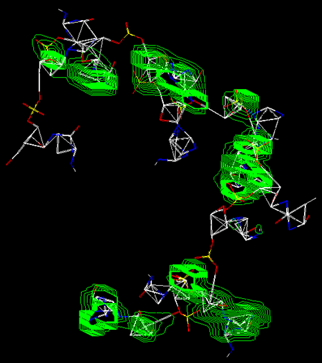
Figure 4: Image of [1bna]Si DNA macromolecule. The Figure represents a second structure obtained after molecular dynamics, for the [1bna]Si DNA molecule, in 2D contours of its electrostatic potential.

Figure 5:Image of [1bna]Si DNA macromolecule. The Figure represents a second structure obtained after molecular dynamics, for the [1bna]Si DNA molecule, in 3D Mapped Isosurface with the distribution of electrical charges. Represented in color variation from pink (positive charges) to green (negative charges), in charge units.
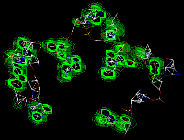
Figure 6: Image of [1bna]Si DNA macromolecule. The Figure represents a third structure obtained after molecular dynamics, for the [1bna]Si DNA molecule, in 2D contours of its electrostatic potential.
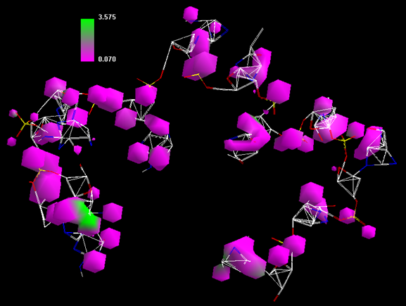
Figure 7: Image of [1bna]Si DNA macromolecule. The Figure represents a third structure obtained after molecular dynamics, for the [1bna]Si DNA molecule, in 3D Mapped Isosurface with the distribution of electrical charges. Represented in color variation from pink (positive charges) to green (negative charges), in charge units.
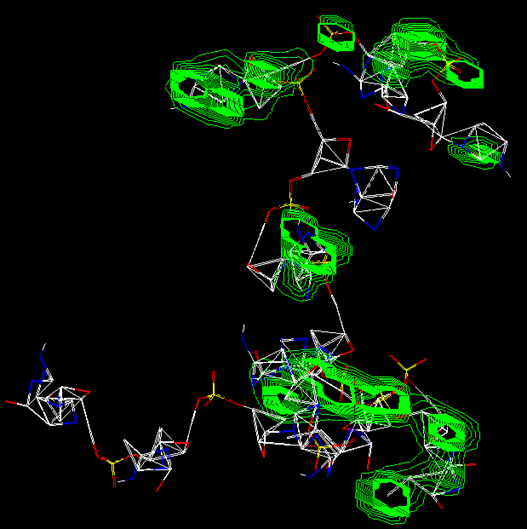
Figure 8: Image of [1bna]Si DNA macromolecule. The Figure represents a fourth structure obtained after molecular dynamics, for the [1bna]Si DNA molecule, in 2D contours of its electrostatic potential.

Figure 9: Image of [1bna]Si DNA macromolecule. The Figure represents a fourth structure obtained after molecular dynamics, for the [1bna]Si DNA molecule, in 3D Mapped Isosurface with the distribution of electrical charges. Represented in color variation from pink (positive charges) to green (negative charges), in charge units.}

Figure 10: Image of [1bna]Si DNA macromolecule. The Figure represents a structure obtained after molecular dynamics, for the [1bna]Si DNA molecule, in 2D contours of its electrostatic potential.
The study has so far been limited to computational Molecular Mechanics methods, as well a via ab initio RHF in the set of basis used ECP minimal basis, on a STO-3G simple basis set. The results are compatible with the theory of quantum chemistry, for simple functionals but a deeper study is necessary to verify the real conditions for the formation of such a macromolecule. Also a proof experimental verification depends on advanced techniques for their synthesis, obtaining in laboratory for experimental biochemical.
It was obtained a cluster of G-quadruplex DNA, forming a cocoon, with great possibility for the pharmaceutical industry in capturing molecules foreign to human DNA.
Dear Editorial Team, Clinical Medical Reviews and Reports. My experience with the journal was highly positive. The peer-review process was rigorous, constructive, and completed in a timely manner. The reviewers provided valuable comments that helped improve the quality and clarity of our manuscript. The editorial office was professional, responsive, and supportive throughout all stages of the publication process. Communication was clear and efficient, and any questions were addressed promptly. Overall, I found the journal to maintain high scientific standards and an excellent publication workflow. I would be pleased to consider submitting future work to this journal. Best wishes from, Elena Popa.

It was my pleasure to submit my testimonial concerning the Reviewer Board of our Scientific Journal “Brain and Neurological Disorders”. The Reviewers focused on some modifications and their contribution was helpful. The ladies of our Editorial Office were also supported my efforts. It was my honor to have such a co-operation and I am looking forward for more collaboration.

Dear Grace Pierce, Editorial Coordinator of Journal of Clinical Research and Reports, Thank you for the speedy and efficient peer review process. I appreciate the fact that your peer reviewers do not take months to respond like with some other journals. I would also like to thank the editorial office for responding quickly to my questions. It is an excellent journal. I plan to submit more manuscripts in the future. Best wishes from, Robert W. McGee

Dear Grace Pierce, Editorial Coordinator of Journal of Clinical Research and Reports, Working with you and your team on our recent publication in JCRR has been a truly wonderful and enjoyable experience. The responses were prompt, and the reviewers were patient, constructive, and highly professional. One reviewer in particular gave me the feeling that a professor was carefully reading and commenting on my coursework, which was deeply touching. The entire process was straightforward and hassle‑free, with no tedious online forms to complete. I highly recommend this journal. Best wishes from, DR Aibing Rao, Head of R&D

I Appreciate the Opportunity to Share my Experience with the Journal of Clinical Research and Reports. The peer review process was timely and constructive, and the feedback provided helped improve the quality of our manuscript. The editorial office was professional, responsive, and supportive throughout the process, ensuring smooth communication and efficient handling of the submission. Overall, it was a positive experience collaborating with your team.

Dear Mercy Grace, Editorial Coordinator of Obstetrics Gynecology and Reproductive Sciences, We would like to express our gratitude for your help at all stages of publishing and editing the article. The editors of the magazine answer all the necessary questions and help at every stage. We will definitely continue to cooperate and publish other works in the Obstetrics Gynecology and Reproductive Sciences! Best wishes from, Alla Konstantinovna Politova,
