
Case Report | DOI: https://doi.org/10.31579/2690-4861/229
1 Department of High risk pregnancy and Perinatology.
2 Department of Obstetrics and Gynaecology.
3 Department of Hematology.
4 Department of Rheumatology, KIMS Health, Trivandrum.
*Corresponding Author: Nina Navakumar, Department of High risk pregnancy and Perinatology.
Citation: Navakumar N, Retnamma V, Rafeekha P, Ranganayaki V, Thomas M, Mahendran B . (2022) Evans Syndrome in pregnancy – Case report of two successful pregnancies in a woman; Review of Literature. A Case Report. International Journal of Clinical Case Reports and Reviews. 11(4); DOI: 10.31579/2690-4861/229
Copyright: © 2022 Nina Navakumar, This is an open-access article distributed under the terms of the Creative Commons Attribution License, which permits unrestricted use, distribution, and reproduction in any medium, provided the original author and source are credited
Received: 20 May 2022 | Accepted: 25 June 2022 | Published: 30 June 2022
Keywords: evans syndrome; autoimmune haemolytic anaemia; immune thrombocytopenia
Evans syndrome is a rare autoimmune disease in which an individual's antibodies attacks the body's own red Autoimmune blood cells and platelets. There is a coexistence of Immune thrombocytopenia (ITP) with haemolytic anaemia (AIHA) with immune neutropenia sometimes in the absence of known underlying etiology.1Association of Evans syndrome with pregnancy is very rare, and only a few cases have been published in medical literature. No definite treatment protocols are defined. Treatment options during pregnancy are further limited due to concerns of teratogenic effect of pharmacological agents. Evans syndrome can be diagnosed with a full blood count film and a direct Coombs test. We describe here a rare case that was diagnosed as secondary Evans syndrome with SLE complicating pregnancy that resulted in two live births in a woman. We have also briefly discussed the pathophysiology, clinical features, diagnosis and the possible treatment options and outcome of Evans syndrome in pregnancy
Evans syndrome is an uncommon condition characterized by Immune thrombocytopenia (ITP) and Autoimmune haemolytic anaemia (AIHA) that can coexist or one follows the other and it is diagnosed with a positive Direct antiglobulin test (DAT). The underlying aetiology is unknown. It runs a chronic course with frequent remissions and relapses 1. It has a more benign state during pregnancy than in non-pregnant state. The foetal outcome may become less favourable as it is affected by transplacental passage of antibodies. It is suggested that Evans syndrome may be a stage of a broader spectrum, generalized immune dysregulation due to high incidence of quantitative serum immunoglobulin abnormalities, lymphoid hyperplasia and associated systemic manifestations.
Dr. Robert Evans was the first one to describe the syndrome in 1951, and the first case of its occurrence during pregnancy was published in 1966. Since then very few cases of Evans syndrome during pregnancy have been reported.
Thirty year old second gravida ; case of Evans syndrome (Gravida 2 Para 1 Live 1 ) booked at 26 weeks gestation managed with a multi-disciplinary team consisting of senior Obstetrician, Haematologist ,Rheumatologist and Maternal – fetal medicine specialist .
She had easy bruising and bleeding gums at 16 years of age with menorrhagia which on investigation swas found to have severe anemia and thrombocytopenia (platelet count of 13000 cells/mm3) which was corrected with multiple transfusion of packed red cell and platelet concentrate. On further evaluation her DAT and positive anti SS-A Ab (autoimmune association). Bone marrow study showed megakaryocytes proliferated with normal hypo lobulated and micro megakaryocytes consistent with ITP. She was managed with pulse dose of intravenous methylprednisolone 1 gm for three days followed by oral Prednisolone 60 mg which improved her Hb and platelet count. She was on irregular follow up.
She was booked with us at 25 weeks in her first pregnancy. She had recurrent episodes of syncope, seizure like activity and loss of consciousness at 26 weeks of gestation for which neurology evaluation was done and found to have a normal electroencephalogram. She was started on oral levetiracetam. At booking haemoglobin was 8.8 gm g/dl, TC of 12000 cells/mm3 and platelet count of 38000 cells/mm3 . Red blood cell indices and serum ferritin were low; peripheral smear showed microcytic hypochromic anaemia with thrombocytopenia. DCT was
negative with a normal lactate dehydrogenase ( LDH ) with positive anti SS-A Ab . Antiphospholipid ( APLA ) work up showed positive lupus anticoagulant ( LA ) with prolonged activated partial thromboplastin time ( APTT )
She was managed as iron deficiency anemia and secondary ITP . She was started on oral iron therapy. Her haemoglobin improved to 12.3 gm/dl with a platelet count of 97000 cells / mm. She developed purpuric rash at 36 weeks with a platelet count of 19000 cells/ mm3 . She was started on oral prednisolone 60 mg od and platelet improved to 38000 cells / mm3. She had induction of labor at 39 weeks of gestation which ended in an emergency LSCS for failed induction with male baby of weight 2.88 kg . She was lost to follow up postpartum .Baby ‘s haemo globin was 15.1 gm / dl TC being 10000 cell/mm3 with Platelet count of 263 cell/ mm.
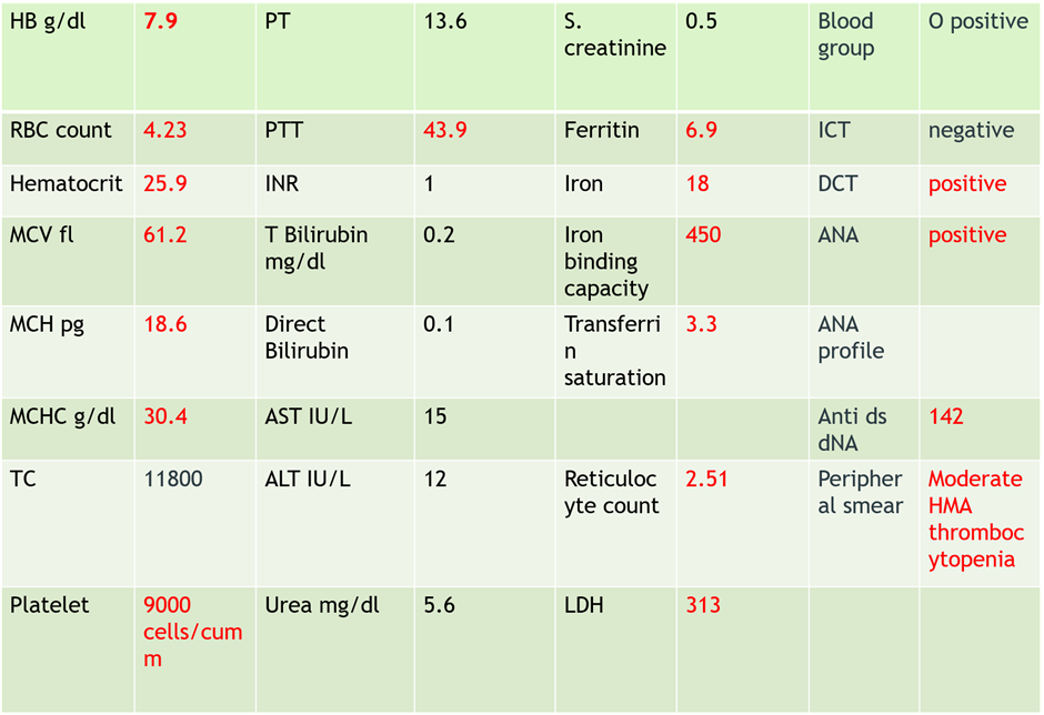
In her second pregnancy she was booked at 26 weeks of gestation . Her lab parameters(Table:1) were suggestive of autoimmune hemolytic anemia ,iron deficiency anemia and thrombocytopenia and was started on oral prednisolone 40 mg , oral iron therapy and folic acid . She was lost to follow up and discontinued steroid herself.
She reported back at 38 weeks of gestation with pain abdomen and decreased fetal movements. She was hemodynamically stable and ultrasound examination showed fetal growth restriction (3rd centile) with oligamnios. Middle cerebral artery peak systolic velocity ( MCA PSV ) was normal and there was no evidence of fetal anemia. Hb was 10.8 gm /dl with a platelet count of 26000 cells / mm3 and was started on oral prednisolone Mean while she had one episode of tonic clonic convulsion with frothing from mouth and up rolling of eyes which was controlled with IV levetiracetam 1 gm and loading dose of magnesium sulphate 4 gm IV followed by 1g / hour. This episode was followed by fetal bradycardia up to 60 for 5 minutes. She had category 1 LSCS under general anaesthesia and delivered a male baby of weight 1.82 kg with normal Apgar score. Her blood pressure during the intra operative period was 160/110 mm of Hg which was bought down with intravenous labetalol. She was given 4 units of random donor platelet transfusion one unit of single donor platelet transfusion
She was given tranexamic acid 1 gm IV 3 doses every 8 hours , Hydrocortisone 100 mg IV 3 doses every 8 hours as stress dose intraoperatively and postpartum. She was electively ventilated post operatively until neuroimaging and was extubated on day1. EEG showed generalized interictal epileptiform discharge with normal magnetic resonance imaging brain ( MRI ) with magnetic resonance venogram ( MRV ). Maternal echo cardiogram and arterial blood gas were normal. She was continued on IV levetiracetam 500 mg IV twice daily and IV magnesium sulphate for 24 hours postpartum. Seizure could be primary seizure disorder with differential diagnosis of eclampsia or neurological manifestation of autoimmune disease. Her immediate platelet count was 34000 cells / mm3 ;hence low molecular weight heparin was not started but intermittent pneumatic compression stockings were given for thromboprophylaxis.
On second postoperative day her BP was 120/80 mm of Hg , Hb - 9.7 g/ dl , Platelet – 70000 cells/ mm3 and started on Enoxaparin 40 mg s/c once daily. Rheumatologist started her on Tacrolimus 0.5 mg two times a day , hydroxychloroquine 200 mg once daily . On fifth postoperative day she was discharged on Tacrolimus 0.5 mg two times a day , levetiracetam, oral prednisolone, hydroxy chloroquine. She was reviewed after 2 weeks with haemoglobin of 12.5 gm / dl and platelet count of 18000 cell/ mm 3 and continued on prednisolone.
Neonate had a Hb of 16.6 gm / dl , TC – 7800 cells/mm3 and platelet of 27000 cell / mm3.Baby was transfused with one unit of random donor platelet which improved the platelet to 1.54 lakhs cell / mm3. Neurosonogram of the baby was normal 15-33% risk of venous thrombo embolism. Thrombocytopaenia is due to antibodies usually IgG against platelet surface glycoproteins, especially Ib/IX, IIb/IIIa.

Laboratory studies that may be considered include the following:
Complete blood count (CBC) , Reticulocyte count , Peripheral blood smear , Coombs test (direct antiglobulin test) , Tests for antierythrocyte, antineutrophil, and antiplatelet antibodies , Lupus antibody (lupus like inhibitor) and antinuclear antibody (ANA) tests. Thus, autoantibody testing for platelets and granulocytes may be positive but a negative result does not exclude the diagnosis and routine testing at presentation may not be helpful. [4]
Features of haemolysis should be sought including a raised reticulocyte count, unconjugated hyperbilirubinaemia and decreased haptoglobins. The direct antiglobulin test (DAT) is almost invariably positive (although often weakly so), even in the absence of haemolytic anaemia, and may be positive for IgG and/or complement (C3) 5. Bone marrow aspiration helps reveal aplastic anemia or an infiltrative disorder. It is usually indicated for excluding infiltrative processes in patients who present with pancytopenia. Otherwise it is not usually helpful as the findings are nonspecific and may be normal or show trilineage increased cellularity. 1It is advisable to measure serum immunoglobulins and immunoglobulin subclasses in all patients; not only to exclude differential diagnoses, such as common variable immunodeficiency (CVID) and IgA deficiency, which have been reported to develop acquired cytopenias, and also as a baseline prior to immunomodulatory therapy. The most important differential diagnosis is ALPS ( auto immune lympho prolifarive syndrome . Therefore measurement of peripheral blood T-cell subsets by flow cytometry is essential in all cases of Evans syndrome. The presence of double negative (CD4) / CD8) , CD3+ , TCRab+ ) T cells has been found to be the most sensitive first-line screening test for ALPS (and allows differentiation from cases of Evans syndrome [5].
ES during pregnancy is not frequent and usually the diagnosis is established previously. Other causes of thrombocytopaenia has to be ruled out .The main differential diagnoses in this circumstance are HELLP syndrome, thrombotic thrombocytopenic purpura and haemolytic uremic syndrome. Therefore, before accepting a diagnosis of Evans syndrome other causes of acquired immune cytopenia should be excluded, in particular SLE, IgA deficiency, COVID, acquired immunodeficiency syndrome and ALPS as all require different management. Other conditions that cause concurrent haemolytic anaemia and thrombocytopenia and may mimic Evans syndrome include paroxysmal nocturnal haemoglobinuria (PNH) [1]
Multi displinary care which involves Obstetrician ,maternal-fetal medicine specialist , Haematologist , Rheumatologist Critical care specialist , Neonatologist and Anaesthetist. ICU care and adequate blood bank facilities should be there. Secondary Evans syndrome due to autoimmune etiology warrants ecospirin for preeclampsia prophylaxis but have to consider associated ITP and severity of thrombocytopenia while starting it in pregnancy. Steroid such as prednisone is the initial treatment of choice for Evans syndrome. Intra venous immuno globulin,chemotherapeutic agents, splenectomy and plasmapheresis are other therapies for refractory cases. It is our practice to use steroids as initial therapy and to add IVIG if patients fail to respond or are steroid dependent. Most of pateints respond to combination of steroids and immuno globulin which is the first line therapy1. But relapses are frequent with first line therapy.Glucocorticoids decrease the destruction of platelets and red blood cells (RBCs) by reducing sequestration. Immuno globulin act by decreasing the level of antibodies crossing the placenta and decreasing maternal IgG antibodies by down regulation with a reduction of these in fetal circulation. Intravenous immunoglobulin for those patients for whom steroids are ineffective or who require unacceptably high doses to remain in remission or in whom toxicity results, the most commonly used first-line therapy is IVIG.1 IVIG is for ES thrombocytopenia but are not recommended for ES-anemia. Second line agents are immunosuppressive agents such as azathioprine and cyclosporine.Azathioprine is used in both ES-thrombocytopenia and ES anaemia .It is maintained in case of ES prior to pregnancy due to its long delay of action, of poor interest in case of ES emerging during pregnancy6. Azathioprine has proven to be safe during pregnancy and lactation.
Second line agents outside pregnancy include immunosuppressive agents Cyclosporin, Mycophenolate mofetil, Vincristine Cyclophosphamide Danazol ,splenectomy, therapeutic antibodies like Rituximab , Alemtuzumab. Other uncommonly used modalities are Azathioprine Antilymphocyte globulin 6-thioguanine Tacrolimus Anti-D and Plasmapheresis in very severe and refractory cases stem cell transplantation (SCT) offers the only chance of long-term cure. The limited data available suggest that allogeneic SCT may be superior to autologous SCT but both carry risks of severe morbidity and of transplant related mortality.
In the acute setting, blood and/or platelet transfusions may also be required to alleviate symptoms although their use should be minimised. Splenectomy may also be considered a second-line treatment1. Splenectomy is done for both ES-anemia and thrombocytopenia. Laparoscopic splenectomy is considered acceptable in patients with refractory Evans syndrome after the second trimester of pregnancy. Splenectomy is useful as it entails removing a primary site of antibody production and sequestrations. But long term remissions are less compared with uncomplicated ITP1 .With Plasmapheresis, antibody bound platelets and RBCs are replaced with unbound cells without affecting the IgG concentration. On the other hand chemotherapeutic agents inhibit the immune system thus affecting anti body production especially Azathioprine [7] . Azathioprine or splenectomy are used exclusively in refractory third trimester cases. Forceps or vacuum extractor delivery is contraindicated; cesarean delivery only if preceded by obstetric recommendation [3].
During pregnancy there are no reliable parameters that can predict fetal platelet status or fetal outcome, and even maternal response to treatment may not end in a desired outcome. The only outcome prediction parameter is the patient's previous history of neonatal outcome8,9. The platelet antibody level should be measured in these cases as the platelet antibodies can pass through the placenta and bind with the fetal platelets, resulting in fetal thrombocytopenia.
In general, pregnant women with ES have a good outcome if appropriate treatment is administered .
The diagnosis of Evans syndrome in pregnant women does not affect the mode of delivery, which depends on obstetric indications. As in ITP vacuum extractor, FBS , fetal scalp electrode for fetal monitoring in active labor are contraindicated; cesarean delivery only if preceded by obstetric recommendation3. Patients with ES requires cautious use of regional anaesthesia to balance patient desire for pain control with bleeding concerns. A collaborative multidisciplinary approach including consultation with anesthetist is needed to develop an institutional protocol for pre-anesthesia evaluation.
Warm AIHA is associated with a 15-33% risk of venous thromboembolism (VTE). VTEs are associated with IVIG, with added risk conferred to pregnancy and the postpartum period. The use of prophylactic anticoagulation after discharge and systematic screening for VTE for patients with ES is an area that may benefit from further studies. The relationship of respiratory infections to ES as potential triggers or as adverse risks from treatment should be further delineated.
Warm AIHA is associated with a 15-33% risk of venous thromboembolism (VTE). VTEs are associated with IVIG, with added risk conferred to pregnancy and the postpartum period. The use of prophylactic anticoagulation after discharge and systematic screening for VTE for patients with ES is an area that may benefit from further studies. The relationship of respiratory infections to ES as potential triggers or as adverse risks from treatment should be further delineated.
In a literature search of cases of ES in pregnancy, a 2010 review article identified a total of 14 pregnancies, with data available for 9 cases 10.Case reports of an additional 5 cases have since been published 10.Of these 14 pregnancies for which data is available, 5 were complicated by preeclampsia, 3 by postpartum hemorrhage, and 1 with placental abruption. Two pregnancies were associated with stillbirth, one of these with a fetal intracranial subdural hematoma and the other with an erythroblastic fetus. One neonate showed evidence of hemolysis 2 months postpartum that spontaneously improved. According to The Confidential Enquiry into Maternal and Child Health (CEMACH) reports in United Kingdom there are no maternal deaths due to Evans syndrome in last 10 years.
Association of Evans syndrome with pregnancy is a very rare disorder and it should be kept in mind for differential diagnosis in patients presenting with unexplained thrombocytopenia during pregnancy. Close follow up, early management, careful planning and preparation for delivery in such women would enhance the chances of a favorable outcome.
None
None
None
Dear Editorial Team, Clinical Medical Reviews and Reports. My experience with the journal was highly positive. The peer-review process was rigorous, constructive, and completed in a timely manner. The reviewers provided valuable comments that helped improve the quality and clarity of our manuscript. The editorial office was professional, responsive, and supportive throughout all stages of the publication process. Communication was clear and efficient, and any questions were addressed promptly. Overall, I found the journal to maintain high scientific standards and an excellent publication workflow. I would be pleased to consider submitting future work to this journal. Best wishes from, Elena Popa.

It was my pleasure to submit my testimonial concerning the Reviewer Board of our Scientific Journal “Brain and Neurological Disorders”. The Reviewers focused on some modifications and their contribution was helpful. The ladies of our Editorial Office were also supported my efforts. It was my honor to have such a co-operation and I am looking forward for more collaboration.

Dear Grace Pierce, Editorial Coordinator of Journal of Clinical Research and Reports, Thank you for the speedy and efficient peer review process. I appreciate the fact that your peer reviewers do not take months to respond like with some other journals. I would also like to thank the editorial office for responding quickly to my questions. It is an excellent journal. I plan to submit more manuscripts in the future. Best wishes from, Robert W. McGee

Dear Grace Pierce, Editorial Coordinator of Journal of Clinical Research and Reports, Working with you and your team on our recent publication in JCRR has been a truly wonderful and enjoyable experience. The responses were prompt, and the reviewers were patient, constructive, and highly professional. One reviewer in particular gave me the feeling that a professor was carefully reading and commenting on my coursework, which was deeply touching. The entire process was straightforward and hassle‑free, with no tedious online forms to complete. I highly recommend this journal. Best wishes from, DR Aibing Rao, Head of R&D

I Appreciate the Opportunity to Share my Experience with the Journal of Clinical Research and Reports. The peer review process was timely and constructive, and the feedback provided helped improve the quality of our manuscript. The editorial office was professional, responsive, and supportive throughout the process, ensuring smooth communication and efficient handling of the submission. Overall, it was a positive experience collaborating with your team.

Dear Mercy Grace, Editorial Coordinator of Obstetrics Gynecology and Reproductive Sciences, We would like to express our gratitude for your help at all stages of publishing and editing the article. The editors of the magazine answer all the necessary questions and help at every stage. We will definitely continue to cooperate and publish other works in the Obstetrics Gynecology and Reproductive Sciences! Best wishes from, Alla Konstantinovna Politova,
