
Research Article | DOI: https://doi.org/10.31579/2641-0419/428
Department of cardiac rehabilitation, Centro Médico Nacional 20 de Noviembre Instituto de Seguridad y Servicios Sociales de los Trabajadores del Estado Facultad Mexicana de Medicina Universidad La Salle.
*Corresponding Author: Lara-Vargas Jorge A, Department of cardiac rehabilitation, Centro Médico Nacional 20 de Noviembre Instituto de Seguridad y Servicios Sociales de los Trabajadores del Estado Facultad Mexicana de Medicina Universidad La Salle.
Citation: Diaz-Zepeda Jennifer S, Lara-Vargas Jorge A, Cárdenas-Beltrán Luis C, Machuca-Loeza Maricruz G, Pineda-Juárez Juan A., et al, (2024), Evaluation of the Effects of a Cardiac Rehabilitation program with Combined Training on Left Ventricular mass in Patients with Heart Failure, J Clinical Cardiology and Cardiovascular Interventions, 17(13); DOI: 10.31579/2641-0419/428
Copyright: © 2024, Lara-Vargas Jorge A. This is an open access article distributed under the Creative Commons Attribution License, which permits unrestricted use, distribution, and reproduction in any medium, provided the original work is properly cited.
Received: 28 November 2024 | Accepted: 06 December 2024 | Published: 13 December 2024
Keywords: cardiac rehabilitation; combined training; left ventricular mass; heart failure
Background and Aim: Heart failure (HF) is a chronic, multisystemic, heterogeneous, and progressive syndrome where sustained activation of compensatory neurohormonal mechanisms leads to maladaptive left ventricular remodeling. There is evidence that regular exercise stimulates physiological cardiac growth, increases mitochondrial biogenesis, mitophagy, and improves mitochondrial dynamics in healthy hearts. However, its effects on left ventricular mass and the relationship with peak VO2 gains, specifically with combined training in HF patients, have not been elucidated. Therefore, the aim of the study is to assess whether a cardiac rehabilitation program (CRP) with combined training can increase left ventricular mass and thereby peak oxygen consumption (VO2p) in patients with HF.
Materials and Methods: A quasi-experimental, non-controlled study was conducted in chronic HF patients who completed the supervised phase 2 of the CRP. Participants underwent transthoracic echocardiography and a cardiopulmonary exercise test with gas analysis (CPET) where echocardiographic variables such as mass, end-diastolic volume, posterior wall thickness and interventricular septal thickness of the left ventricle were measured, as well as cardiopulmonary exercise variables such as VO2p, VT1, VAT, VT2, FATmax and crossover, at admission and discharge.
Results: A total of 50 patients diagnosed with HF were included, 70% men, with a mean age of 63.9 ± 11.7 years. In the inferential analysis between baseline parameters and after completing the CRP, significant differences were found in LVEF (p <0.001), end diastolic volume (p <0.001), left ventricular mass indexed to total body surface area (p <0.001), oxygen pulse (p <0.001), Mets-C (p <0.001), VT1 (p = 0.013), VAT (p = 0.002), VT2 (p <0.001), FATmax (p = 0.003), and crossover (p <0.001). VO2 peak behavior was analyzed in patients who had an increase in ventricular mass (n=17) compared to those who had a decrease in ventricular mass (n=33), finding both groups had statistically significant changes in VO2 peak (p = 0.04).
Conclusions: CRPs based on combined physical training are effective and safe in patients with chronic HF, improving peak VO2 in clinically stable patients regardless of changes in left ventricular geometry measured through left ventricular mass. While the most frequently presented adaptation at the end of the intervention was reverse remodeling, this condition does not contravene the gains in cardiorespiratory fitness, as this ventricular mass could generate greater fitness and aerobic power.
Chronic heart failure is one of the most common cardiovascular disorders in the world, with an annual incidence of 0.1-0.5% and a prevalence of 1-3% [1,2]. This heterogeneous and progressive clinical syndrome initially activates various compensatory mechanisms that modulate ventricular function within physiological limits, which later become deleterious [3]. Among the most important adaptations are the generalized increase in sympathetic nervous tone, attenuation of parasympathetic tone and activation of the renin-angiotensin-aldosterone system, leading to loss of heart rate variability and increased peripheral vascular resistance [3]. As HF progresses, sustained activation of neurohormonal systems generates hypertrophy, fibrosis, stiffness, and structural and geometric alterations known as left ventricular remodeling [4].
Evidence demonstrates that regular exercise induces structural and molecular changes, specifically through signaling pathways that stimulate physiological cardiac growth in healthy hearts and hypertrophy of skeletal muscle [4,5,6,7]. Exercise also increases mitochondrial biogenesis, mitophagy, and improves mitochondrial dynamics by activating the AMPK protein and upregulating sirtuins 1/3, factors that promote the synthesis of new mitochondrial proteins and muscle growth [4,8]. Cardiac rehabilitation based on aerobic training (AT) can increase maximal oxygen consumption (VO2max) and decrease left ventricular mass in hypertensive patients (9). In patients with reduced left ventricular ejection fraction (LVEF), this training modality increases LVEF, decreases end diastolic dimensions of the left ventricle, and improves motion abnormalities [10,11,12,13,14]. Additionally, this has been developed in a diverse manner with varying frequencies of strength training (ST) intended to promote muscle expression and mitochondrial biogenesis [15].
Multiple studies and meta-analyses, such as HF-ACTION [42], CROS-HF [16], and the most recent meta-analysis by Taylor et al. [17] have clearly demonstrated that exercise is safe, improves exercise tolerance, ventricular remodeling, endothelial function, health-related quality of life, and reduces hospital readmissions in patients with HF, both with reduced and preserved ejection fraction [18]. However, its effects on left ventricular mass and the relationship with peak VO2 gains, specifically with combined training, have not been elucidated. Our hypothesis is based on evidence of the ability of ST (peripheral and inspiratory) combined with ET to activate physiological pathways of cardiac muscle growth in a pathological environment which will generate an increase in left ventricular mass with a possible increase in VO2max in patients with HF. Therefore, the objective of the study is to evaluate the effect of a combined CRP on left ventricular mass in patients with HF.
Materials And Methods
A quasi-experimental, non-controlled study was conducted with a convenience sampling of patients who completed phase II of the supervised CRP. Among the inclusion criteria were patients of any gender older than 18 years known to have HF with reduced and preserved LVEF due to any ischemic etiology, infiltrative cardiomyopathies, cardiotoxic agents, arrhythmias, and mild to moderate repaired or unrepaired valve disease. They were required to have a transthoracic echocardiogram and a cardiopulmonary exercise test with gas analysis (CPET) at the beginning of the program and within 6 months after discharge. The exclusion criteria included pregnant patients, those with decompensated HF, coexisting orthopedic disease that prevented participation in a physical training program, missed over 80% of sessions, had aortic dissection, symptomatic severe aortic stenosis, acute pulmonary embolism, recent myocardial infarction (less than 2 days), uncontrolled cardiac arrhythmias with evidence of low output, uncontrolled hypertension (greater than 200/100), high-grade atrioventricular block, presence of intracardiac thrombus, acute pericarditis or myocarditis, hypertrophic cardiomyopathy, or advanced HF on a transplant protocol, with or without intracardiac devices and complex congenital heart disease.
Measurement of Echocardiographic and Cardiopulmonary Variables
Left ventricular measurements were performed using a Phillips EPIQ1, 4, and CVx ultrasound system. Linear measurements were taken from the left parasternal long axis for interventricular septal and left ventricular posterior wall thickness. The end diastolic volume of the left ventricle was obtained from apical four- and two-chamber views, considering the upper limits of the corresponding normal range: 74ml/m² for men and 61 ml/m² for women, values indexed to total body surface area (19). LVEF was calculated using the biplane modified Simpson method, with a threshold of abnormality <52 xss=removed>
At admission and discharge from the supervised phase II of the CRP, all patients underwent a symptom-limited CPET for assessment of dyspnea, angina, nausea, vomiting, muscle fatigue, and/or patient request. After calibrating volumes and gases (O2 and CO2) and fasting for at least 4 hours before the test, a standardized protocol was executed, either a modified Bruce Ramp or a modified Naughton Ramp protocol depending on the patient's DASI greater or less than 5 METs, performed with incremental loading. Oxygen consumption (VO2), carbon dioxide production (VCO2), and ventilation per minute were continuously evaluated using expired gas analysis, indirect calorimetry, and continuous cardiopulmonary variables. The test was deemed maximal if one of the following two criteria was met: RER ≥1.15 and/or ≥85% of the predicted maximum heart rate for age. As part of the CPET, all patients underwent baseline spirometry in a standing position. Once the test was completed, a physical quality assessment was conducted, including strength, balance, flexibility, and coordination, as well as a cardiovascular risk stratification consultation by a specialist.
Cardiac Rehabilitation Program Protocol
Combined physical training was carried out through 3 weekly sessions for 4 or 6 weeks depending on the cardiorespiratory fitness level obtained from the initial exercise test. Each session lasted 60 minutes, alternating aerobic training (AT) on a cycle ergometer or treadmill, which included a 5-minute warm-up at 40-50% of the heart rate reserve (HRR) and/or Borg scale 10-11, followed by a moderate intensity phase at least 70% of the HRR and/or Borg scale 12-13 for 20 minutes, and finally a 5-minute cool-down, with progression of 2.5-5% per session based on patient tolerance. The ST consisted of a warm-up, an active phase with 3 sets of 10, 12, and 15 repetitions per muscle group (starting with 30% of maximum repetition in upper limbs and 40% in lower limbs), and finally cooling down with relaxation exercises. Blood pressure and heart rate were measured at rest before both types of exercise, at maximum effort, and during recovery. All sessions were carried out in the designated area for cardiac rehabilitation and/or gym under the supervision of a cardiologist, physical rehabilitation physician, physiotherapist and nurse.
Patients also received virtual sessions of diaphragmatic re-education and online classes on various topics for managing cardiovascular risk factors, such as medication adherence, transition to phase III of the program, anxiety and stress management, depression and sleep disorder management, and finally, heart-healthy dietary habits, twice a week. Participants were also evaluated and given nutritional counseling at the beginning and end of phase II, where body composition was assessed using bioimpedance and skinfold measurements, and a dietary plan was established according to their needs.
Based on the results of the study by Edelman et al. in 2011 [18], which reported an increase of 0.7 in left ventricular mass following an AT exercise program and taking into account the characteristics of the population, a 95% confidence interval was considered, with 80% statistical power and a 10% margin of error, resulting in a sample size of 21 patients. The distribution of quantitative variables was analyzed for normality using the Kolmogorov-Smirnov test, those with a parametric distribution were reported as means ± standard deviation, while qualitative variables were presented as percentages. The descriptive analysis of demographic characteristics, comorbidities, and HF etiology was carried out using measures of central tendency (means) ± standard deviation, absolute values (n) and percentages. Echocardiographic characteristics were established as qualitative and quantitative variables. Qualitative variables were studied with absolute values (n) and percentages. Quantitative variables were analyzed using means ± standard deviation and percentage change. For inferential analysis and comparison of quantitative variables, the paired Student’s t-test was used, and for categorical variables, the McNemar test. Statistical significance was defined with a p-value of less than 0.05. Data analysis was performed using SPSS statistical software version V23 (IBM 2020).
A total of 50 patients with a diagnosis of heart failure were analyzed, with a predominance of male sex at 70% and an average age of 63.9 ± 11.7 years. Among the patients, 15 had a body mass index in the overweight range and 12 were obese; 58% of patients were diabetic and 66% were hypertensive (Table 1).

Table 1. Characteristics of the Study Population.
Data are presented as means ± standard deviation and in number (n) and percentage (%). CABG: coronary artery bypass grafting. PCI: percutaneous coronary intervention. TAVI: transcatheter aortic valve implantation.
Among the etiologies of HF 56% was ischemic, 14% valve disease, 8% dilated cardiomyopathy, 4% amyloidosis, 10% congenital heart disease, 8% pulmonary arterial hypertension, 4% arrhythmias, 2% viral myocarditis, and 2% atrial myxoma (Table 1).
LVEF was analyzed initially and at the end of the program and was classified as reduced <40>p = 0.68). Basally, 32 patients had left ventricular mass indexed to total body surface area (BSA) within normal limits, while 18 had increased ventricular mass (Table 2).
| Parameter | n = 50 | |
| Pre intervention n (%) | Post-intervention n (%) | |
| LVEF | ||
| Preserved | 20 (40) | 24 (48) |
| Intermediate | 11 (22) | 13 (26) |
| Reduced | 19 (38) | 12 (26) |
| LV dimension | ||
| LV dilatation | 14 (28) | 11 (22) |
| Left ventricular mass | ||
| Normal | 32 (64) | 34 (68) |
| Hypertrophic | 18 (36) | 16 (32) |
Table 2. Morphological and Functional Characteristics of the Left Ventricle Before and After the CRP Program
Data are presented in absolute values (n) and percentages (%). LVEF: left ventricular ejection fraction. LV: left ventricle. LVEF reduced <40>74ml/m² for men and >61 ml/m² for women. Normal ventricular mass: <95g>
In the inferential analysis between baseline parameters and following the completion of the CRP, significant differences were found in LVEF (p <0>p <0>p <0>p=0.14 and p=0.17, respectively), there was a 10.8% change in the thickness of the LV posterior wall, which is clinically significant (Table 3).
The variables assessing maximum cardiorespiratory fitness included peak oxygen consumption (VO2p) measured through gas analysis and expressed in metabolic equivalents (METs), METs obtained from estimated treadmill load (METs-c), and oxygen pulse (PO2). All three variables exhibited statistically significant differences with percentage changes greater than 10%, rendering them clinically significant (Table 4). VO2p increased by 17%, from a baseline of 4.7 ± 1.3 to 5.5 ± 1.5 at the end of the program, with p<0>p<0>p<0>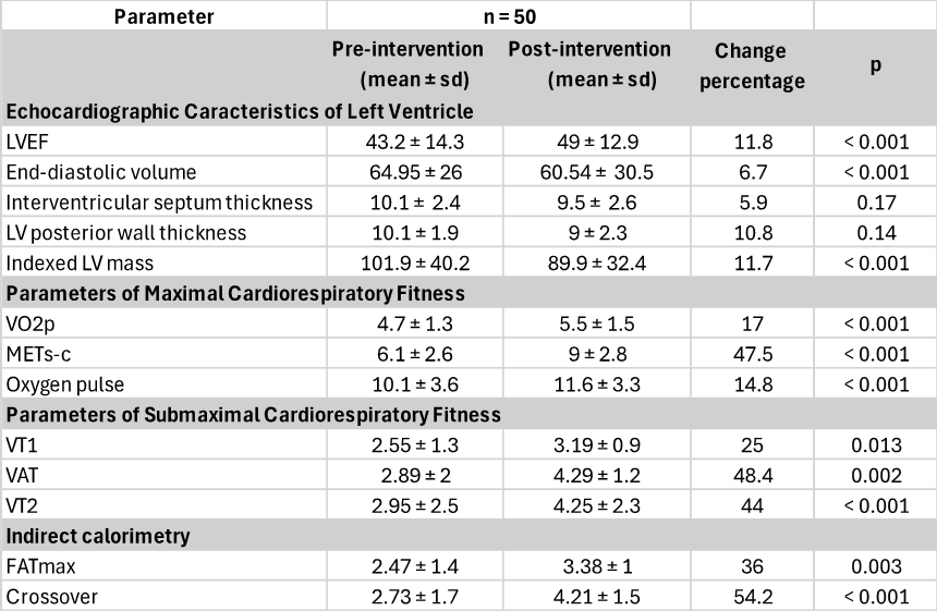
Table 3. Echocardiographic Characteristics of the Left Ventricle, Cardiorespiratory Fitness Parameters and Indirect Calorimetry Before and After the Cardiac Rehabilitation Program
Data are presented as means ± standard deviation and percentage change. LVEF: left ventricular ejection fraction. LV: left ventricle. VO2p: peak oxygen consumption expressed in METs. METs: metabolic equivalents. METs-c: estimated treadmill load. VT1: first ventilatory threshold or aerobic threshold. VAT: respiratory exchange ratio equal to 1. VT2: second ventilatory threshold or anaerobic threshold. FATmax: maximal fat oxidation point. Crossover: exercise intensity at which the energy supplied by carbohydrates exceeds that provided by fats.
The submaximal cardiorespiratory fitness variables analyzed included ventilatory thresholds from the cardiopulmonary exercise test, which showed significant increases: VT1 had a 25% increase, from a baseline of 2.55 ± 1.3 to 3.19 ± 0.9 at the end of the program, with p=0.013. VAT shifted from a baseline of 2.89 ± 2 to 2.29 ± 1.2, with an increase of 48.4% and p=0.002. VT2 showed a significant increase (p<0>
Indirect calorimetry assessed FATmax and crossover (point of interchange between fat and carbohydrate oxidation rates), both showing significant differences. The initial FATmax was reported at a mean of 2.47 ± 1.4 and improved to 3.38 ± 1 post-program, with a percentage change of 36% and p=0.003. Crossover also showed an increase of 54.2%, with a baseline mean of 2.73 ± 1.7 and ending at 4.21 ± 1.5, p <0>
The behavior of left ventricular mass was analyzed (Table 4), revealing that patients with an increase in ventricular mass (n=17) had an 18.2% increase, from a baseline of 92.3 ± 33.3 to 109.1 ± 33.6, p<0 n=33)>
Table 4. Left Ventricular Mass Behavior
Data are presented as means ± standard deviation and change percentage. LV: left ventricle.
The behavior of VO2p, expressed in METs, was studied in patients who increased their ventricular mass compared to those who decreased it. Patients with an increase in LV mass achieved a mean gain of 4.9 ± 1.5
MET and the ones that decreased LV mass 5.9±1.4 METs. Both groups showed significant increases in METs ( p = 0.04), however, the group in which LV mass decreased presented greater gains (graph 1).
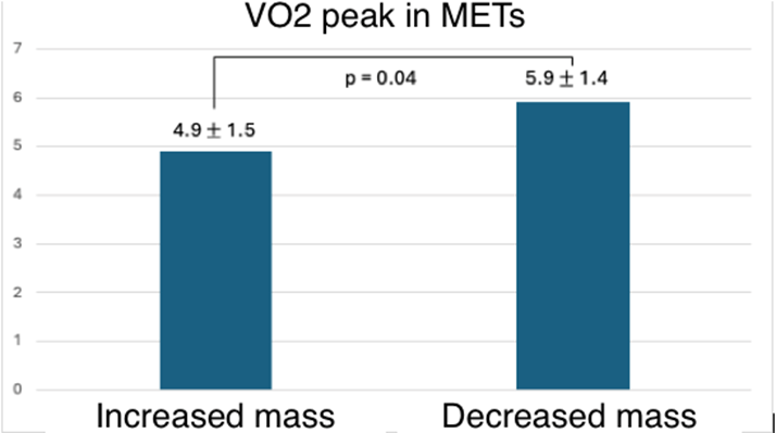
Graph 1. Peak Oxygen Consumption (VO2p) Expressed in Metabolic Equivalents (METs) in the Group of Patients Who Increased Left Ventricular Mass and in the Group Who Decreased Left Ventricular Mass
Data are presented as means ± standard deviation. LV: left ventricle. In the group that increased LV mass, the mean gain was 4.9 ± 1.5 METs (VO2p measured in METs), while the group that experienced a decrease in mass achieved an increase of 5.9 ± 1.4 METs (VO2p measured in METs). Both groups showed significant increases in METs (p = 0.04).
The geometry of the LV was assessed at the beginning and end of the program, and the changes observed are detailed in Table 5.
| Geometry | n = 50 | |
| Pre-intervention n (%) | Post-intervention n (%) | |
| Normal | 15 (30) | 15 (30) |
| Concentric remodeling | 14 (28) | 11 (22) |
| Concenric hypertrophy | 4 (8) | 6 (12) |
| Eccentric hypertrophy | 5 (10) | 8 (16) |
| Physiological hypertrophy | 3 (6) | 4 (8) |
| Dilated hypertrophy | 6 (12) | 1 (2) |
| Eccentric remodeling | 2 (4) | 4 (8) |
| Mixed hypertrophy | 1 (2) | 1 (2) |
Table 5. Pre-Intervention and Post-Intervention Left Ventricular Geometry
Data are presented as absolute values (n) and percentages.
Patients underwent supervised training with an overall mean training volume of 329.26 METs/week and 395.56 Kcal/week. The group that exhibited an increase in ventricular mass had a mean training volume of 319.41 compared to 334.33 in the group that experienced a decrease in LV mass. Kcal/week in the group that increased ventricular mass was 381.11 vs. 403 Kcal/week in the group that decreased LV mass. There were no reported adverse events during any of the exercise training sessions.
This research is the first specifically designed to investigate the effects of combined training on left ventricular mass in patients with HF of varying pathogenesis and independent of LV systolic function. Our findings demonstrate that combined training induces a reverse remodeling effect, leading to a decrease in end-diastolic volumes, an increase in LVEF and changes in left ventricular mass, primarily reductions, which are associated with greater oxidative potential (evidenced by the indirect calorimetry and ventilatory threshold variables), VO2p, METs-c, and PO2 significantly.
Exercise offers multiple physical and health benefits to individuals with chronic HF. Training modality, frequency, duration, and intensity are key factors that influence the degree of adaptations achieved. Aerobic training (AT) has been the primary exercise modality in cardiac rehabilitation programs (CRPs), with predominant evidence in the literature; however, strength training (ST) provides additional and complementary benefits to ET [25].
AT characterized by sustained increases in cardiac output with reduced peripheral vascular resistance induces morphological and functional adaptations, both central and peripheral. Centrally, it generally increases stroke volume and improves cardiac contractility, coupled with cardiac remodeling, specifically eccentric hypertrophy, enables greater venous return, ventricular filling, and cardiac output. Peripherally, it enhances vascular capacitance, permeability, and capillary density, which in turn improves muscle perfusion and contributes to an increased oxygen supply [27,28]. ST characterized by an increase in peripheral vascular resistance and slightly in cardiac output, during short episodes, produces concentric hypertrophy, increases lean mass and muscle strength [21,22,23,24]. Both training types achieve morphological and functional gains that collectively yield greater cardiovascular and musculoskeletal benefits; however, their effects on left ventricular mass have not yet been fully elucidated, particularly considering the interference effect on combined adaptations [44,45,46].
Training volume, a byproduct of frequency, duration, and intensity, as well as program length, will influence the gains achieved, but this volume quantifies the ET-dependent effects [30]. In our study, we implemented a program lasting 4 to 6 weeks, as regular training over at least six weeks is minimally sufficient for developing central and peripheral adaptations, as evidenced in our results [31,33]. Additionally, we improved patient adherence to the program due to its hybrid constitution.
Various international guidelines recommend exercise-based CRPs for patients with HF, whether with preserved or reduced LVEF, with a class I indication and level of evidence A [20]. Generally, the recommendation is to perform 30 minutes of moderate-intensity AT (starting with 40 and progressing up to 80% VO2p) at least 5 days per week, along with strength training, performing 10-15 repetitions for each muscle group, 2 to 3 times a week at 30-60% of 1RM, totaling 500 METs/week or 1500 Kcal/week, which is quite similar to the prescription we carried out in our study, except for the weekly METs and Kcal, which were lower in our sample. However, it is important to note that only supervised AT was quantified, as the contribution of ST is not feasible to quantify using the method employed by Kaminski [43]. This recommendation is derived from numerous trials, systematic reviews, and meta-analyses that have documented gains in exercise tolerance, quality of life, and reduced hospital readmissions, without adverse effects in patients with HF [15,16,17,26,27,28,31].
We know that regular exercise can restore autonomic, neurohormonal, and abnormal hemodynamic function, but several studies have demonstrated that it can reverse or attenuate LV remodeling [17,33,37]. The importance of this lies in the fact that left ventricular remodeling plays a key role in the progression of HF and is associated with increased morbidity and mortality. Moderate-intensity AT is capable of decreasing end-diastolic volumes, improving ejection fraction and increasing VO2peak by approximately 2.98 ml/kg/min or between 12% and 31% in patients with HF by enhancing contractility, preload, and vascular reserve [27,28,29,30,33,37]. However, combined training has not been conclusive in improving EF or reducing end-diastolic volumes [27,38,39,40,41] but has shown improvements in VO2peak [38,39,40]. Nevertheless, our study found significant gains in EF and VO2peak, as well as a reduction in end-diastolic volume with combined training.
Although the complex changes occurring in the heart during remodeling have traditionally been described in relation to anatomy, the remodeling process also affects cardiac myocyte biology and energy systems. In HF the concentrations of ATP and myocardial phosphocreatine are decreased, which, along with mitochondrial dynamics anomalies, compromises ATP generation. It has been shown that AT in patients with HF leads to improvements in VO2max by increasing the number of mitochondria and enzyme activity, thereby enhancing energy substrate utilization [12,25,28]. In our study, we hypothesize that the increase in mitochondrial biogenesis, along with greater capillary supply, led to improved cardiorespiratory fitness measured by submaximal variables and VO2p [12], meaning that training was able to enhance oxidative systems by generating metabolically active myocardial mass, which resulted in higher VO2p independent of changes in ventricular geometry. Indirect calorimetry supports these findings by showing a shift in the loading rates of FATmax and crossover to a higher VO2 at the final exercise test compared to the initial, suggesting a more developed oxidative base, despite the predominant reduction in ventricular mass.
At the same time, this increase in mitochondrial biogenesis is likely to result in lower lactate production during submaximal exercises and stricter control of the acid-base state, which is related to a positive impact on performance in daily living activities. This adaptation can be observed in the ventilatory thresholds (VT1, VAT, and VT2), which appeared at a higher VO2 at the end of the program, indicating a better submaximal cardiorespiratory fitness that directly impacts quality of life in this population [27,29,32]. Unfortunately, we could not perform histopathological studies of the myocardium to corroborate these intracellular findings.
It is important to mention that peripheral adaptations in circulation and skeletal muscle caused by exercise may also contribute to VO2p modifications [34]. Maximum cardiorespiratory fitness expressed as VO2max or VO2peak reflects the integrated capacity to transport oxygen from the atmosphere to the mitochondria to obtain the energy necessary for living [2]. Therefore, by quantifying an individual's functional capacity, it depends on a linked chain of processes that include ventilation and pulmonary diffusion, biventricular function, the ability of the vasculature to transport blood, and the competence of muscle cells to receive and utilize oxygen for ATP generation, it is considered a reflection of overall health. VO2max is determined by many factors, including the heart [2]. A change in cardiac geometry could modify the ability to increase ventricular volume and thus VO2peak, but we deem it necessary to clarify whether it was the increase or decrease in ventricular mass that was responsible for functional adaptation during exertion.
The primary objective of this study was to evaluate whether a cardiac rehabilitation program with combined training could increase left ventricular mass and thus peak VO2 in patients with HF. We know that cardiac hypertrophy is essential for maintaining pump function in HF; however, as it progresses, cellular organization breaks down, fibrosis occurs, contractile elements are lost, and energy metabolism is altered, leading to pathological hypertrophy. Within our results, we observed that the majority of the participants showed a significant decrease in left ventricular mass, likely due to the reduction in cardiac fibrosis. Conversely, one third of participants showed a significant increase in left ventricular mass, which may be secondary to the activation of signaling pathways promoting cardiac growth in a pathological environment and likely involves mass capable of generating significant VO2p gains.
Campos et al. demonstrated that 8 weeks of moderate-intensity AT improved left ventricular function associated with gains in mitochondrial oxidative capacity and reduced cardiac fibrosis in rodents with HF [5]. Schaible et al. found that an 8 to 10 weeks swimming program reversed pathological hypertrophy in rodents [35]. Based on these findings, we deduce that combined training can decrease left ventricular mass by reducing cardiac fibrosis, lowering afterload, triggering a sympathovagal balance and decreasing vasoconstrictive neurohormones that ultimately ended reducing the imposed hemodynamic load.
However, the most relevant result shows that the increases in peak oxygen consumption (VO2peak) after cardiac rehabilitation program (CRP) were independent of changes in left ventricular mass. We believe that this discordant effect between left ventricular mass and its ambiguous gain in cardiorespiratory fitness variables largely depends on the diversity of the pathophysiology of heart failure and that this reverse remodeling is independent of the increase in mitochondrial oxidative capacity, generating a metabolically active ventricular mass that leads to gains in VO2p and other variables associated with cardiorespiratory fitness, in which we can also notice an increase in oxygen pulse (PO2) which is recognized to be its main cardiac component measured by cardiopulmonary exercise test.
In our study, we established a multidisciplinary CRP with combined exercise and inspiratory diaphragmatic re-education sessions due to the evidence showing significant gains in health-related quality of life, VO2peak, and cardiac remodeling. Additionally, by including specialists in nutrition, psychology, nursing, physiotherapy, and physical rehabilitation, we contributed to managing associated cardiovascular risk factors that also improve the afore mentioned outcomes.
During the study there were no significant complications. Evidence has shown that the incidence of major cardiovascular complications during outpatient CRPs is 1 in every 60,000 hours [42]; however, the incidence is lower in supervised programs, as was the case in our study.
Many limitations and methodological biases exist. Most participants were men with heart diseases of various etiologies, and their pathophysiological heterogeneity influenced the results. Due to the sample size, it was not possible to assess whether specific changes in ventricular geometry were significant after the intervention, nor to conduct sub-analyses only on ventricular mass. It was not possible to study the morphological changes induced by combined training at a histological level because myocardial biopsies were not performed. This was a quasi-experimental study, as it was uncontrolled and unblinded, and the definition of the intervention largely had to be according to the characteristics of the patients due to the high risk of the population. Although adherence to the program was over 80%, the exercise frequency was one supervised session per muscle group per week, so we do not know if patients complied with the recommendations of two sessions per week per muscle group in a non-supervised manner, nor if increasing exercise sessions would generate greater gains in ventricular mass. Lastly, the transthoracic echocardiograms performed before and after the program depended on the operator's experience for the accuracy of the measurements, alongside inter-observer and intra-observer variability, as well as patient characteristics to allow for high-quality imaging. Despite this and the limitations of the population studied, the results were significant enough to draw conclusions.
CRPs based on combined physical training are effective and safe in patients with heart failure, improving VO2p in clinically stable patients independently of changes in left ventricular geometry measured through left ventricular mass. Although the most frequently presented adaptation at the end of the intervention was reverse remodeling, this condition does not contradict gains in cardiorespiratory fitness, as this ventricular mass can generate greater resistance and aerobic power.
Dear Editorial Team, Clinical Medical Reviews and Reports. My experience with the journal was highly positive. The peer-review process was rigorous, constructive, and completed in a timely manner. The reviewers provided valuable comments that helped improve the quality and clarity of our manuscript. The editorial office was professional, responsive, and supportive throughout all stages of the publication process. Communication was clear and efficient, and any questions were addressed promptly. Overall, I found the journal to maintain high scientific standards and an excellent publication workflow. I would be pleased to consider submitting future work to this journal. Best wishes from, Elena Popa.

It was my pleasure to submit my testimonial concerning the Reviewer Board of our Scientific Journal “Brain and Neurological Disorders”. The Reviewers focused on some modifications and their contribution was helpful. The ladies of our Editorial Office were also supported my efforts. It was my honor to have such a co-operation and I am looking forward for more collaboration.

Dear Grace Pierce, Editorial Coordinator of Journal of Clinical Research and Reports, Thank you for the speedy and efficient peer review process. I appreciate the fact that your peer reviewers do not take months to respond like with some other journals. I would also like to thank the editorial office for responding quickly to my questions. It is an excellent journal. I plan to submit more manuscripts in the future. Best wishes from, Robert W. McGee

Dear Grace Pierce, Editorial Coordinator of Journal of Clinical Research and Reports, Working with you and your team on our recent publication in JCRR has been a truly wonderful and enjoyable experience. The responses were prompt, and the reviewers were patient, constructive, and highly professional. One reviewer in particular gave me the feeling that a professor was carefully reading and commenting on my coursework, which was deeply touching. The entire process was straightforward and hassle‑free, with no tedious online forms to complete. I highly recommend this journal. Best wishes from, DR Aibing Rao, Head of R&D

I Appreciate the Opportunity to Share my Experience with the Journal of Clinical Research and Reports. The peer review process was timely and constructive, and the feedback provided helped improve the quality of our manuscript. The editorial office was professional, responsive, and supportive throughout the process, ensuring smooth communication and efficient handling of the submission. Overall, it was a positive experience collaborating with your team.

Dear Mercy Grace, Editorial Coordinator of Obstetrics Gynecology and Reproductive Sciences, We would like to express our gratitude for your help at all stages of publishing and editing the article. The editors of the magazine answer all the necessary questions and help at every stage. We will definitely continue to cooperate and publish other works in the Obstetrics Gynecology and Reproductive Sciences! Best wishes from, Alla Konstantinovna Politova,
