
Research Article | DOI: https://doi.org/10.31579/2768-0487/031
1Specialist general surgery and lecturer general surgery Dubai Medical College Dubai UAE.
2Specialist internal medicine Dubai hospital, UAE.
3Assistant Professor GI department SIUT.
4Medical officer Sindh children services hospital.
5Associate professor GI department SIUT.
6Senior specialist general surgery Dubai Hospital UAE.
7Consultant general surgeon, Liaquat National Hospital Karachi Pakistan.
8Consultant and head of general’s surgery department LiAQUAT National hospital Karcahi, Pakistan.
*Corresponding Author: Aliya Ishaq, Specialist general surgery and lecturer general surgery Dubai Medical College Dubai UAE.
Citation: A Ishaq, MJH Khan, Muhammad s khan, M Ishaq, A Parveen, et al. (2021) Enterocutaneous fistula; Management challenges, Areterospective review of 11 patients treated in our institute. Journal of Clinical and Laboratory Research. 3(1); DOI:10.31579/2768-0487/031
Copyright: ©2021 Aliya Ishaq. This is an open-access article distributed under the terms of the Creative Commons Attribution License, which permits unrestricted use, distribution, and reproduction in any medium, provided the original author and source are credited.
Received: 04 June 2021 | Accepted: 11 June 2021 | Published: 16 June 2021
Keywords: enterocutaneous fistula; enteroatmospheric fistula; spontaneous closure; mortality; timeline
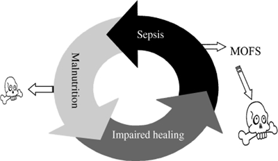
Enterocutaneous fistula is a local pathology and systemic disorder.
OBJECTIVES:
To analyze postoperative outcomes, morbidity, and mortality in patients treated for enterocutaneous fistula in our institute for past 18 months.
DESIGN, SETINGS AND PATIENTS:
Reterospective review of records of patients presented to Liaquat national university hospital Karachi, Pakistan between Jan 2010 to June 2011 with diagnosis of EC fistula.
RESULTS:
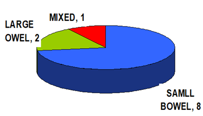
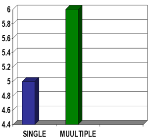
A total of eleven patients presented with diagnosis of enterocutaneous fistula in our institute in specified duration. Mean age at presentation was 33 years with amle to female ratio of 3:1.72.7 % had high output fistula and 27.2 % had low output fistula. Small bowel was involved in 72.7%, large bowel in 18.18% and 9.0% had both small and large bowel fistula.45.45% patients had single fistula while 54.55 had multiple fistula. Total length of stay varied between 22-150 days .6/11 (54.54%) had nosocomial infection, 3/11(27.27%) had bed sores.2/11(18.18%) had TPN related complications.Spontaneous closure occurred in 8/11(72.77) patients and definitive surgical closure was performed in one patient. Mortality rate was 18.8 %.
CONCLUSION:
Enterocutaneous fistula is a devastating outcome for both surgeons and patients, sytemetic timely multidisciplinary approach can save lives.
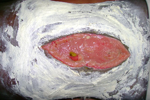
An enterocutaneous fistula (ECF) is an abnormal connection between the gastrointestinal tract and the skin or atmosphere (enteroatmospheric fistula [1] [EAF]) .Estimated 75-85% are iatrogenic, occur after an abdominal surgery.
There are several ways in which ECF has been classified, including by output, etiology, and source [1, 2, 3]. Most often, a high-output ECF is characterized as one with >500 mL/24 hours, low output <200 mL/24 hours, and a moderate output fistula between 200 and 500 mL/24 hours [1].
Historical reported mortality rates as high as 10-30%. Sepsis is the leading cause of death. Other factor include high output and comorbidity.Recent reported series show a declining mortality (6-33 or less) with improving supportive care, especially nutrition [3].
There is consensus among authorities that the management of a patient with EC fistula should proceed in an ordered sequence [3].
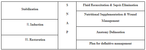
Treatment phases [4]
Management:
Management can be divided in to three phases
Acute phase:
Window period:
Subacute phase:
Repair and reconstructive phase:
OBJECTIVE:
INCLUSION CRITERIA:
All patients who presented to hospital with enterocutaneous fistula for previous 2 years were included in study.
They were all primarily treated in peripheral hospitals.
Management:
We adopted the three phase approach as described above for management of all patients with ECF (entero cutaneous fistula)
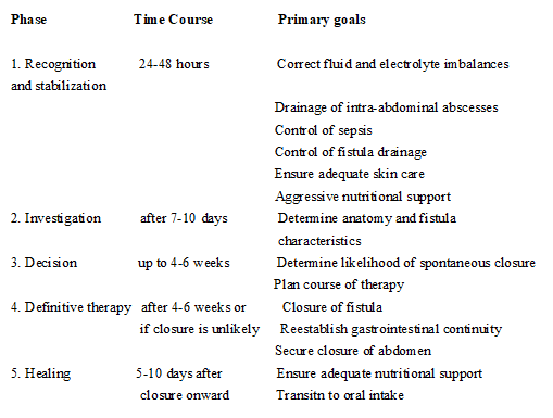
In the first phase (the acute phase) recognition and stabilization of ec fistula was done. The goal of this phase was to correct fluid and electrolyte imbalances, malnutrition, sepsis,abscess formation and wound infection. These problems were addressed within first 24-48 hrs of admission.
DVT prophylaxis.
The next phase was phase of anatomical delieniation.
Other had multiple fistulae in both small and large gut (post gun shot, multiple laparotomies.
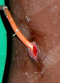
Different ways used for enteral feeding. For all low output and distal fistulas enteral feeding was started immediately after delineation and predigested dietary supplements were uses. In two patients with high output proximal fistula fistuloclysis was tried that is fistula contents taken from proximal limb through a feeding tube and reinserted distally using a Foley catheter and balloon inflated with 15 ml of water, It was very difficult and messy and was not totally successful requiring loss as well as multiple time cleaning because of leakage but was continued.
One patient feeding jejunostomy was made during initial surgery while controlling the sepsis and was used for enteral feeding.
Different types of dressings and drains were used to prevent skin complications related to fistula

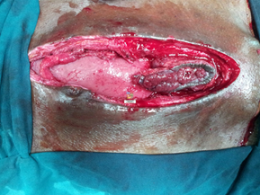
DRESSINGS USED:
In the final phase, definitive operation was done including fistula resection and resection anastomosis and biological mesh was used to reconstruct abdominal wall (one patient)
(Range: 15 - 45)
Type of fistula:
High output: 72.7%
Low output: 27.2%

Location of fistula:
No of fistulae:

Except for 3 patient all presented with sepsis and were nutritionally depleted.
Morbidity:
Recurrence: 1 out of 2 in surgically treated patient
Readmissions: 1
Length of hospital stay: 22 – 150 days.
Nosocomial infections: 6
Bed sore: 3
TPN related complications: 2
Line sepsis: 2
CVP Line: Out of 6, 2 had sepsis
PICC Line: Out of 5 none had sepsis
Mortality: 2 (18.18%)
Cause of mortality include multiple proximal high output fistulae and severe sepsis.
Spontaneous closure: 8 out of 11(72.7%) in 20 days to 3 months period.
Defenitive procedure after sub-acute phase: 1 out of 11 after 6 months of conservative treatment.
Patients died in sub-acute phase: 2 out of 11
The enterocutaneous fistula (ECF) is a devastating complication for both surgeons and patients alike. Prior to the advent of sophisticated critical care support and parenteral nutrition, the development of an ECF nearly equated to a death sentence. In the current era, the mortality rate has been reduced to 5 to 20% [5, 6]. However, the development and management of an ECF remains a chronic, debilitating condition [5].
We used the standard approach described by Schecter et al [6] for the management of EC fistula that is divided in 3 phases,first phase includes recognition and stabilization ,so patients after being diagnosed with having EC fistula were admitted in high dependency unit and correction of electrolytes and fluid balances were done along with control of fistula and sepsis source control. Patients with severe sepsis underwent ct scan abdomen with contrast in initial phase and those having intrabdominal collection and sepsis were taken to operation theater and drainage of abcess with controleed fistula formation was done as well as feeding jejunostomy was made in 2 cases, specialized vacuum dressings with laparostomy and vicryl mesh placement in presence of controlled fistula was performed in 2 patients. Broad spectrum antibiotics were started as per pus culture and blood transfusion was started when indicated. Parenteral nutrition was started in almost all patients and few of them with low output fistula were started on enteral feeding as well. Daily electrolytes and weight measurements were taken and strict in put out put charting was done along with chest and body physio and DVT prophylaxis
The provision of total parenteral nutrition has been associated with an increased rate of spontaneous closure of fistulas in several series [9, 10, 11]. Parenteral nutrition has long been recognized to be an integral part of the management of enterocutaneous fistulas [9, 10].We started TPN in all patients after insertion of PICC line /central line in acute phase. We don’t have enough number of patients to determine that correlation.
After 7 to 10 days, the patient has generally stabilized, and the fistula has matured to the point of supporting intubation of thin catheters in all orifices. At this point, the patient should undergo fluoroscopic fistulography with water-soluble contrast under the direct supervision of a senior radiologist and the senior surgeon responsible for the patient’s care. The information gained by such a study includes (1) the source of the fistula; (2) the nature (length, course, and relationships) of the fistula tract; (3) the absence or presence of bowel continuity (end vs. side fistula); (4) the absence or presence of distal obstruction; (5) the nature of the bowel adjacent to the fistula (inflammation, stricture); and (6) the absence or presence of an abscess cavity in communication with the fistula. The fistulogram provides information not obtainable through any other study, and early films can be particularly useful in defining anatomy and relationships. As previously discussed, water-soluble contrast may also be injected into abscesses at the time of drainage as a type of early fistulogram [9]. In our study. After the phase of stabilization and defining fistula output fistulogram was done to delineate the site of fistula and bowel anatomy and then same management with dressings ,TPN ,enteral feeding ,different dressings ,wound and skin care using zinc oxide was continued including fistuloclysis and patients were observed for a period of 3- 6 months .
We did not have a large enough sample to determine which factors determine good outcomes for enterocutaneous fistulas. However, several studies from other centers have looked at this. Using multiple logistic regression analysis, Visschers et al [15]. Found that intact abdominal walls and administration of parenteral nutrition were independent predictors of spontaneous closure of enterocutaneous fistulas. In our study Spontaneous closure was achieved in 3 months in 72.7 % cases without need for surgery and surgical intervention was done in 1 patient after completing 6 months of conservative treatment with mature fistula and repeat imaging using ct scan and fistulogram was done prior to surgery and excision of fistula followed by reconstructiuon and diversion ileostomy and abdominal wall reconstruction was done. 2 of our patients died in sub-acute phase because of hight output fistula and sever sepsis .Our mortality rate is 18.8% which is comparable to most institutes specialized in management of ECF.
Enterocutaneous fistula is a local pathology and systemic disorder with major impact on patient’s psychology,finance emotions and wellbeing . Given that most are iatrogenic, the most effective means of treatment is prevention with sound surgical judgment and meticulous technique. However, when faced with the development of an ECF, early recognition with systemic orderly approach by a multidisciplinary team specialized in treatment of these challenging patients can save lives.
Dear Editorial Team, Clinical Medical Reviews and Reports. My experience with the journal was highly positive. The peer-review process was rigorous, constructive, and completed in a timely manner. The reviewers provided valuable comments that helped improve the quality and clarity of our manuscript. The editorial office was professional, responsive, and supportive throughout all stages of the publication process. Communication was clear and efficient, and any questions were addressed promptly. Overall, I found the journal to maintain high scientific standards and an excellent publication workflow. I would be pleased to consider submitting future work to this journal. Best wishes from, Elena Popa.

It was my pleasure to submit my testimonial concerning the Reviewer Board of our Scientific Journal “Brain and Neurological Disorders”. The Reviewers focused on some modifications and their contribution was helpful. The ladies of our Editorial Office were also supported my efforts. It was my honor to have such a co-operation and I am looking forward for more collaboration.

Dear Grace Pierce, Editorial Coordinator of Journal of Clinical Research and Reports, Thank you for the speedy and efficient peer review process. I appreciate the fact that your peer reviewers do not take months to respond like with some other journals. I would also like to thank the editorial office for responding quickly to my questions. It is an excellent journal. I plan to submit more manuscripts in the future. Best wishes from, Robert W. McGee

Dear Grace Pierce, Editorial Coordinator of Journal of Clinical Research and Reports, Working with you and your team on our recent publication in JCRR has been a truly wonderful and enjoyable experience. The responses were prompt, and the reviewers were patient, constructive, and highly professional. One reviewer in particular gave me the feeling that a professor was carefully reading and commenting on my coursework, which was deeply touching. The entire process was straightforward and hassle‑free, with no tedious online forms to complete. I highly recommend this journal. Best wishes from, DR Aibing Rao, Head of R&D

I Appreciate the Opportunity to Share my Experience with the Journal of Clinical Research and Reports. The peer review process was timely and constructive, and the feedback provided helped improve the quality of our manuscript. The editorial office was professional, responsive, and supportive throughout the process, ensuring smooth communication and efficient handling of the submission. Overall, it was a positive experience collaborating with your team.

Dear Mercy Grace, Editorial Coordinator of Obstetrics Gynecology and Reproductive Sciences, We would like to express our gratitude for your help at all stages of publishing and editing the article. The editors of the magazine answer all the necessary questions and help at every stage. We will definitely continue to cooperate and publish other works in the Obstetrics Gynecology and Reproductive Sciences! Best wishes from, Alla Konstantinovna Politova,
