
Case Report | DOI: https://doi.org/10.31579/2690-1897/274
Candidate of biological science, assistant professor of pathophysiology department named D.A. Maslakov, Grodno State Medical University; Grodno, Belarus.
*Corresponding Author: Ramachandran Muthiah., Candidate of biological science, assistant professor of pathophysiology department named D.A. Maslakov, Grodno State Medical University; Grodno, Belarus.
Citation: Ramachandran Muthiah., (2025), End stage Endomyocardial Fibrosis- Echocardiographic Features, J, Surgical Case Reports and Images, 8(8); DOI:10.31579/2690-1897/274
Copyright: © 2025, Ramachandran Muthiah, this is an open access article distributed under the Creative Commons Attribution License, which permits unrestricted use, distribution, and reproduction in any medium, provided the original work is properly cited.
Received: 08 September 2025 | Accepted: 16 September 2025 | Published: 20 September 2025
Keywords: endomyocardial fibrosis; burnt-out stage; pericardial effusion; endocardial calcification; ‘cobra-head’ fibrosis
Tropical endomyocardial fibrosis (EMF) is a public health problem affecting the children, young adSSults and elderly individuals in an epidemic fashion in the coastal districts of south India. Due to lack of resources for research in these endemic areas, its etiology remains elusive and hypotheses ranging from infections and allergic causes to malnutrition and toxins have not been tested rigorously. The disease is characterized by endocardial fibrosis and the right ventricle is the cardiac chamber most frequently affected. Patients may present clinically with heart failure and an associated AV (atrioventricular) valve regurgitation is common. Several features of the advanced disease called as ‘burnt-out’ or “End stage” endomyocardial fibrosis (EMF) are not fully understand. Background of these case studies described the clinical presentation, echocardiographic features and management of this late stage of the disease.
Endomyocardial fibrosis (EMF) is an active and progressive disease, characterized by patchy fibrosis of endocardial surface of the heart. It was first described in West African troops serving in the Middle East and in indigenous African subjects in Uganda. It can be thought of in the same general pattern of behavior as rheumatic heart disease and increasingly recognized in the tropical and subtropical regions of the world within 15 degrees of the equator. The epidemiology of EMF is a ‘vanishing mystery’ in the southern districts of India and the coastal belt of Thoothukudi in Tamil Nadu state is the “hot spot” for this enigmatic disease. It is the commonest cause of restrictive cardiomyopathy in Africa and worldwide and discussed as a neglected tropical disease [1].
In 1938, Arthur Williams, the foundation professor of medicine at Makerere University, Kampala, Uganda described two cases of mitral incompetence and correlated with large patches of fibrosis affecting the ventricular walls at necropsy and it is the earliest documentation of EMF in the literature. A pathologist, Jack N.P.Davis first coined the term ‘ endomyocardial fibrosis’(EMF) while working at Mulagu hospital in Uganda and said that “he had met a new disease” [2] and it accounted for 15% of the deaths due to congestive heart failure in that hospital and delineated the clinico-pathologic features of this new restrictive cardiomyopathy and its distribution in Africa [3] in 1954 at a Royal society meeting, still called as “Davies disease” by some authors[4]. Endomyocardial fibrosis (EMF) was first reported from India at Christian Medical college, Vellore by JD Ball. The highest prevalence of the disease remains in the region of sub-Saharan Africa and a study in rural area of Mozambique showed that 20% of a random sample of 1063 subjects of all age groups had echocardiographic evidence of this disease with a male preponderance [5].The natural history of EMF is not completely understood and the disease usually come to clinical attention in the late, chronic, burnt-out phase after remaining asymptomatic for long periods and so these cases have been reported.
Case 1 (Right ventricular Endomyocardial Fibrosis in Psoriasis)
A 52-year old male was admitted with symptoms of cough and breathlessness for 6 months duration. He had sudden onset of itchy skin lesions on the trunk and extremities for one-month duration following the respiratory infection. Blood chemistry, ECG and X-ray chest were normal. Serum ASO (anti-streptolysin O) titer was negative. Physical examination revealed numerous, itchy, drop-shaped small salmon-pink papules, 1-10 mm in diameter, scaly and covered with silvery-white ‘micaceous scales’, predominantly seen on the trunk and limbs as shown in Figures 1 and 2, suggesting the “Guttate Psoriasis”. It appears suddenly, 2-3 weeks after an episode of upper respiratory infection caused by group A beta- hemolytic streptococcal infection. On scratching, pinpoint bleeding occurs when the scales are removed (Auspitz’s sign) and the lesio ns induced by trauma to the skin (Koebner phenomenon). It is an immune-mediated inflammatory disease that causes an incre ase in epidermal cell turnover with hyperproliferation of keratinocytes. The environmental, genetic and immunological factors play a role in its pathogenesis.
Figure 20: Apical four chamber view showing right ventricular EMF with apical fibrosis and tricuspid regurgitation in a 62-year old female. Fibrotic lesion is characterized by rugose border with a mushroom appearance and thus differentiating from Apical right ventricular hypertrophic cardiomyopathy as shown in Figure-26.
Figure 1: Guttate Psoriasis on the trunk and extremities in a 52 –year old male (Photo image taken with consent)
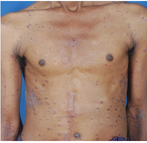
Figure 2: Guttate Psorisis in the back of trunk and extremities in a 52-year old male.(Photo image taken with consent)
Transthoracic echocardiography revealed strong fibrous strands appearing as ‘finger like projections’ or ‘cobra-head’ appearance in the right ventricular apex due to fibrosis of muscular trabeculae, suggesting right ventricular endomyocardial fibrosis as shown in Figures 3 to 7.
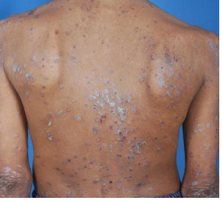
Figure 3: Subcostal view showing the ‘finger like’ projections of fibrous strands in the RV (right ventricular) apex suggesting Right ventricular endomyocardial fibrosis in a 52-year old male with psoriasis.
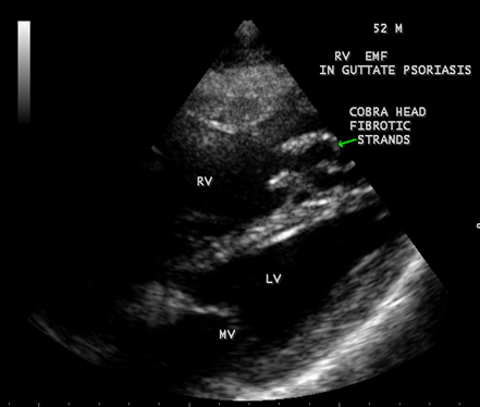
Figure 4: Tilted apical view showing the ‘cobra-head’ appearance of fibrous strands suggesting right ventricular endomyocardial fibrosis in a 52-year old male with Psoriasis.
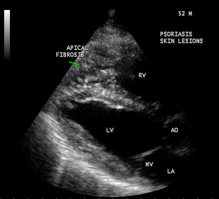
Figure 5: Parasternal long axis view showing the RV (right ventricular) apical fibrosis in a 52-year old male with psoriasis
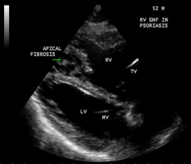
Figure 6: Dense fibrosis in RV apex in Psoriasis in a 52-year old male.

Figure 7: Thick fibrotic stands in RV apex due to fibrosis of muscular trabeculae in Psoriasis.
The patient was advised daily sun exposure, sea bathing (balneotherapy), topical moisturizers such as petrolatum jelly and a combination therapy with vitamin D analog (calcipotriol and calcipotriene) or retinoid (tazarotene) and topical steroid ointments. Psoralen is a photosensitizer for either topical (bath) or systemic use. Systemic therapy for severe cases includes non-biological (retinoids (acetretin), immunosuppressives (cyclosporine or azathioprine)) and biological (protein with pharmacological activity) agents. Guttate Psoriasis is responsive to phototherapy and therapies such as UVB (short wave ultraviolet B) and PUVA (psoralen and ultraviolet A (long wave) radiation) have low efficacy and may increase the risk of malignancies such as squamous cell carcinoma and malignant melanoma. Drugs such as chloroquine, beta-blockers, aspirin or other NSAIDS should be avoided. Drugs targeted on T cells (since psoriasis is related to excess T cell activity) such as efalizumab and alefacept were withdrawn since they result in progressive multifocal leukoencephalpathy. The respiratory infection was treated with azithromycin 500 mg daily for 5 days. Psoriasis is a manifestation of EMF or both conditions result from the same etiopathological mechanisms are yet to be evaluated.
Case 2 (Right ventricular endomyocardial fibrosis in Tuberculosis) A 23-year old male was admitted with sudden onset of ascites for one-month duration. He was treated for pulmonary tuberculosis 3 years back with a positive sputum AFB (acid fast bacilli). Blood chemistry revealed as ( Total count-7400 cells/cu.mm of blood ( normal-4000 to 11000 cells/cu.mm of blood), polymorphs -70% (normal- 40 to 75 %), lymphocytes-22%(normal-20 to 40%), eosinophils-8% (normal- 1 to 4%), ESR (erythrocyte sedimentation rate)-10 to 22 mm/hour ( normal- 0 to 15mm/hour), platelets-2.5 lakhs/cu.mm of blood and a mild elevation of serum bilirubin-total-2mg/dl(normal---up to 1.2 mg/dl) direct-1.2mg%(normal—upto—0.3 mg/dl), indirect-0.8mg%(normal – upto 0.9 mg/dl). Total serum proteins 5.2gm% (normal -6.6 to 8.3 gm/dl), albumin-3.2gm% (normal- 3.5 to 5.0 gm/dl), globulin -2.0gm%(normal 2.5 to 3.5 gm/dl ), urea-39 mg%(normal 15-50 mg/dl), creatinine-0.1mg%(normal- 0.7 to 1.4mg/dl), sugar-112 mg/dl random (normal – 80 to120 mg/dl- random sample). Ascites fluid tapping revealed an exudate (protein-3 gm%) and cytology revealed no malignant cells. Ascites fluid adenosine deaminase (ADA)activity revealed 10.4 U/L (normal < 40>Figure 8. His pulse rate was 108 bpm and blood pressure 100/70 mmHg. Auscultation revealed clear lung fields and no cardiac abnormalities. ECG revealed no arrhythmias and X-ray chest showed right- sided pleural effusion and extensive calcification over the cardiac shadow as shown in Figure 9. Transthoracic echocardiography revealed apical fibrosis of right ventricle, moderate pericardial effusion, right atrial dilatation as shown in Figures 10 and 11, suggesting right ventricular endomyocardial fibrosis and severe tricuspid regurgitation as in Figure 12 indicates coexisting pulmonary hypertension due to pulmonary damage caused by tuberculosis as shown in Figure 13 and free RV outflow tract as in Figure 15. Patient was treated with antituberculous drugs, antifailure measures such as digoxin and diuretics, ascites fluid tapping and antibiotics. He showed mild improvement in his symptoms.
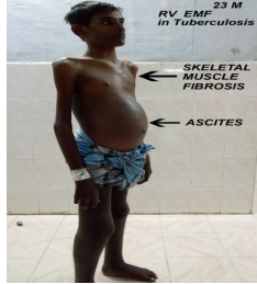
Figure 8: Physical appearance of RV EMF in burnt-out stage with heart failure, ascites and skeletal muscle wasting due to fibrosis in a 23 –year old male in tuberculosis (Photo image taken with consent)
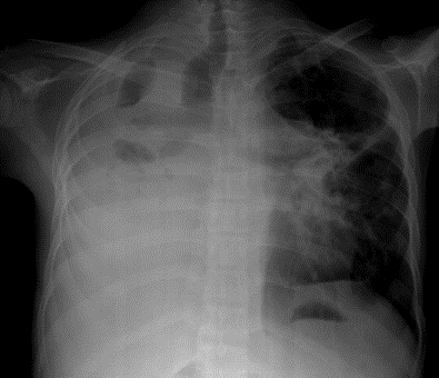
Figure 9: X-ray chest PA (postero-anterior) view showing right-sided pleural effusion and endocardial calcification over the left ventricle in burn- out stage of EMF (endomyocardial fibrosis).
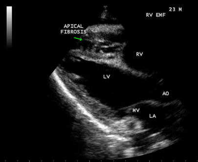
Figure 10: Parasternal long axis view showing RV (right ventricular) apical fibrosis suggesting EMF (endomyocardial fibrosius) in a 23-year-old male with tuberculosis
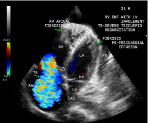
Figure 11: Apical four chamber view suggesting RV (right ventricular) apical fibrosis, RA dilatation and severe tricuspid regurgitation suggesting right ventricular endomyocardial fibrosis with an extension of fibrosis in the LV apex
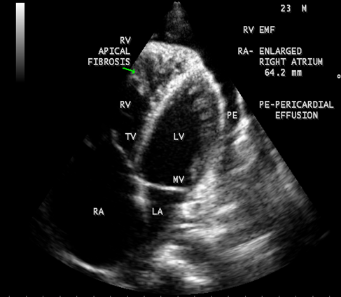
Figure 12: Apical four chamber view showing the RV apical fibrosis with mild pericardial effusion in a 23-year old male with tuberculosis.
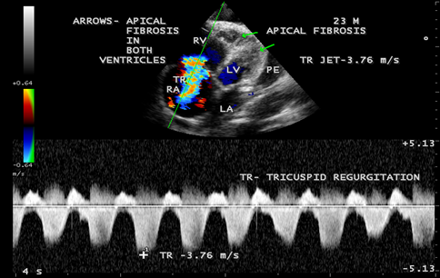
Figure 13: CW (continuous wave Doppler) showing the tricuspid jet velocity 3.76 m/s with Pulmonary artery systolic pressure of 67 mmHg ((4 x 3.76 m/s)2 + RA pressure (10 mmHg), suggesting pulmonary hypertension due to coexisting lung disease (tuberculosis) in RV EMF with fibrosis extending to LV apex.
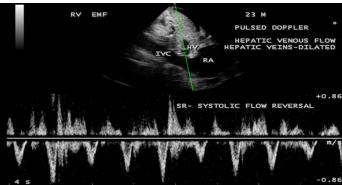
Figure 14: Pulsed Doppler showing systolic flow reversal in hepatic venous flow due to severe tricuspid regurgitation and dilated hepatic veins.
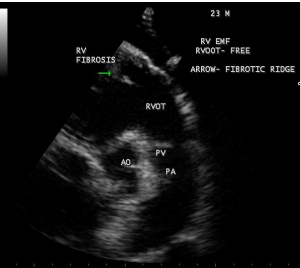
Figure 15: RV outflow tract is free of fibrosis and fibrous strands stand as ridge in Endomyocardial fibrosis with tuberculosis.
Case 3. (Right ventricular endomyocardial fibrosis with massive pericardial effusion in a 85 –year old male)
A 85-year old obese female was admitted in the cardiac intensive care unit with breathlessness. X-ray chest revealed massive pericardial effusion with calcification in the right ventricular region as shown in Figure 16. ECG revealed low voltage complexes. Blood chemistry revealed normal. Transthoracic echocardiography revealed large pericardial effusion with Right ventricular apical fibrosis, suggesting right ventricular endomyocardial fibrosis as shown in Figure 17 with tamponade physiology as in Figure 18 and treated with pericardiocentesis and pericardial fluid revealed an exudate on biochemical analysis.
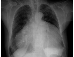
Figure 16. X-ray chest PA view showing massive pericardial effusion in a 85-year old female with endocardial calcification in the right ventricle suggesting the burnt-out stage of endomyocardial fibrosis.
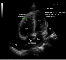
Figure 17: Apical four chamber view suggesting RV apical fibrosis with massive pericardial effusion
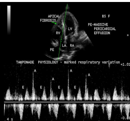
Figure 18: Pulsed-doppler imaging showing tamponade physiology.
Case 4. Right ventricular endomyocardial fibrosis presented with atrial fibrillation in a 62-year old female as shown in Figures 19 and 20.
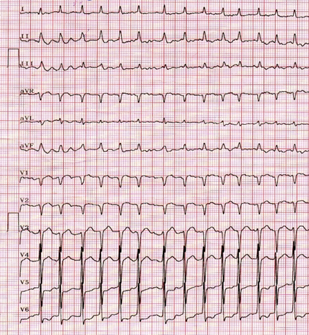
Figure 19. ECG Showing atrial fibrillation in a 62-year old female with Right ventricular endomyocardial fibrosis.
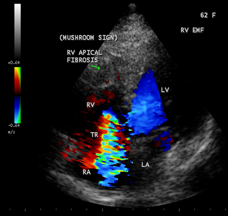
Figure 20: Apical four chamber view showing right ventricular EMF with apical fibrosis and tricuspid regurgitation in a 62-year old female. Fibrotic lesion is characterized by rugose border with a mushroom appearance and thus differentiating from Apical right ventricular hypertrophic cardiomyopathy as shown in Figure-26.
Case 5. Right ventricular EMF presented with complete heart block in a 75- year old female as shown in Figures 21 and 22.
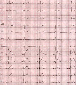
Figure 21: ECG showing complete heart block in a 75-year old female with right ventricular EMF.
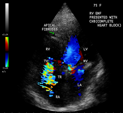
Figure 22: Apical four chamber view showing right ventricular EMF with apical fibrosis in a 75 –year old female
Case 6. Left ventricular endomyocardial fibrosis mimicking as Apical left ventricular hypertrophic cardiomyopathy in a 2- year old male child as shown in Figures 23 , 24 and 25.
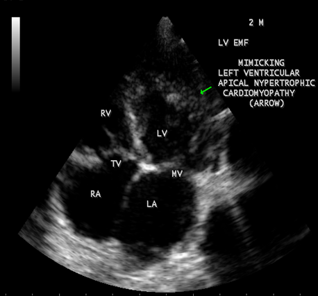
Figure 23: (Left ventricular endomyocardial fibrosis mimicking as apical left ventricular hypertrophic cardiomyopathy in a 2- year old male child}
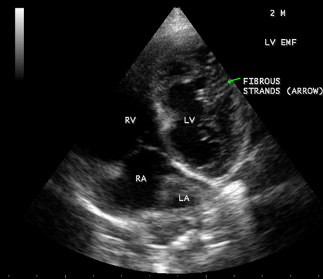
Figure 24: Apical four chamber view showing the endocardial fibrosis of the left ventricle in a 2-year old male child
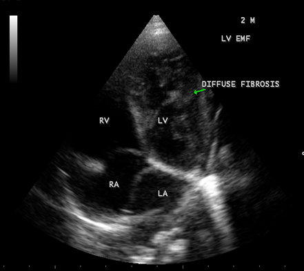
Figure 25 Apical four chamber view showing diffuse endocardial fibrosis of left ventricle in 2-year old male child.
Case 7 Apical right ventricular hypertrophic cardiomyopathy in a 60- year old female mimicking as right ventricular EMF as shown in Figure 26.
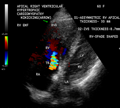
Figure 26: Apical Right ventricular hypertrophic cardiomyopathy (asymmetric lateral wall hypertrophy with a thickness of 30 mm) mimicking as right ventricular endomyocardial fibrosis in a 60- year old female. (rugose border, a characteristic feature of EMF is absent and mild tricuspid regurgitation)
Case 8. Left ventricular endomyocardial fibrosis coexisting with RHD (rheumatic heart disease) in a 60-year old male as shown in Figures 27 and 28.

Figure 27: showing the thickening and calcification of mitral leaflets with flail Posterior mitral leaflet suggesting rheumatic involvement in a 60-year old male.
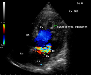
Figure 28: showing the mitral regurgitation due to PML (posterior mitral leaflet) involvement in a 60-year old male.
The other echocardiographic manifestations of burnt-out endomyocardial fibrosis are shown in Figures 29 to 44 as given below
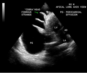
Figure 29: 2D Echocardiographic imaging showing massive pericardial effusion with ‘cobra-head’
fibrous strands in the pericardial sac suggesting the burnt- out stage of endomyocardial fibrosis in a 60-year old male.
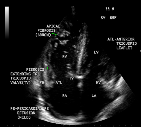
Figure 30: showing thick fibrous strands in the right ventricle with mild pericardial effusion suggesting right ventricular endomyocardial fibrosis in a 33-year old male.
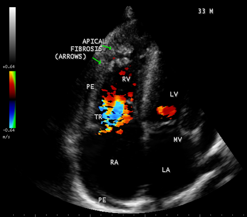
Figure 31: showing tricuspid regurgitation with thick fibrous strands in the Right ventricle in a 33-year old male in RV EMF.
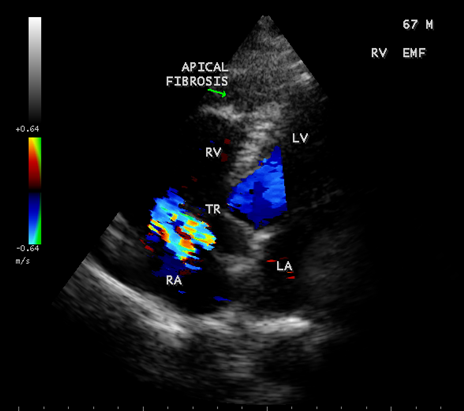
Figure 32. Endomyocardial fibrosis with aneurysmal right ventricle in a 67-year old male with RV apical fibrosis.
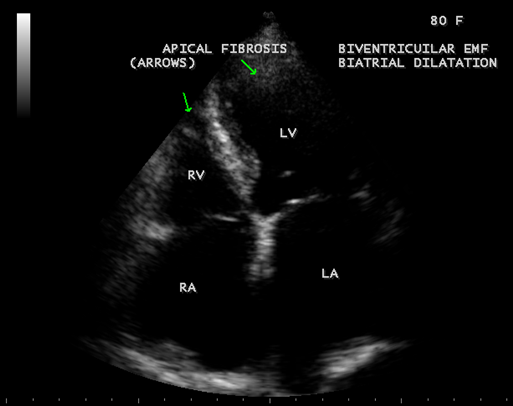
Figure 33: Endomyocardial fibrosis with biatrial enlargement in a 80-year old female suggesting biventricular EMF.
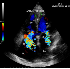
Figure 34: Biventricular EMF showing AV (atrioventricular) valve regurgitation in a 67- year old male.
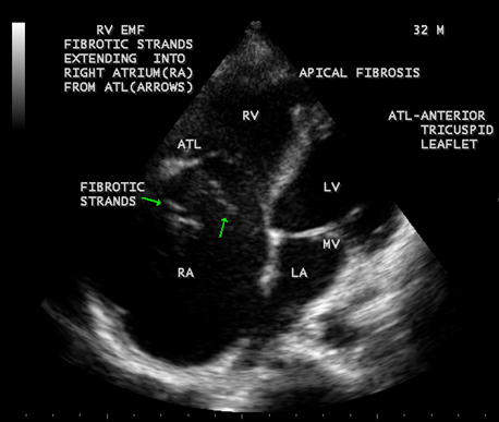
Figure 35: Endomyocardial fibrosis showing thick fibrous strands in the right atrium in a 32 -year old male and a dilated right atrium
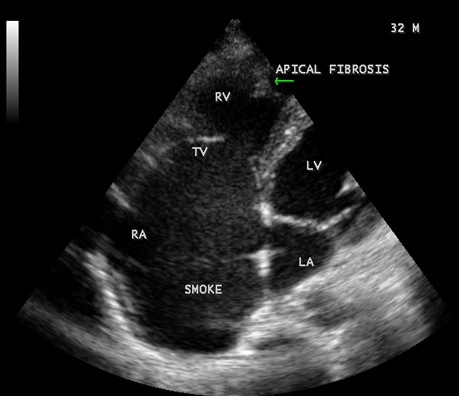
Figure 36: Endomyocardial fibrosis showing SEC (spontaneous echo contrast or smoke) in the right atrium in a 32 -year old male.
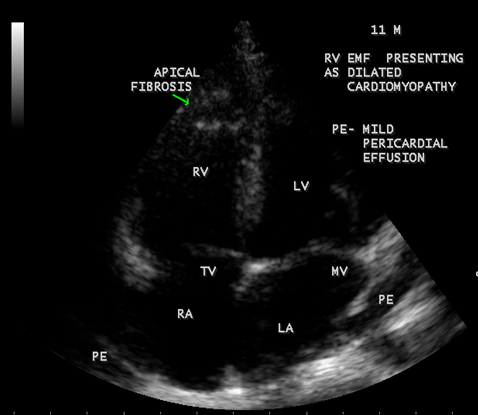
Figure 37: Endomyocardial fibrosis presented with dilated cardiomyopathy with RV apical fibrosis in a 11- year old male child.
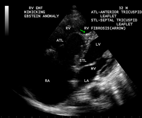
Figure 38: Right ventricular Endomyocardial fibrosis mimicking as Ebstein anomaly in a 32- year old male.
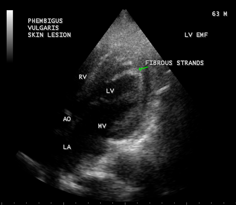
Figure 39: Left ventricular endomyocardial fibrosis in Pemphigus skin lesions in a 63-year old male.
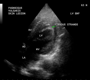
Figure 40: Endomyocardial fibrosis with biventricular enlargement in a 50-year old female and tricuspid annular retraction.
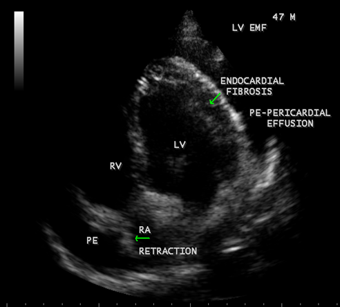
Figure 41: showing Endomyocardial fibrosis with moderate pericardial effusion and right atrial notch (RA retraction) in a year 47-year old male
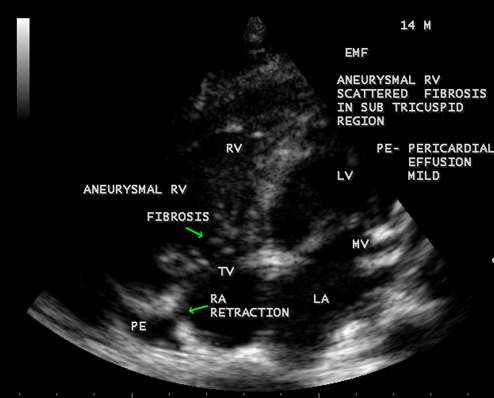
Figure 42: Endomyocardial fibrosis showing subtricuspid fibrosis, aneurysmal right ventricle, mild pericardial effusion and right atrial notch (RA retraction) in a 14-year old male.

Figure 44: Endomyocardial fibrosis showing thickening of LV tendons seen as fibrous ridges as an initial manifestation of LV EMF in a 23-year old male.
Etiopathogenesis
The majority of EMF cases are reported from low-lying humid parts of tropical countries. In Tanzania and Mozambique, cases have clustered along the coastal forests [6], the coastal districts in south India and in China, the largest number of case reports from southern province of Guangxi [7]. The underlying etiology and its unique geographic distribution remain a great mystery in cardiology [8]. The proposed causes of endomyocardial fibrosis are listed in Table-1 given below, but none of these causes could establish itself as the reason for this enigmatic disease [9].
| Infections | Allergy | Malnutrition | Toxic agents |
Toxoplasmosis Rheumatic fever Malaria Myocarditis Helminth parasites Psoriasis Pemphigus Tuberculosis | Esoninophila Auto-immunity | Protein deficiency Magnesium deficiency | Cerium Cassava Thorium Serotonin Plant toxins Vitamin D |
Table 1: Proposed causes of endomyocardial fibrosis)
Researches in African populations showed a high prevalence of anti-heart antibodies in EMF patients when compared to those with rheumatic heart disease and it is not clear whether these autoantibodies are the cause or the result of EMF. In Mozambique, when the serum of patients was tested for the presence of anti-myocardial proteins [10], an increased immunoglobulins G and M reactivity were detected in EMF patients [11]. The role of infectious agents appears as possible causes or triggers for this disease [12], however, no specific organism has been consistently associated with endomyocardial fibrosis.
Presence of interstitial fibrosis, myohypertrophy, and calcification suggest a role of cytokines in its genesis [13],[14]. The inflammatory response occurring in the younger age group could manifest as calcification in the later years. Dominant right ventricular involvement, argues for toxic factors which are removed in the lungs or a factor which affects the heart when the right ventricle is dominant or predisposed, as during in utero life since the right ventricle receives most of the umbilical venous return.
Davies described three phases of the disease in his patients from Uganda. The initial phase is an acute carditis phase, starts as a febrile episode with facial swelling and in severe cases with heart failure, progress into subacute and chronic burnt-out phase. The condition is associated with fibrosis of endocardium of right and left ventricles, involving predominantly the apices and inflow regions, resulting in impaired filling, valvular regurgitation owing to frequent involvement of papillary muscles with tethering to ventricular walls. Endocardial lesions in EMF may evolve into necrotic, thrombotic and fibrotic stages as proposed by Olsen in Uganda. Most patients have rapidly progressive heart failure, leading to death within 2 years of the initial insult [15]. However, some patients may have a steady period without any clinical deterioration and recently a high prevalence of predominantly asymptomatic cases occurred in Mozambique [16], others may experience remissions of the clinical signs with no further progression.
When the endocardium is replaced by collagenous fibrosis (consist of collagen deposition and fibroblast proliferation), the final fibrotic stage is reached after several years of disease activity. Fibrotic obliteration of the apices of the affected ventricles is the hallmark of the disorder and fibrosis involving the papillary muscles and chordae tendineae leading to atrioventricular valve distortion and regurgitation. In the left ventricle, the fibrosis extends from the apex to the posterior mitral leaflet, usually sparing the anterior mitral leaflet and outflow tract and cause PML (posterior mitral leaflet) distortion and regurgitation. Like the peculiar geographical distribution, the fibrotic endomyocardial involvement stops short of the ventricular outflow tract like a ridge [17] as shown in Figures 15,29,31,35 and 44. The fibrotic tissue often creates a nidus for thrombus formation, which can be extensive. Atrial thrombi also occur and the right atrium may be aneurysmally dilated. Aneurysmal right atrium with spontaneous echo contrast was detected in a 32- year old male as shown in Figure 36 [18- Figure 3]. In addition, there are fibrosis and granular septation extending into the underlying myocardial tissue and myocyte hypertrophy is common [19]. Fibrotic process causes tethering of leaflets into ventricular walls and may mimic Ebstein’s malformation as shown in Figure 38[20],[21]. Fibrosis increases the stiffness of the heart, resulting restrictive physiology, AV (atrioventricular) valve regurgitation which has been linked to atrial arrhythmias such as atrial fibrillation as shown in Figures 19 and 20 in a 62-year old female. Atrial fibrillation has been reported in more than 30% of patients with EMF. Fibrosis impairs activation patterns of the conduction system and may provide substrate for wave breaks and reentry [22]. Fibrosis reduces conduction velocity and cause conduction abnormalities like junctional rhythms, heart blocks as shown in Figure 21 and 22 in a 75-year old female and atrioventricular conduction delay [23].
Endocardial calcific deposits can be present involving diffuse areas of the ventricles and Cockshott et al described this feature in 1967. Calcification, an impressive finding on imaging denotes a burnt-out phase of endomyocardial fibrosis (EMF) and confirming the malignant nature of the disease. Chest X-rays show varying degrees of cardiomegaly and at times typical endocardial calcifications in the left and right ventricles as shown in Figure 9 (left ventricular endocardialcalcification) [24- Figure 2(a)] and in Figure 16 (right ventricular endocardial calcification)[25- Figure 2 (c)- shows calcification in both ventricles], [26].
A large pericardial effusion is often present and noted as another peculiar feature of this disease [27]. Pericardial effusion and ascites dominate the clinical picture of right ventricular EMF [28], [29], [30]. Etiology of pericardial effusion is possibly inflammatory and EMF is to be considered as ‘pancarditis’ since all the layers are involved. Adhesions between the parietal and visceral layers of the pericardial sac may develop and visible as strong fibrotic strands as shown in Figures 29. A right ventricular EMF presented with massive pericardial effusion was detected in a 85 –year old female as shown in Figure 16, 17 and 29[31]. Cardiomegaly can be exaggerated by pericardial effusion, and pleural effusion is also a common finding as shown in Figure 9 [16-Figure 2]. Giant ascites in EMF is not fully explained by congestion alone and it is due to peritoneal inflammation and reduced reabsorption of peritoneal fluid, caused by fibrosis since the fluid is an exudate with predominant lymphocytes. The triad of elevated JVP (Jugular venous pressure), ascites and hepatomegaly formed the hallmark of right ventricular EMF.
Progression to generalized edema, hypoalbuminemia, cachexia, malnutrition, skeletal muscle fibrosis (fibrosis is also present in skeletal muscles, suggestinga generalized fibrotic process that would explain remarkable skeletal muscle atrophy present in some patients) [ 32] in advanced disease with hepatic dysfunction as shown in Figure 8. [33 –Figure 3 E].
Endomyocardial Fibrosis may present as dilated cardiomyopathy in a child as shown in Figure 37 [34]. In some cases, scattered areas of fibrosis in the submitral and subtricuspid regions may cause valvular regurgitation as shown in Figure 42. The valvular regurgitations occur in rheumatic heart disease and the differential features are given in Table 2. It may coexist with RHD (rheumatic heart disease) rarely as shown in Figures 27 and 28 [35].
| EMF (endomyocardial fibrosis) | RHD (rheumatic heart disease) |
Starts as a febrile illness with facial swelling May occur in neonatal period and infancy AV (atrioventricular) regurgitation mild to moderate Stenosis of AV valves uncommon Semilunar valves are unaffected Pancarditis like picture is present Antiheart antibodies are found Infectious and immunological mediated mechanism is suggested Endocardial and apical fibrosis. Fibrosis is a diffuse process and may affect skeletal muscles | Starts as a febrile illness with joint swelling Uncommon in neonatal period and infancy Moderate to severe
Common Commonly affected Present Found Suggested Commissural fusion and chordal fibrosis. Localized to valvular apparatus only. |
Table 2: Differential features of EMF (endomyocardial fibrosis) and RHD (rheumatic heart disease) Echocardiographic Features
Today echocardiography is used as the screening tool at the community level as the diagnosis of EMF could be confirmed at the bedside. Echocardiography accurately assesses the pathological abnormalities of chronic disease and it is the gold standard technique for the diagnosis of EMF [36]. It reveals dense endomyocardial echocardiograms along different parts of the mural and valvular endocardium and AV valve dysfunction [37] as shown in Figures 3 to 44. The typical feature of EMF is the obliteration of trabecular portion of the ventricle and in advanced cases, there is shrinkage of the cavities creating an apical notch, regurgitation, slow flow with spontaneous echo contrast as in Figure 36 and considerable pericardial effusion. Similar to apical notch of right ventricle, a right atrial notch is well seen as contraction (or retraction) of tricuspid annulus as in Figure 40 and right atrial notch as in Figure 41 and 42, indicating the retraction of rightatrial cavity as a peculiar feature of right ventricular EMF. Biventricular enlargement as shown in Figure 40 and biatrial enlargement as in Figure 32 are the characteristic features of advanced stage of EMF. The fibrosed muscular trabeculae extending into the cavities from the walls of the chambers in the right ventricle visible as ‘cobra heads’ as in Figure 4 and in pericardial sac as in Figure 29, in the left ventricle. Aneurysmal right ventricle with scattered areas of fibrosis in the sub tricuspid region and a notch in the right atrium is well seen in a 14 –year old boy as in Figure 42 . Right atrial notch is frequently noticed in EMF patients as shown in Figure 41 in a 47- year old male with left ventricular EMF and moderate pericardial effusion.
The features of burnt-out EMF (endomyocardial fibrosis) is summarized in Table 3 given below.
| Clinical | Radiology | Echocardiography |
Features of heart failure
Skin lesions
Skeletal muscle atrophy Giant exudative ascites | Exudative pericardial effusion
Cardiomegaly
Pleural effusion rarely Endocardial calcification | Notch or retraction of right sided cardiac chambers (never in left sided chambers) Prominent fibrous strands and may appear as ‘cobra head’ in cardiac chambers and pericardial space Aneurysmal right atrium Biatrial enlargement Biventricular enlargement Dilated cardiomyopathy Apical and endocardial fibrosis with apical obliteration and tethering of AV valve leaflets to the ventricular walls |
Table 3: Features of burnt-out EMF (endomyocardial fibrosis)
Screening of population
A left ventricular EMF mimicking apical left ventricular hypertrophic cardiomyopathy in a year-old boy as shown in Figures 23, 24 and 25 in a 2-year old male child and an apical right ventricular cardiomyopathy in a year-old female as shown in Figure 26 mimicking as right ventricular EMF have been found by Transthoracic echocardiographic screening. A right ventricular EMF associated with Psoriasis as shown in Figure 3 to7 in a 52- year old male and a left ventricular EMF associated with pemphigus in a 63- year old male as in Figure 39 were detected in this region of Thoothukudi.
Outcome the EMF is usually progressive and the time course of decline varies [38]. Since majority of cases come to attention in the advanced stage, survival rate is averaging about 2 years after the onset of symptoms [38]. Ascites and atrial fibrillation are the poor prognostic indicators [39]. Patients with right sided disease may remain asymptomatic for several years and the relative longevity is attributed to the capacity to maintain cardiac output with a mild increase in right atrial pressure [40] and the prognosis of patients with EMF depends on the extent and distribution of the disease in the cardiac chambers.

Figure 45: (D–F) Late gadolinium enhancement sequences showing the ‘double V sign’ (red arrows) with endocardial enhancement and superimposed apical thrombus [59].
RF (Rheumatic fever and EMF may coexist in the same patient as shown in Figures 46 & 47.
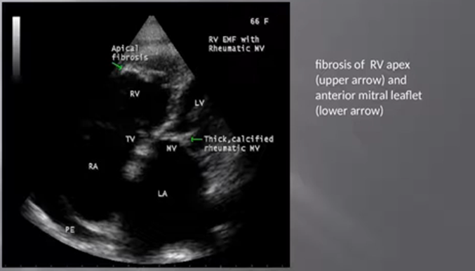
Figure 46: showing fibrosis of RV apex (upper arrow- EMF) and anterior mitral leaflet (lower arrow- RF)
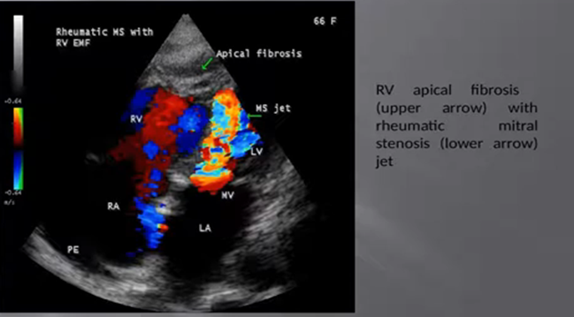
Figure 47 showing RV apical fibrosis (upper arrow) and rheumatic mitral stenosis (lower arrow) [60]
Medical Treatment
Medical therapy is aimed to control the symptoms with amelioration of acute illness and to prevent and treat heart failure, arrhythmias and thromboembolism. In endemic areas, small doses of diuretics and antibiotics are helpful to treat the initiating febrile illness with facial swelling. Steroids have little or no influence on the natural course of EMF [41] and antifailure measures such as digitalis, diuretics, ACE (angiotensin converting enzyme) inhibitors are utilized to treat heart failure. Patients with advanced disease may require invasive procedures to alleviate effusion and arrhythmias. Recurrent pericardial effusion can benefit from pericardio-pleural windows or pericardio-peritoneal shunts. Management of ascites relies on frequent evacuation of fluid by paracentesis with intravenous replacement of albumin at the time of procedure is used to compensate the protein loss [42],[43]. Patients with atrial fibrillation and thrombus or SEC (spontaneous echo contrast) may need anticoagulant therapy with warfarin and antiplatelet agents (aspirin or clopidogrel) and AV blocks may require pacemaker implantation
Surgical Therapy Surgery improves survival and must be performed before the occurrence of irreversible cardiac and hepatic damage [44]. In advanced stage of EMF, surgical decortications (conservative endocardiectomy) through a relatively well-preserved cleavage plane to excise the thick fibrotic endocardial lining and valve repair or replacement. This procedure causes reduction in ventricular filling pressures, improvement in hemodynamic status, but the operative mortality is high (15 to 25%) [45]. Long-standing ascites, extensive fibrosis and calcification, impaired myocardial function are relative contra-indications for surgery. Recurrence of fibrosis may occur after surgery, but excellent long-term survival was observed in certain cases [46]. Cardiac transplant was done in one patient [47]. Investigational Therapy The presence of inflammatory markers such as C-reactive protein, cytokines (IL-5, TNF-α) suggest that anti-inflammatory agents can be used to control the febrile illness at the initial stage and to prevent the progression of the disease similar to the role of acetyl salicylic acid in rheumatic fever. EMF is an inflammatory disorder and the fusion protein, a tyrosine kinase created by fusion of PPGFRA and FIPILI genes, found as a therapeutic target for imatinib in idiopathic hypereosinophilic syndrome has been investigated in the treatment of EMF [48]. The use of Dopamine agonists in Parkinson’s disease induce valvular fibrosis through its action on 5HT (serotonin)2B receptors and polymorphism in this receptor may cause EMF [49]. 5HT2B receptor blockers such as ‘ terguride’may be helpful to inhibit the fibrotic process of EMF [50].
Endomyocardial fibrosis continues to be an enigmatic disease [51]. The final common pathway causing the endothelial damage and fibrosis is to be evaluated [52]. It has been found that ‘burnt-out’ stage of EMF was associated with skin disease such as Psoriasis, Pemphigus and pulmonary infection caused by tuberculosis, and concluding that EMF is an immunologically mediated disorder similar to rheumatic heart disease, following an infectious origin and the nature of infection varies in different geographical areas [53] Endomyocardial fibrosis, first described >75 years ago, is a cause of restrictive cardiomyopathy with an unclear aetiopathogenesis that is most commonly found in children and adolescents from tropical regions of Africa, Asia and South America. However, more cases are being identified in Kerala, a north Indian state [54]. The epidemiological trends of this cardiomyopathy are difficult to ascertain. The characteristic hallmark of endomyocardial fibrosis is ventricular fibrosis [55],[56] that causes diastolic dysfunction and atrioventricular regurgitation. The outcomes in affected patients remain poor. Cases of EMF may emerge in non-endemic areas, posing a diagnostic challenge for healthcare professionals unfamiliar with this condition [57]. A major focus of research is the identification of biomarkers of preclinical disease and new therapeutic targets. Collaborative multidisciplinary research and cross-learning from other fibrotic conditions should impart knowledge and help to improve the survival rates and the quality of life of patients with endomyocardial fibrosis [58].
Multimodality imaging is essential for the initial and definitive diagnosis of this disease. Cardiac magnetic resonance, through late gadolinium enhancement, allows tissue characterization and identifies the ‘double V sign’, which is pathognomonic of the disease, allowing confirmation of the diagnosis as shown in Figure 45.
The worsening heart failure despite successful valve repair, attributed to unexplained restrictive filling patterns [61] and its reason remains a vanishing mystery. Whether the same strains of rheumatic fever may also cause apical fibrosis is not clearly understood and so the rends are going to be difficult to eradicate these global epidemic problems in the tropics
Dear Editorial Team, Clinical Medical Reviews and Reports. My experience with the journal was highly positive. The peer-review process was rigorous, constructive, and completed in a timely manner. The reviewers provided valuable comments that helped improve the quality and clarity of our manuscript. The editorial office was professional, responsive, and supportive throughout all stages of the publication process. Communication was clear and efficient, and any questions were addressed promptly. Overall, I found the journal to maintain high scientific standards and an excellent publication workflow. I would be pleased to consider submitting future work to this journal. Best wishes from, Elena Popa.

It was my pleasure to submit my testimonial concerning the Reviewer Board of our Scientific Journal “Brain and Neurological Disorders”. The Reviewers focused on some modifications and their contribution was helpful. The ladies of our Editorial Office were also supported my efforts. It was my honor to have such a co-operation and I am looking forward for more collaboration.

Dear Grace Pierce, Editorial Coordinator of Journal of Clinical Research and Reports, Thank you for the speedy and efficient peer review process. I appreciate the fact that your peer reviewers do not take months to respond like with some other journals. I would also like to thank the editorial office for responding quickly to my questions. It is an excellent journal. I plan to submit more manuscripts in the future. Best wishes from, Robert W. McGee

Dear Grace Pierce, Editorial Coordinator of Journal of Clinical Research and Reports, Working with you and your team on our recent publication in JCRR has been a truly wonderful and enjoyable experience. The responses were prompt, and the reviewers were patient, constructive, and highly professional. One reviewer in particular gave me the feeling that a professor was carefully reading and commenting on my coursework, which was deeply touching. The entire process was straightforward and hassle‑free, with no tedious online forms to complete. I highly recommend this journal. Best wishes from, DR Aibing Rao, Head of R&D

I Appreciate the Opportunity to Share my Experience with the Journal of Clinical Research and Reports. The peer review process was timely and constructive, and the feedback provided helped improve the quality of our manuscript. The editorial office was professional, responsive, and supportive throughout the process, ensuring smooth communication and efficient handling of the submission. Overall, it was a positive experience collaborating with your team.

Dear Mercy Grace, Editorial Coordinator of Obstetrics Gynecology and Reproductive Sciences, We would like to express our gratitude for your help at all stages of publishing and editing the article. The editors of the magazine answer all the necessary questions and help at every stage. We will definitely continue to cooperate and publish other works in the Obstetrics Gynecology and Reproductive Sciences! Best wishes from, Alla Konstantinovna Politova,
