
Case Report | DOI: https://doi.org/10.31579/2641-0419/418
President of all Nations Morning star hospital, Enayam Thoppu, Kanyakumari District, Tamil Nadu state, India.
*Corresponding Author: Ramachandran Muthiah President of all Nations Morning star hospital, Enayam Thoppu, Kanyakumari District, Tamil Nadu state, India.
Citation: Ait Yahya AE, Ztati Simohammed, Noussaiba Malhabi, Imane Katif, Mohammed EL Jamili, et al, (2024), “End-stage” Constrictive Pericarditis- Stem cell Therapy, J Clinical Cardiology and Cardiovascular Interventions, 7(12); DOI: 10.31579/2641-0419/418
Copyright: © 2024, Noussaiba Malhabi. This is an open access article distributed under the Creative Commons Attribution License, which permits unrestricted use, distribution, and reproduction in any medium, provided the original work is properly cited.
Received: 16 September 2024 | Accepted: 30 October 2024 | Published: 29 October 2024
Keywords: ‘end-stage’ constrictive pericarditis; engorged neck vein; septal bounce; waffle procedure; amniotic stem cell therapy
Aim: To present a case of ‘end-stage’ constrictive pericarditis with clinical manifestations such as ascites mimicking as cirrhosis of liver.
Introduction: ‘End-stage’ constrictive pericarditis has been readily confused with cirrhosis of liver in which there may be ascites, but venous pressure is normal and the neck veins are not engorged. There may be cardiac enlargement in other causes of heart failure. Etiology remains unknown in majority of case and inflammatory process play a central role in its development.
Case Report: A 67-year old male presented with sudden onset of tachycardia. Clinical examination revealed right-sided heart failure, ‘Egg-shell’ calcification in Chest X-ray and a characteristic echocardiographicfeatures of pericardial constriction such as septal bounce and dynamic respiratory changes in mitral inflow velocity. The patient was advised medical measures since it is in advanced stage.
Conclusion: When clinical signs of right heart failure become unresponsive to increased doses of diuretics, constrictive pericarditis is more likely the underlying disease since severe, right-sided failure develops in very advanced, the end-stage of the disease.
The normal pericardium is a fibroelastic sac surrounding the heart and consists of two layers. The visceral pericardium (serous pericardium) is a single layer of mesothelial cells contiguous with epicardium and reflects on itself over the origin of great vessels up to 1 to 2 cm and pulmonary veins. A tough, fibrous layer as a parietal pericardium and the sac created by these layers contain a small amount of fluid (< 25>
A 67-year old male was admitted with sudden onset of palpitations in the emergency room. ECG revealed tachycardia with a heart rate of 150 bpm as in Figure 4 and blood pressure 110/70 mmHg. Blood chemistry revealed normal. Physical examination showed an engorged neck vein as shown in Figure 2 which fails to decrease with inspiration (Kussmaul’s sign) with a deep Y descent (Freidreich’s sign) reflecting the predominant ventricular filling during early diastole, ascites and pedal edema as shown in Figure 1 suggesting a right-sided heart failure. Auscultation revealed pericardial knock, an early diastolic sound occurs due to cessation in diastolic filling and retraction of apical impulse in systole. X-ray chest revealed “egg-shell’ calcification as shown in Figure 3. Transthoracic echocardiography revealed the features of constrictive pericarditis as in Figures 7 to 12. Since the patient is in end-stage disease, he was given conservative medical measures such as diuretics, antibiotics and anti-inflammatory drugs and the rhythm was controlled with calcium channel antagonist, verapamil 40 mg three times daily as shown in Figures 5 and 6.
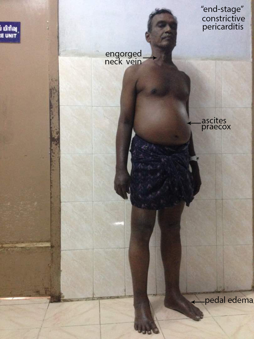
Figure 1: Showing the clinical features of ‘end-stage’ Constrictive pericarditis.
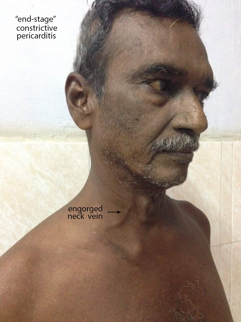
Figure 2: Showing the ‘engorged neck vein’ as a feature of elevated venous pressure in ‘end-stage’ constrictive pericarditis
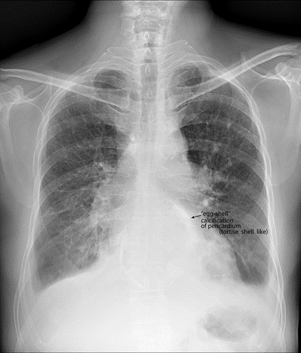
Figure 3: X-ray chest PA (postero-anterior) view showing the ‘egg-shell’ calcification’ – ‘tortoise-shell’ like and flattening of right heart border in ‘end-stage’ constrictive pericarditis.
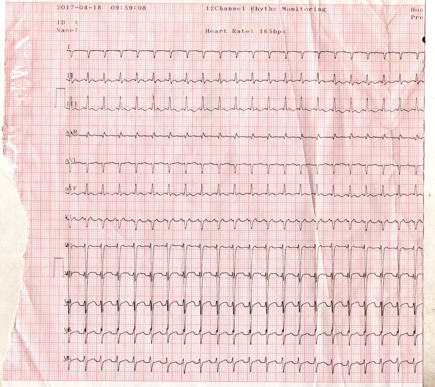
Figure 4: ECG showing tachycardia (rate 150 bpm) in a 67 year old male with ‘end-stage’ constrictive pericarditis.
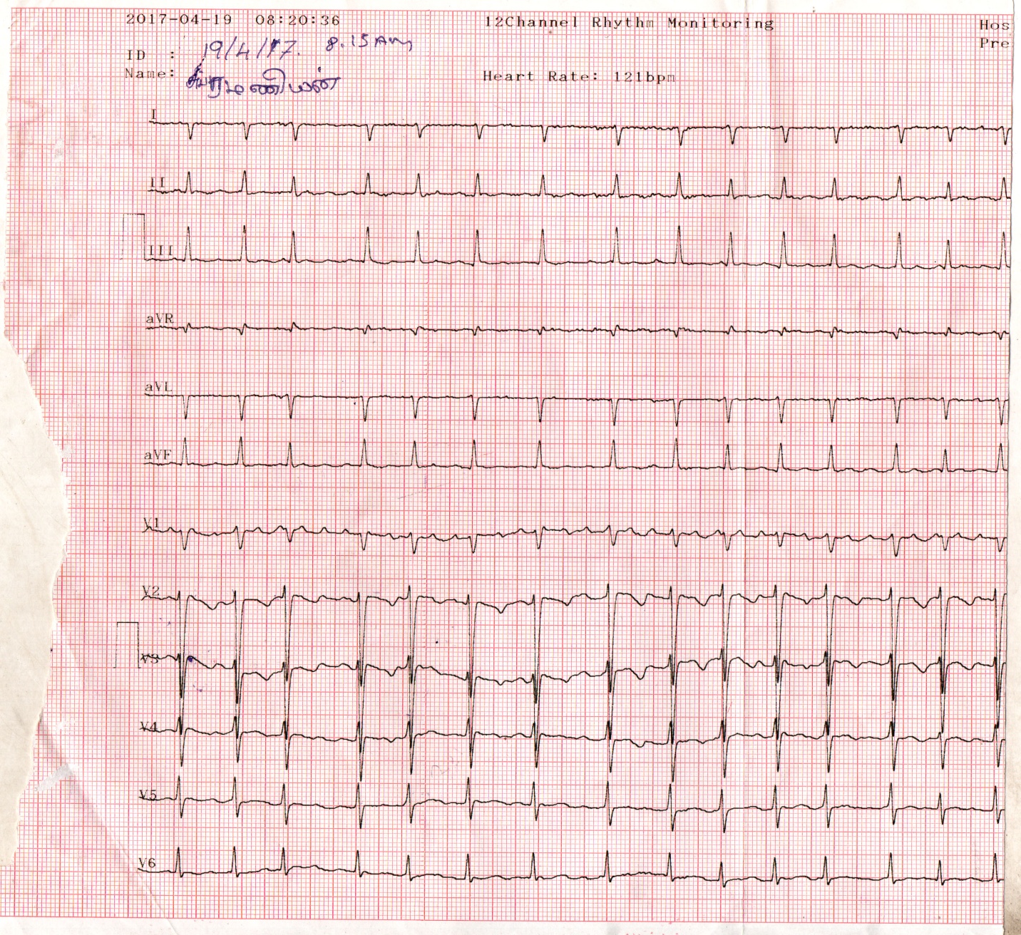
Figure 5. ECG showing atrial fibrillation after controlling the heart rate with verapamil

Figure 6: ECG normalizing on continuation of verapamil

Figure 7: Showing the ‘ acute angle’ between the LA (left atrium) and LV(left ventricle) posterior walls and a pericardial thickness of 8 mm.

Figure 8: Apical view showing the ‘septal bounce’ as a sign of ventricular interdependence and bulging of IAS (interatrial septum) towards LA (left atrium).

Figure 9: M-mode LV study showing the ‘septal notch’ and ‘ dip and flattening’ of LV posterior wall

Figure 10: Pulsed Doppler imaging showing ‘the dynamic respiratory change’ of the mitral inflow velocity of constrictive pattern.


Figure 11: Pulsed Doppler imaging showing the expiratory increase in diastolic IVC flow reversal in both phases of respiration, but more prominent in expiration

Figure 12: Subcostal view showing the dilated IVC (inferior vena cava- plethoric ) with no respiratory variation.

Figure 13: Pulsed Doppler imaging showing the mitral inflow velocity with no respiratory variation of restrictive pattern.

Figure 14: Showing the IVS (interventricular septum) calcification in effusive-constrictive pericarditis as evidenced by mild pericardial effusion with an associated disease of Endomyocardial fibrosis.
Review of literature
In 1669, Lower [2] described the clinical effects of interference of cardiac diastole by a constricting fibrous pericardium. In 1756, Morgagni [3] contributed to the understanding of pathophysiology of constrictive pericarditis. In 1828, Lancisi described the characteristic syndrome of constrictive pericarditis and in 1842 [4], Chevers was the first to present clearly the clinical picture of chronic constrictive pericarditis. In 1870, Wilks [5] further emphasized this syndrome. In 1896, Pick [6] described the postmortem evidence of adhesive pericarditis and an atypical fibrosis (pseudocirrhosis) of the liver and its capsule in patients with constrictive pericarditis. The thickened peritoneum over the liver is not to be the engorgement of liver itself in so-called Pick’s disease, but rather to the occurrence of acute peritonitis at the time of acute pericarditis followed by residual fibrosis. Concato described the effusion in serous cavities (polyserositis) in patients with constrictive pericarditis is due to the result of cardiac compression, and inflammation of serous membranes is absent or occur secondarily.
Etiopathogenesis
Constrictive pericarditis is most commonly caused by conditions or events that cause inflammation to develop around the heart. Inflammatory process of the pericardium typically causes pain and fluid accumulation and more chronically results in fibrosis and calcification of pericardium with pericardial constriction, the process that inhibit diastolic filling of the heart. The most common antecedents are idiopathic and tuberculosis. In many cases, the etiology is not identified, however, pericardial fibrosis and calcification are often idiopathic in origin [7]. The tuberculosis accounted for 49% of cases of constrictive pericarditis in a series reported in 1962 [8] and it was found to be most common cause in third-world countries such as India [9],[10]. Viral pericarditis is more common in the west and in Europe and North, it is often a sequelae of cardiac surgery and mediastinal irradiation.
Constrictive pericarditis can occur after many pericardial disease process. All causes of pericarditis can lead to subsequent constriction [11]. Rheumatic fever, although frequently accompanied by pancarditis, does not result in chronic constrictive pericarditis and may have pericardial adhesion which are not maximally constricting. The pericarditis associated with uremia and with myocardial infarction is not of the constricting type and most cases of effusive-constrictive pericarditis are often idiopathic, can occur in malignancy of breast and lung, tuberculosis and hypothyroidism (cholesterol pericarditis or ‘gold paint’ pericarditis) The causes of constrictive pericarditis are shown in Table 1
| Most common | Less common |
Idiopathic -42 to 49 % [12] Mediastinal irradiation (5-10 years duration) Following Cardiac surgery-11 to 37 % (post-pericardiectomy-10 to 40 %, previous cardiac surgery-0.3 %) Radiotherapy- 9 to 31 % Post-infectious- 3 to 6 % Tuberculosis-49 % (the most common cause In developing countries) Viral infections (coxsackie virus A and B, Adenovirus, echovirus Pyogenic infections
| Other infections Neoplasms -5-17 % (lung-33 %, breast-25 %, adenocarcinoma of intestine [13]) Connective tissue disorders (rheumatoid Arthritis, systemic lupus erythematosis, Scleroderma) Drugs(procainamide,hydralazine,methysergide) Trauma Hereditary-Mulibrey nanism(Mu-muscle, Li-liver, br-brain, ey-eyes, nanism-dwarf) in Finland and United states [14] |
Table 1: showing the causes of constrictive pericarditis
Hemodynamic changes
The normal pericardium can stretch to accomodate the physiological changes in cardiac volume. In constrictive pericarditis, the visceral and parietal pericardium are fibrosed and fused together [15], although not necessarily always thickened [16], prevent the heart from expansion and resulting in minimal ability to adapt to volume changes and significant dynamic respiratory variation in blood flow in the chambers of the heart attributed to isolation of the cardiac chambers from intrathoracic respiratory pressure changes, ie, dissociation between intrathoracic and intracardiac pressures with enhanced ventricular interaction as reported by Hatle et al in 1989 [17].
In the heart with a normal pericardium, inspiration causes a decrease in intrathoracic pressure, which is reflected in the cardiac chambers as decrease in intracardiac pressures simultaneously and there is no change in the driving pressure from the lungs across the pulmonary veins into the left atrium and across the mitral valve into the left ventricle. There is some increase in the filling of the right ventricle because of enhanced venous return, but filling of the left ventricle is unaffected throughout the cardiac cycle. In patients with constrictive pericarditis, the rigid pericardium does not allow the decrease in intrathoracic pressure to be transmitted to the left -sided chambers and there is a lower driving force from the lungs into the left side of the heart and the left ventricle becomes underfilled with a reciprocal increase in the filling of the right ventricle and therefore a septal shift occurs [18]. Conversely, during expiration, there is decreased filling of the right ventricle and increased filling of the left ventricle. As both ventricles are sharing the same limited space, the chamber size and function of one ventricle affect the other ventricle and this interaction is known as ‘ventricular interdependence’ since the amount of blood flow into one ventricle is dependent on the amount of blood flow into the other ventricle and it is enhanced in constrictive pericarditis with a discordance in right and left heart fillings.
Once the ventricular diastolic filling reaches the limitations of the pericardial restraint, the pressure and volume in the cavity rise, filling ceases, and congestion occurs [19]. If the right heart chambers are predominantly constricted, there is naturally an engorgement of neck veins as shown in Figure 2, which is constantly engorged in 86% of patients with constrictive pericarditis. The decreased compliance of right ventricle causes a rise in right atrial pressure that is greater than the fall in pleural pressure, ultimately leading to distended neck veins during inspiration [20], called as ‘Kussmaul’s sign’, which may be seen in right ventricular failure, right ventricular infarction, tricuspid stenosis and restrictive cardiomyopathy. It is nonspecific for constrictive pericarditis and reflects an elevation of Jugular venous pressure (JVP) on inspiration rather than the expected decrease in JVP. When the right heart fails because of constriction of left heart chambers, it may simulate the effect of tricuspid valve disease. With more severe constriction, the peripheral edema and ascites occur. In constrictive pericarditis, the ascites occurs early, disproportionately prominent as well as recurrent and appropriately called as ‘ascites praecox’, followed by minimal edema as a manifestation of later part (end-stage) of the disease and it is usually confined to lower extremities and sacrum whereas in congestive heart failure, the edema appears first and ascites much later.
In isolated constrictive pericarditis, the myocardium is unaffected and therefore the systolic function and early diastolic filling are normal. In the mixed form (constrictive-restrictive- mainly due to radiation –induced, post cardiac surgery), atrophy of myocardial cells and fibrosis may develop during long-term compression by the pericardium. Both the irritation of the heart by the actual process involving the myocardium and the constricting effect of left heart chambers on the right ventricle and right atrium result in atrial arrhythmias such as atrial fibrillation as shown in Figure 5 and less commonly atrial flutter as complications in chronic constrictive pericarditis. With diminution in the output of heart, the blood pressure, especially the pulse pressure tends to be low and the blood pressure decreasing even to the point of disappearance during inspiration, manifested as absence of pulse, an important sign called as ‘paradoxical pulse’ in some of the more advanced cases. Pulsus paradoxus is an exaggeration of normal decrease in systolic blood pressure during inspiration and is formally defined as an inspiratory decrease in systolic blood pressure greater than 10 mmHg during quiet breathing (readily detected by sphygmomanometer, when the reduction is >20 mmHg- it can be detected by simple palpation of brachial artery, severe cases-inspiratory disappearance of pulse) and it is commonly associated with cardiac tamponade, but can be seen occasionally in other conditions such as effusive-constrictive pericarditis, acute severe asthma, acute pulmonary embolism and right ventricular infarction. Normally, the systolic pressure varies with respiratory cycle, but not to the extent seen in pulsus paradoxus. During inspiration, the right ventricle distends due to increased venous return and causes the interventricular septum to bulge into the left ventricle, decreasing the capacity for left ventricular filling and causing a pooling of blood into the pulmonary vessels, which in turn results in a decrease in left ventricular stroke volume, manifested as an exaggerated decrease in the systolic blood pressure [21]. ‘Reversed pulsus paradoxus’(an inspiratory rise in arterial pressure) may occur in hypertrophic obstructive cardiomyopathy [22] and aortic regurgitation tends to prevent the development of pulsus paradoxus despite the presence of cardiac tamponade and it may be absent when the LV and RV diastolic pressure is high or decompression of respiratory changes in pressures as in atrial septal defects. The venous pressure, on the other hand, is very much elevated and frequently exceeding 200 mm of H2O, even exceeding 300 mm of H2O. The salient features of end-stage constrictive pericarditis are shown in Table 2.
High venous pressure Diminution of blood pressure Paradoxical pulse Atrial fibrillation Ascites Low cardiac output (cardiac index = ≤ 1.2 L/m2/minute) Pseudocirrhosis Pedal edema |
Table 2. showing the salient features of constrictive pericarditis
Diagnostic Methods
Radiological The plain radiograph is frequently abnormal in patients with hemodynamically significant constrictive pericarditis [23]. A typical X-ray chest of a patient with constrictive pericarditis shows a normal sized heart (47%) or only mildly enlarged (16%) and moderate to marked enlargement (37%) in effusive-constrictive pericarditis [24]. Cardiac contour abnormalities, particularly the flattening of right cardiac border is a characteristic feature of constrictive pericarditis, but infrequently present. The left atrium, which is covered only partly by the pericardium may be enlarged.
Calcification of pericardium on chest X-ray strongly suggests constrictive pericarditis in patients with features of heart failure ( especially right heart failure) and it is more obvious in regions where the normal fat is found, namely in atrioventricular and interventricular grooves. A localized form of constriction in the mid ventricular segments as a result of localized severe calcification resembling a ‘napkin’ ring shape is termed as “ napkin-ring” constrictive pericarditis [25]. Once calcification has developed, it represents chronic pericarditis and it was present in 40
“End-stage” constrictive pericarditis has posed a diagnostic dilemma since it presented with features of right-sided heart failure such as dyspnea, ascites, edema and elevated JVP (Jugular venous pressure) [95]. Approximately 9% of patients with acute pericarditis go on to develop constrictive physiology. The most important diagnostic tool is clinical suspicion and cardiac catheterization is no longer performed to diagnose it [96]. Two-dimensional echocardiography is used mainly to rule out other causes of right-sided heart failure and Doppler echocardiography may provide additional diagnostic information and confirm the presence of constrictive physiology. Medical therapy may be used as a palliative measure to control symptoms and to optimize hemodynamics in ‘end-stage’ disease, who are not candidates for surgery [97].
Dear Editorial Team, Clinical Medical Reviews and Reports. My experience with the journal was highly positive. The peer-review process was rigorous, constructive, and completed in a timely manner. The reviewers provided valuable comments that helped improve the quality and clarity of our manuscript. The editorial office was professional, responsive, and supportive throughout all stages of the publication process. Communication was clear and efficient, and any questions were addressed promptly. Overall, I found the journal to maintain high scientific standards and an excellent publication workflow. I would be pleased to consider submitting future work to this journal. Best wishes from, Elena Popa.

It was my pleasure to submit my testimonial concerning the Reviewer Board of our Scientific Journal “Brain and Neurological Disorders”. The Reviewers focused on some modifications and their contribution was helpful. The ladies of our Editorial Office were also supported my efforts. It was my honor to have such a co-operation and I am looking forward for more collaboration.

Dear Grace Pierce, Editorial Coordinator of Journal of Clinical Research and Reports, Thank you for the speedy and efficient peer review process. I appreciate the fact that your peer reviewers do not take months to respond like with some other journals. I would also like to thank the editorial office for responding quickly to my questions. It is an excellent journal. I plan to submit more manuscripts in the future. Best wishes from, Robert W. McGee

Dear Grace Pierce, Editorial Coordinator of Journal of Clinical Research and Reports, Working with you and your team on our recent publication in JCRR has been a truly wonderful and enjoyable experience. The responses were prompt, and the reviewers were patient, constructive, and highly professional. One reviewer in particular gave me the feeling that a professor was carefully reading and commenting on my coursework, which was deeply touching. The entire process was straightforward and hassle‑free, with no tedious online forms to complete. I highly recommend this journal. Best wishes from, DR Aibing Rao, Head of R&D

I Appreciate the Opportunity to Share my Experience with the Journal of Clinical Research and Reports. The peer review process was timely and constructive, and the feedback provided helped improve the quality of our manuscript. The editorial office was professional, responsive, and supportive throughout the process, ensuring smooth communication and efficient handling of the submission. Overall, it was a positive experience collaborating with your team.

Dear Mercy Grace, Editorial Coordinator of Obstetrics Gynecology and Reproductive Sciences, We would like to express our gratitude for your help at all stages of publishing and editing the article. The editors of the magazine answer all the necessary questions and help at every stage. We will definitely continue to cooperate and publish other works in the Obstetrics Gynecology and Reproductive Sciences! Best wishes from, Alla Konstantinovna Politova,
