
Research Article | DOI: https://doi.org/10.31579/2766-2314/126
Equipe d’Electrochimie Moléculaire et Matériaux Inorganiques, Faculté des Sciences et Techniques de Béni Mellal, Université Sultan Moulay Slimane, Maroco
*Corresponding Author: Abdelilah Chtaini, Equipe d’Electrochimie Moléculaire et Matériaux Inorganiques, Faculté des Sciences et Techniques de Béni Mellal, Université Sultan Moulay Slimane, Maroco.
Citation: Amine Smaini, Rajaa Maallah, Salah Eddine El Qouatli, Rachida Najih and Abdelilah Chtaini, (2023), Enzymatic Degradation of Aflatoxin Accumulation in Dry Maize Kernels as a Promising Post-Harvest Mitigation Strategy, J, Biotechnology and Bioprocessing, 4(7); DOI:10.31579/2766-2314/126
Copyright: © 2023, Abdalrauf M.A.B. This is an open access article distributed under the Creative Commons Attribution License, which permits unrestricted use, distribution, and reproduction in any medium, provided the original work is properly cited.
Received: 02 October 2023 | Accepted: 13 October 2023 | Published: 27 October 2023
Keywords: anaerobic; digestion; biogas; micromycetes; waste
Agricultural and agro-industrial activities generate large quantities of waste which are harmful to the environment. This waste, rich in organic matter, can be recycled and transformed by biotechnological processes which constitute a solution of choice for remedying pollution problems. The aim of this study is the treatment of the organic fraction of waste by anaerobic digestion, which consists of degradation in the absence of oxygen of the organic matter into a mixture of methane (CH4) and carbon dioxide (CO2) called biogas. For this we have chosen the following samples (manure; manure mixed in bananas, oranges, lemons, potatoes and tomatoes; manure mixed in breads, zucchinis, carrots, cucumbers and strawberry). We used a biogas plants based on simple tools. Among these samples, the T container (manure) the most productive mixture of biogas and the T (manure) and F2 mixture (manure + breads + zucchinis + carrots + cucumbers + strawberry) degraded faster than the F1 manure + mixture (bananas, oranges, lemons, potatoes and tomatoes). The micromycetes that these samples contain are Penicillium italicum, Penicillium digitatum, Rhizopus sp, Mucor, Aspergillus sp, Cladosporium. Micromycetes give good biogas yield. The genus Aspergillus accelerates the degradation of organic matter. Anaerobic digestion not only prevents pollution, but also produces energy, compost and replenishes nutrients. Anaerobic digestion can turn a waste problem into a source of wealth. This technology is becoming essential in the process of reducing waste and producing biogas, a source of renewable energy.
In recent years, a great deal of attention has been paid to virology, with the objective to develop appropriate and affordable methods for the detection of whole viruses or their fragments. Conventional methods commonly used for the detection of antibodies against viruses are related to enzyme-linked immunosorbent assays (ELISA) [1], hemagglutination inhibition (HI) [2-3] and Western blot assay (WB) [4]. However, they are often laborious and time-consuming or require expensive instruments and are only available in experienced laboratories. Furthermore, emergency cases require a rapid portable detection system.
Consequently, an important need still exists to create simple, sensitive and inexpensive diagnostic methods for the detection of antibodies against different types of viruses. Immunosensors incorporating specific antigen are promising alternative systems for the direct detection of biomolecules such as immunospecies. Low sample consumption, reasonable instrument cost and good possibility for miniaturization and minimization of analysis time in immunoassay are the main reasons for the extensive development of electrochemical immunosensors for immunoassay.
Several different types of immunosensors have been successfully developed for the detection of various types of antibodies [5], his-tagged proteins [6] or antigens [6], [7], [8].
In this paper, we present a sensitive and selective immunosensor for the detection of antibodies against West Nile virus which could also be used to detect the presence of corvid-19 antibodies.. The basis is the species-induced electrical changes in the biological species, in particular the specific interaction between the peroxidase-secondary antibody conjugate (HRP-conjugate) and West Nile virus-specific antibodies (antibody/secondary antibody binding) evaluated mainly by cyclic voltametry (CV) and square wave voltametry (SQW) and electrochemical impedance spectroscopy in the presence of NaCl as electrolyte media. Finally, the immunosensor was used for the detection of West Nile virus-specific antibody response in quail serum samples and the results were compared with those obtained with ELISA.
Experimental Apparatus and Software
Voltammetric experiments were performed using a voltalab potentiostat (model PGSTAT 100, Eco Chemie B.V., Utrecht, The Netherlands) driven by the general-purpose electrochemical systems data processing software (voltalab master 4 software) run under windows 2007. The three-electrode system consisted of a chemically modified carbon paste electrode as the working electrode a saturated calomel electrode (SCE) serving as reference electrode, and platinum as an auxiliary electrode.
Electrodes
Modified electrodes were prepared by mixing a carbon powder and the desired weight of natural phosphate. The body of the working electrode for voltammetric experiments was a PTFE cylinder that was tightly packed with carbon paste. The geometric area of this electrode was 0.1256cm2. Electrical contact was made at the back by means of a bare carbon. The modified electrodes were immersed in a peroxidase-secondary antibody conjugate (PAS) solution. The effect effect of time of cumulation and concentration of solution were studied. Then the modified electrodes (CPE-NP modified with PAS) were immersed in a cell 20 ml of distilled water, 0.1M NaCl and the antibodie suspensions and then characterized as a function of time by cyclic voltammetry and by electrochemical impedance spectroscopy.
Figure 1 represents the cyclic voltammograms recorded at the CPE-NP electrode modified with PAS, with a scanning speed equal to 100 mV / S in the NaCl solution (0.1 mol / l) containing or not antibodie in a range of potential between -2 V and 2 V. It can be seen that the presence of antibodie is manifested by the appearance of a reduction peak at around 0.2V.

Figure 1: Overlay of cyclic voltammograms of CPE-NP / PAS (a) without antibodies and CPE-NP / PAS with antibodies (b), in NaCl at 0.1 M; v = 100mV/s; pH = 7
The morphology of the electrode surface of CPE-NP modified with PAS, before incubation in antibodies (curve a) and after 20 min incubation in antibodies (curve b), was observed by optical scanning microscopy (Figure 2). It can be seen that the presence of the antibody is manifested by the appearance of three-dimensional reliefs, deposited over the entire surface of the electrode. The antibodies have attached to the electrode surface probably via redox properties.
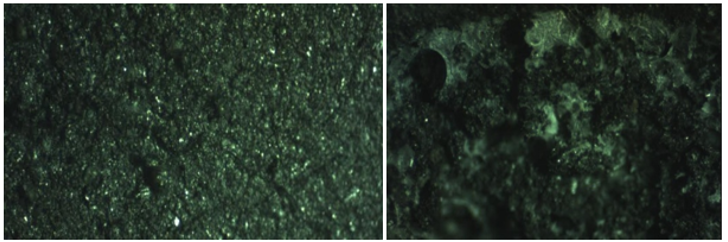
Figure 2 : Micrograph of the CPE-NP / PAS Electrode before incubation in antibodies (a) and after 20 min incubation in antibodies (b)
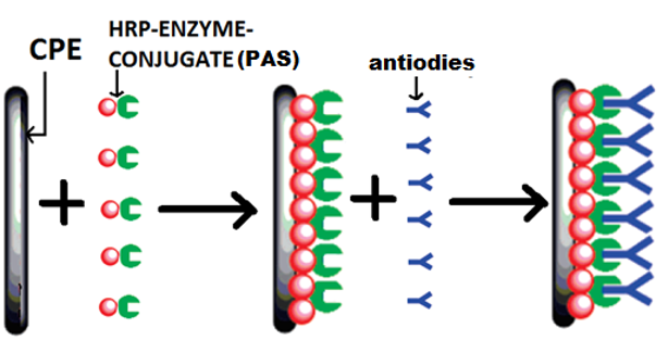
Figure 3 : illustre the overall scenario reflecting the proposed mechanism from the development of the PAS-modified CPE-NP electrode to the adsorption of the antibody using the redox properties of the PAS and the biological analyte (Antibody).
Effect of antibody incubation time
The CPE-NP / PAS immunological sensor was incubated with a solution containing antibodies at different times. The results are shown in Fig. 4, by cyclic voltammetry. During the first 20 minutes of antibody contact with the CPE-NP electrode modified with PAS, the cathode peak current density increases rapidly with the incubation time, which indicates a good electroactivity on the electrode surface due to the reaction between the
antibodies and the secondary peroxidase-antibody, more precisely to the formation of the bond between the antibodies and the secondary antibodies, After 20 min, it becomes almost constant and the tendency to saturate, due to the probable saturation of the antibody-secondary antibody binding sites conjugated to peroxidase-conjugated peroxidase.
Thus, 20 minutes was selected as the optimal incubation time to maximize the signal and minimize the assay time.
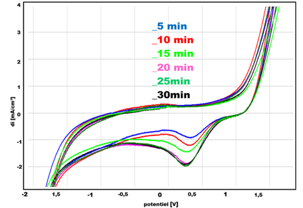
Figure 4: Superimposition of cyclic voltammograms of CPE-NP / PAS at different deposition times, in NaCl at 0.1 M; v = 100mV/s, from -2V to 2V, pH = 7
After each incubation time of the PAS-modified CPE-NP electrode, samples of the solution containing the antibodies were taken to measure the optical density of the sample after the Elisa test. The results are shown in Table 1 and in Figure 5.

Table 1: Optical density as a function of contact time of CPE-NP / PAS with antibodies S/N is the percentage given by the following equation:

- Samples with S/N less than or equal to 50% are considered negative.
- Samples with an S/N greater than 50% are considered positive.

Figure 5: Optical density as a function of contact time of CPE-NP / PAS With the antibodies
Effect of antibody dilution factor (the concentration effect)
The immunosensor was incubated in different media containing increasing concentrations of antibodies for 20 min. An increase in current density was recorded as the concentration of antibodies increased. As shown in Figure 6. The detection power of the electrode therefore increases with increasing antibody loading up to C2 and then the tendency to saturate :
- At low concentrations of adsorbed antibodies, the signal is very low but not zero. Antibodies immobilized on the surface do not ensure complete coverage of all active sites.
- At higher concentrations, it is evident that the signal increases, revealing the specific adsorption of antibodies to the adsorbed conjugate. A slight tendency to saturation is shown at the highest concentration (> C2); this tendency is attributed to saturation of the surface binding sites.
This electrochemical behavior of the electrode is confirmed by Electrochemical impédance spectroscopy (Fig. 7), EIS curves have the shape of a half-loop, the diameter of which corresponds to the electron transfer resistance.

Figure 6: Superimposition of cyclic voltammograms of CPE-NP / PAS at different antibody concentrations in NaCl at 0.1 M; v = 100mV/s, from -2V to 2V; pH = 7

Figure 7: Superimposition of electrochemical impedance spectra of CPE-NP / PAS at different antibody concentrations in NaCl at 0.1 M; 100 mHz at pH = 7
Study of the effect of optical density (OD) of antibodies on CPE-NP / PAS
The calibration curve was plotted under the optimised conditions, previously determined, using cyclic voltammetry. The current intensity of the cathodic peak on CPE-NP / PAS is proportional to the optical density (OD) of the antibodies obtained after the classical ELISA method, in the range of C6 to C1 (Fig. 8). This linearity is expressed by the following relationship:
Di = 11,79DO - 5,292 R² = 0,982

Table 2: Influence of antibody concentration on the intensity of reduction peaks Obtained by VC on the surface of CPE-NP / PAS
Determination of the limit of detection and quantification of CPE-NP / PAS
According to Miller and Miller [2], the standard deviation of the measured mean current (SD) can be modeled by the equation:

Where ij is the experimental value of the current identified at manipulation j and Ij is the corresponding value calculated at the same optical density using the calibration equation. n is the number of measurements performed. The calculated SD (standard deviation) value is used to determine the detection limit (LD) and quantitation limit (LQ).
LD= 3 ×SD/slope
LQ= 10 ×SD/slope
For the PAS-modified CPE-NPelectrode, the detection limit is 1.8.10-3 and the quantification limit is 7.2. 10-2.
Analytical Application
The applicability of the immunosensor for the detection of antibodies in quail serum was demonstrated. 5 quail serum samples with different degrees of infection were tested. The results are given in Table 3, which demonstrate that the immunosensor meets the requirements for clinical analysis to assess the degree of West Nile infection in quail serum.
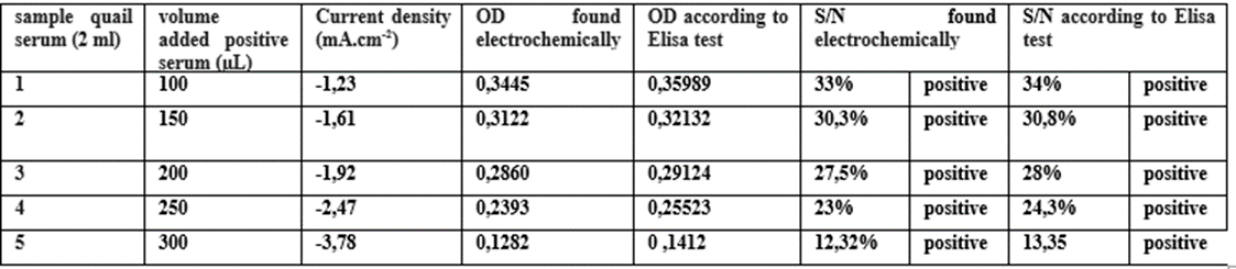
Table. 3 : Analytical Application Results
This work demonstrated that the mixed carbon-phosphate paste electrode modified with PAS is a feasible alternative for the bio analytical determination of various virus. Analytical results show that the proposed is comparable to the ELISA test, but much faster and much less expensive, with good sensitivity and reproducibility.
Dear Editorial Team, Clinical Medical Reviews and Reports. My experience with the journal was highly positive. The peer-review process was rigorous, constructive, and completed in a timely manner. The reviewers provided valuable comments that helped improve the quality and clarity of our manuscript. The editorial office was professional, responsive, and supportive throughout all stages of the publication process. Communication was clear and efficient, and any questions were addressed promptly. Overall, I found the journal to maintain high scientific standards and an excellent publication workflow. I would be pleased to consider submitting future work to this journal. Best wishes from, Elena Popa.

It was my pleasure to submit my testimonial concerning the Reviewer Board of our Scientific Journal “Brain and Neurological Disorders”. The Reviewers focused on some modifications and their contribution was helpful. The ladies of our Editorial Office were also supported my efforts. It was my honor to have such a co-operation and I am looking forward for more collaboration.

Dear Grace Pierce, Editorial Coordinator of Journal of Clinical Research and Reports, Thank you for the speedy and efficient peer review process. I appreciate the fact that your peer reviewers do not take months to respond like with some other journals. I would also like to thank the editorial office for responding quickly to my questions. It is an excellent journal. I plan to submit more manuscripts in the future. Best wishes from, Robert W. McGee

Dear Grace Pierce, Editorial Coordinator of Journal of Clinical Research and Reports, Working with you and your team on our recent publication in JCRR has been a truly wonderful and enjoyable experience. The responses were prompt, and the reviewers were patient, constructive, and highly professional. One reviewer in particular gave me the feeling that a professor was carefully reading and commenting on my coursework, which was deeply touching. The entire process was straightforward and hassle‑free, with no tedious online forms to complete. I highly recommend this journal. Best wishes from, DR Aibing Rao, Head of R&D

I Appreciate the Opportunity to Share my Experience with the Journal of Clinical Research and Reports. The peer review process was timely and constructive, and the feedback provided helped improve the quality of our manuscript. The editorial office was professional, responsive, and supportive throughout the process, ensuring smooth communication and efficient handling of the submission. Overall, it was a positive experience collaborating with your team.

Dear Mercy Grace, Editorial Coordinator of Obstetrics Gynecology and Reproductive Sciences, We would like to express our gratitude for your help at all stages of publishing and editing the article. The editors of the magazine answer all the necessary questions and help at every stage. We will definitely continue to cooperate and publish other works in the Obstetrics Gynecology and Reproductive Sciences! Best wishes from, Alla Konstantinovna Politova,
