
Research Article | DOI: https://doi.org/10.31579/2767-7370/090
Research and Training Center ‘Physical and Chemical Materials Science’ Under Kyiv Taras Shevchenko University and NAS of Ukraine, Kiev, Ukraine.
*Corresponding Author: Yuri Pivovarenko, Research and Training Center ‘Physical and Chemical Materials Science’ Under Kyiv Taras Shevchenko University and NAS of Ukraine, Kiev, Ukraine.
Citation: Yuri Pivovarenko, (2024), Electrical Forces that Build and Recover Neurons, J New Medical Innovations and Research, 5(2); DOI:10.31579/2767-7370/090
Copyright: © 2024, Yuri Pivovarenko. This is an open access article distributed under the Creative Commons Attribution License, which permits unrestricted use, distribution, and reproduction in any medium, provided the original work is properly cited.
Received: 03 February 2024 | Accepted: 12 February 2024 | Published: 19 February 2024
Keywords: nerve; recovery; extracellular filaments; pulse therapy; laser therapy
It was previously shown that it is the electric charge (potential) of water that determines its structuring properties in relation to substances both dissolved in water and in contact with it. In particular, it has been shown that negative electrization of the water used causes the formation of needle-like, thread-like and plant-like crystals and sediments. Taking all this into account, the formation of dendritic structures in aqueous solutions of chlorides exposed to pulsed electromagnetic fields (PEMFs) has been proposed to be explained by the negative electrization of such solutions; accordingly, the regenerative effect of PEMFs on nerve tissue was proposed to be explained in the same way. Over time, it became clear that such explanations are incomplete because they do not take into account the electric fields created by charged surfaces, in particular the outer sides of the cytoplasmic membranes of neurons. To eliminate this incompleteness, a number of additional modeling studies were carried out. In particular, the effect of various charged surfaces on aqueous solutions was studied. As a result, it was shown that in aqueous solutions in contact with charged surfaces, filaments are formed that are collinear to the vectors of electric forces created by such surfaces. It was also shown that these same electrical forces are capable of determining both the integrity of the formed filaments and their mutual separation. Naturally, all the results obtained suggested that it is the electrical forces acting on the outer sides of the cytoplasmic membranes that create all extracellular filaments, including neuronal ones.
It was previously established that positively charged water promotes the formation of compact salt crystals and sediments, and negatively charged water promotes the formation of needle-like or tree-like ones (Figures 1 – 4) [1 – 4].
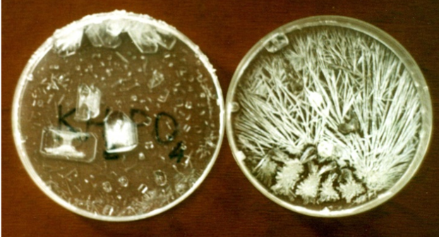
Figure 1: It is the crystals that formed after the drying of solutions of KH2PO4 prepared on the water with potentials of +250 mV (left) and –250 mV (right) [1, 2].
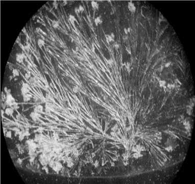
Figure 2: "Bouquets of flowers" are formed in drying NaCl solutions prepared in negatively charged water [1].

Figure 3: Left: these are intense blue prismatic crystals that formed in a copper sulfate solution prepared with positively charged water, which has a high hydrating capacity and high surface tension. Right: these are pale green, grass-like crystals formed in a copper sulfate solution prepared with negatively charged water, which has a low hydrating capacity and low surface tension.
It is worth noting here that fully hydrated copper sulfate, namely CuSO4●5H2O, has an intense blue color (left), while partially hydrated cooper sulfate, for example CuSO4●3H2O, is pale green (right) and completely anhydrous copper sulfate, namely CuSO4, is colorless [6].
All this makes it possible to use this particular salt as a visual indicator of the electrization of water.

Figure 4: It is copper oxide Cu2O dispersed in the water containing a gradient of electric potentials; water with positive potential is above and water with negative potential is below [1].
At the same time, it was found that positive electrization of water increases its surface tension, while negative electrization of water decreases it; in particular, this dependence manifests itself both in the tendency of positively charged water to compress and thereby increase its contact angle (Figure 5, right), and in the tendency of negatively charged water to spread and thereby reduce its contact angle (Figure 5, left)
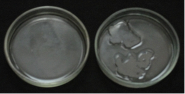
Figure 5: Left: 5 ml of water with an electric potential of –200 mV cover all the bottom of a Petri dish. Right: 5 ml of water with an electric potential of +200 mV do not cover the bottom of a Petri dish; the surface of such water decreases rapidly after mixing [1].
(Apparently, it is worth recalling here that the surface tension of any liquid directly correlates with its contact angle [5].)
Naturally, all this led to the conclusion that the increased surface tension of positively charged water promotes the formation of compact structures, and the reduced surface tension of negatively charged water promotes the formation of needle-like, thread-like and tree-like structures [1].
Apparently, it is worth adding here that the results obtained (Figures 1 – 5) also agree well with the ability of aqueous protons, with which
positively charged water is enriched, to form both oxonium ions, namely H3O+, and very compact symmetrical aggregates, namely H3O+•3H2O [6]. Also consistent with the same results is the ability of aqueous hydroxyl ions, which enrich negatively charged water, to form linear aggregates, namely OH–•3H2O [6].
One way or another, it was the knowledge of the established dependencies (Figures 1 – 5) that made it possible to purposefully create the desired structures, including those characteristic of living organisms (Figure 6).

Figure 6: These crystals, whose shape is similar to plant fragments and bird feathers (top right), formed after CuSO4 solutions prepared in water at various negative potentials dried [7].
One way or another, all the presented results (Figures 1 – 6) ultimately led to the conclusion that it is the electric charge (potential) of water that determines its structuring properties in relation to substances both in contact with water and dissolved in it [1]. All this, in particular, allowed
assuming that the appearance of dendrite-like structures in aqueous solutions of chlorides exposed to pulsed electromagnetic fields (PEMFs) (Figure 7) is also due to the negative electrization of the water contained in these solutions [3].
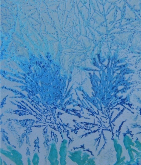
Figure 7: The crystals formed after drying an aqueous solution of CuCl2, which was previously subjected to the action of EMF, pulsing with a frequency of 10 Hz for 10 minutes
For contrast, the crystals formed were treated with ammonia vapors [3].
Considering the obvious similarity of these same salt structures (Figure 7) with nerve dendrites and nerve endings (Figure 8), it was also assumed that the regenerative effect of PEMFs on nervous tissues [8 – 17] is based on their ability to negatively electrify the latter [3]; the predominance of chlorides in nerve tissues [18] was also taken into account.
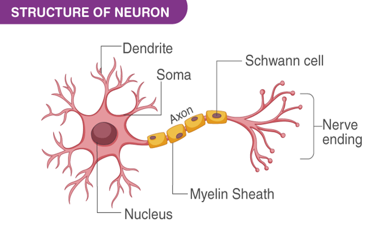
Figure 8: Diagram of a typical neuron.
In fact, it was assumed that the root cause of the regenerative effect of PEMFs and negatively charged electrets on nervous tissues [3] is the same.
Over time, it became clear that all these assumptions do not allow taking into account the contribution of cytoplasmic membranes of neurons, the
outer sides of which form negatively charged surfaces [18], in the formation of nerve dendrites, axons and endings (Figure 8); at the same time, it was assumed that the same electrical forces, the vectors of which are perpendicular to all charged surfaces (Figure 9), determine the direction of growth of neuronal axons and endings (Figure 8).

Figure 9: This particular diagram is used for educational purposes; It is important to note that the location of the electric field vectors created by a negatively charged surface is the same [5, 19].
It should be noted right here that the analysis of the effects of laser exposure on the neural environment (Figure 10) [20] partially confirmed the adequacy of this assumption.
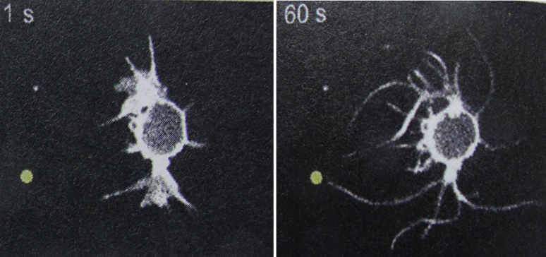
Figure 10: These are enlarged images of a neuron located near a spot illuminated by a laser (light yellow spot).
Comparing both photos allows evaluating the changes in the neuron that occurred within 1 minute [20].
Thus, given that the Poynting vector determines not only the direction of light propagation, but also the direction of movement of positive charges [21], it is very likely that negatively charged fragments of the cytoplasmic membrane of a neuron are drawn to a positively charged spot illuminated by a laser (Figure 10).
One way or another, it was decided to further test the supposed participation of charged surfaces in the formation of filaments; however, it was expected that the results of this test could be extrapolated to the cytoplasmic membranes of neurons.
Waters with the required charges were obtained as in [1].
Since the color and shape of copper salt crystals are particularly sensitive to the electrical potential of the water in which these crystals are formed (Figure 3), these salts were used here as indicators to visualize the electrization of aqueous media, just as in [1].
All salts were purchased from the “Ukrreakhim” (Ukraine).
Initially, it makes sense to analyze some previously obtained results. Thus, it was previously established that in aqueous solutions of salts, which are prepared in negatively charged water and are in contact with glass surfaces, filaments are formed, initially oriented perpendicular to such surfaces (Figures 11, 12) [4].

Figure 11: These are filaments formed in a glass Petri dish after drying a CuCl2 solution prepared in negatively charged water [4].

Figure 12: These are spiral filaments formed at the wall of a glass Petri dish filled with an aqueous solution of CuSO4 prepared in negatively charged water [4].
It is worth noting here that glass sorbs aqueous hydroxyl ions OH– [6], thereby receiving a negative charge, which electrostatically repels the negatively charged components of aqueous-salt solutions, thereby probably orienting the filaments formed from these components (compare Figures 9, 11, 12).
It is also likely that the negative, i.e., identical, charges of these filaments determine their mutual electrostatic repulsion and thereby ensure their separation; at the same time, it is no less likely that the electrical forces created by the glass surface polarize the structural components of such filaments, thereby ensuring their integration due to dipole-dipole interactions [5].
One way or another, all these considerations allowed us to assume that it is the negatively charged surface of the glass that exhaustively provides both the shape and direction of these same filaments (Figures 11, 12).
Given the predominantly negative charge on the outer surfaces of living cells [18], it was concluded that the forces that create filaments on the surface of cells (Figure 13) are of the same electrical nature and direction as the forces that create filaments on glass surfaces (Figures 11, 12); since this conclusion did not fundamentally contradict the previously proposed assumption about the participation of negatively charged media in the formation of neuronal dendrites [3], it seemed completely justified.
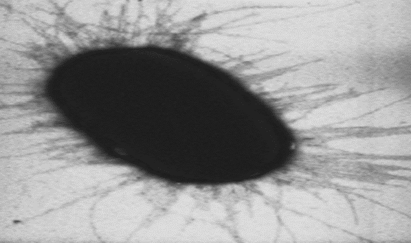
Figure 13: It is a separate cell of E. coli.
However, this conclusion seemed justified only until it was discovered that similar filaments are formed around rhombic crystals immersed in negatively charged water (Figure 14); in this experiment it was assumed that these crystals retained a positive charge on the water in which they formed (Figure 3).

Figure 14: When rhombic crystals are removed from a solution of copper sulfate and introduced into negatively charged water, numerous filaments quickly cover the surfaces of the crystals [2].
When analyzing this result, one should take into account the polymorphism of crystals formed in solutions prepared with differently charged water; in particular, it should be taken into account that rhombic crystals are formed in solutions prepared in positively charged water (Figures 1, 3).
Actually, it was this result (Figure 14) that led to the conclusion that both negatively and positively charged surfaces are involved in the formation of filaments in aqueous media. Over time, this conclusion received experimental confirmation.
Thus, it has been established that the filaments formed in aqueous-salt solutions prepared in negatively charged water and located between the negatively charged surfaces of glass and the positively charged surfaces of corrosion-resistant metals are similar to all previously obtained
filaments (compare Figures 11, 12, 14 and 15); at the same time, the identity, at least visual, of the endings of these same filaments was discovered (Figure 15).
When analyzing the last result (Figure 15), Kyon's rule should be taken into account: upon contact of the two phases, the phase with greater dielectric permeability receives a positive charge and the phase with lower dielectric permeability – negative [6, 22]. Thus, taking into account that the dielectric constant of liquid water varies, depending on temperature, in the range from 55.1 to 88.3 [6], and the dielectric constant of metals is always significantly higher (in theoretical calculations, the dielectric constant of metals is usually taken equal to ꝏ [23]), liquid water upon contact with any metal acquires a negative charge, and this same metal acquires a positive charge.

Figure 15: This is a "copper" coin surrounded by a dried aqueous solution of K2CO3 prepared in negatively charged water.
(Since the dielectric permeability of water changes depending on its temperature [6], the term “dielectric constant” is not applicable to water. In addition, the term "dielectric constant" does not correlate well with the sensitivity of water's dielectric permeability to high-frequency electromagnetic fields [19]; that it is this sensitivity that is important for the topic under discussion will soon become clear.)
Thus, all the results obtained (Figures 11, 12, 14, 15) showed that visually identical filaments are formed in aqueous solutions in contact with both negatively and positively charged surfaces.
Finally, it was discovered that apparently dendritic structures form in aqueous environments around corroding metals (Figure 16).

Figure 16: This is a corroded "copper" coin surrounded by a dried aqueous solution of CuCl2 prepared in negatively charged water.
Since these dendrites are formed from metal corrosion products (compare Figures 4 and 16), their formation is appropriate to compare with the formation of nerve and bacterial filaments, undoubtedly formed from components of cytoplasmic membranes (Figures 8, 10, 13). This, accordingly, suggests that cytoplasmic membranes not only supply “building material” for cellular filaments, but also generate electrical forces that create them.
It is especially worth noting that no auxiliary factors were involved in the formation of these same dendrites (Figure 16); this, accordingly, suggests that electrostatic repulsion is quite sufficient for the formation of any dendrites, including nerve ones (Figure 8).
Perhaps, the effect of skin electrons [23] on salt crystals dendrites should also be mentioned here. So, it is quite obvious that needle-like salt crystals grow from the area where skin electrons enter the studied salt solution (Figure 17).
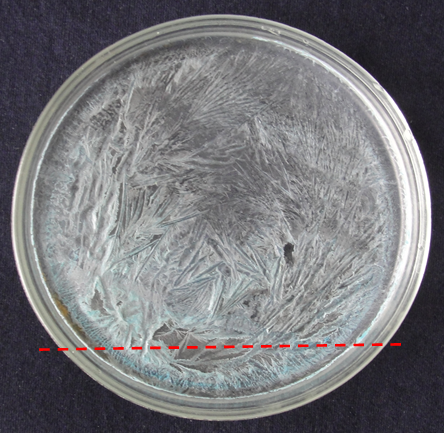
Figure 17: These are needle-shaped crystals formed after the drying of an aqueous solution of CuSO4 located under a wire with a pulsed electric current with a frequency of 2 Hz (dashed red line); it is worth noting that these needles are directed away from the wire.
Apparently, it is also worth noting here that electrons in terrestrial conditions move exclusively downward, like any anions [24]. Therefore, skin electrons fall into the studied salt solution below that part of it, which is located under the wire with an electric current (dashed red line in Figure 17).
Thus, the results obtained (Figures 1 12, 14 – 16) clearly demonstrate the ability of charged surfaces to form filaments in aqueous media that are
collinear to the electric field lines undoubtedly created by these surfaces (Figure 9). At the same time, the obvious similarity of filaments formed on both non-living and living surfaces (Figures 10 – 16) suggests the identity of the electrical forces that create any filaments; this, in turn, suggests that all filaments are visualizations of electric field lines, including extracellular ones.
To ensure the adequacy of these suggestions, it is worth extrapolating them to such a phenomenon as the “corona effect” (Figure 18), characterized by its discoverers as mysterious [25].
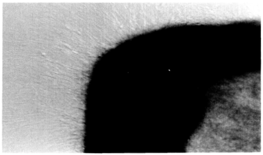
Figure 18: This is a fragment of the cytoplasmic membrane of a neuron with filaments formed under the influence of variable EMFs and creating a “corona effect” [25].
So, it is likely that the clearly visible filaments forming this very “corona”, which apparently consists of negatively charged fragments of the cytoplasmic membrane of the neuron (Figure 18), appear due to their electrostatic repulsion from the negatively charged outer surface of the cytoplasmic membrane; it is also likely that this repulsion is enhanced due to the multiple decrease in dielectric constant of the aqueous environment of the neuron exposed to variable EMFs [19].
Therefore, this very “corona” (Figure 18) can be considered only as a variety of the filaments presented above (Figures 10 – 15), quickly forming in favorable conditions.
At the same time, when analyzing the effect of the laser on the aqueous environment of the neuron (Figure 10), it should also be taken into account that under the influence of visible light, the dielectric constant of water decreases by ~10 times [19].
It should probably be added here that the formation of this very “corona” (Figure 18) can also be considered as a result of the skin effect (Figure 17), which undoubtedly occurs in variable EMFs [23] and manifests itself in this case in the pushing of negatively charged fragments of the cytoplasmic membrane of neuron to the periphery.
One way or another, the proposed analysis of the “corona effect” showed the productivity of the suggestions outlined at the beginning of this discussion, and thus confirmed their adequacy.
It seems appropriate to add here that the proposed explanation of the "corona effect" can also be used to explain the regenerative effect of PEMFs on nervous tissues [8 – 17]. To verify this, two facts should be taken into account:
In view of this, any PEMF that generates individual direct current pulses can reduce the dielectric constant of biological tissues, including nerve tissues (of course, taking into account the undoubted porosity of biological tissues).
Thus, it is likely that it is the electric fields generated by charged surfaces that build both nonliving and living filaments. It is also likely that all these filaments visualize the structures of such electric fields, in fact, their lines of force (Figure 19).

Figure 19: These are visualizations of electric field lines created by spherical (left) and extended (right) bodies used with education aim (compare with Figures 10 – 17).
The structures shown above, both salt and living, appear to be similar to both of these visualizations; this similarity suggests that these same structures are also visualizations of the corresponding electric fields.
This, accordingly, suggests that the natural ability of neurons to self-repair is based on the electrostatic repulsion of neuronal fragments by cytoplasmic membranes. This, in turn, allows anticipating that amplifying this repulsion will contribute to the natural ability of neurons to recreate. Thus, it is very likely that both PEMFs and the laser are means of
amplifying the electrical forces that determine the natural regenerative abilities of neurons.
Despite the unusual nature of the proposed approach, which obviously requires a radical revision of established ideas about the mechanisms of formation of extracellular filaments [30 – 32], it seems to be very productive and therefore requires development.
In particular, it is expected that this approach will make it possible to identify the forces that form networks in water-salt solutions under the influence of PEMFs (Figure 20) with the forces that form neural networks.
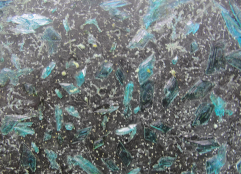
Figure 20: This is a dried aqueous solution of CuSO4, which, until completely dry, was exposed to an EMF pulsating at a frequency of 2 Hz.
The white “beads” connected by white threads are probably quite visible here.
It is clear that these networks were formed in a purely non-living environment, that is, in the absence of any "living force".
Also, developing this same approach, it is worth adding that it can no less successfully be used to explain the restorative effect of PEMFs on bones, the nature of which is considered unclear [33]. Otherwise, the ideas presented both earlier and here can be seen as attempts to respond to Szent-György, who urged biologists not to ignore the existence of "two matrices: water and electromagnetic fields" [29].
Dear Editorial Team, Clinical Medical Reviews and Reports. My experience with the journal was highly positive. The peer-review process was rigorous, constructive, and completed in a timely manner. The reviewers provided valuable comments that helped improve the quality and clarity of our manuscript. The editorial office was professional, responsive, and supportive throughout all stages of the publication process. Communication was clear and efficient, and any questions were addressed promptly. Overall, I found the journal to maintain high scientific standards and an excellent publication workflow. I would be pleased to consider submitting future work to this journal. Best wishes from, Elena Popa.

It was my pleasure to submit my testimonial concerning the Reviewer Board of our Scientific Journal “Brain and Neurological Disorders”. The Reviewers focused on some modifications and their contribution was helpful. The ladies of our Editorial Office were also supported my efforts. It was my honor to have such a co-operation and I am looking forward for more collaboration.

Dear Grace Pierce, Editorial Coordinator of Journal of Clinical Research and Reports, Thank you for the speedy and efficient peer review process. I appreciate the fact that your peer reviewers do not take months to respond like with some other journals. I would also like to thank the editorial office for responding quickly to my questions. It is an excellent journal. I plan to submit more manuscripts in the future. Best wishes from, Robert W. McGee

Dear Grace Pierce, Editorial Coordinator of Journal of Clinical Research and Reports, Working with you and your team on our recent publication in JCRR has been a truly wonderful and enjoyable experience. The responses were prompt, and the reviewers were patient, constructive, and highly professional. One reviewer in particular gave me the feeling that a professor was carefully reading and commenting on my coursework, which was deeply touching. The entire process was straightforward and hassle‑free, with no tedious online forms to complete. I highly recommend this journal. Best wishes from, DR Aibing Rao, Head of R&D

I Appreciate the Opportunity to Share my Experience with the Journal of Clinical Research and Reports. The peer review process was timely and constructive, and the feedback provided helped improve the quality of our manuscript. The editorial office was professional, responsive, and supportive throughout the process, ensuring smooth communication and efficient handling of the submission. Overall, it was a positive experience collaborating with your team.

Dear Mercy Grace, Editorial Coordinator of Obstetrics Gynecology and Reproductive Sciences, We would like to express our gratitude for your help at all stages of publishing and editing the article. The editors of the magazine answer all the necessary questions and help at every stage. We will definitely continue to cooperate and publish other works in the Obstetrics Gynecology and Reproductive Sciences! Best wishes from, Alla Konstantinovna Politova,
