
Case Report | DOI: https://doi.org/10.31579/2690-1897/121
Assistant professor, Radio-diagnosis Pacific Institute of Medical Sciences (PIMS), Umarda, Udaipur, Rajasthan, India-313001
*Corresponding Author: Rajaram Sharma, Assistant professor, Radio-diagnosis Pacific Institute of Medical Sciences (PIMS), Umarda, Udaipur, Rajasthan, India-313001
Citation: Rajaram Sharma. (2022). Efficacy of MRI for detection of frozen pelvis. Journal of Surgical Case Reports and Images 5(5); DOI: 10.31579/2690-1897/121
Copyright: © 2022, Rajaram Sharma, this is an open access article distributed under the Creative Commons Attribution License, which permits unrestricted use, distribution, and reproduction in any medium, provided the original work is properly cited.
Received: 30 June 2022 | Accepted: 05 August 2022 | Published: 28 October 2022
Keywords:
A 26- year old female presented to us with severe pelvic pain. She was adviced follicular study and MRI pelvis. There was a large, multilobulated cyst in her pelvic region appearing hyperintense on both T1 and T2 fat sat sequences. There was also presence of multiple, hypointense peritoneal deposits seen on both T1 and T2 fat sat sequences. The findings concluded the diagnosis of frozen pelvis.
organs get distorted and adhered to each other. It commonly occurs as a result of endometriosis. Pelvic endometriosis is the presence of functional endometrium outside the uterus. It may be present as small microscopic structures to large cyst, known as endometriomas. It is mainly found in women of child-bearing age, mean age of diagnosis being 25-29 years. The patient may be asymptomatic or may have severe pelvic pain, adnexal mass or infertility. It is usually diagnosed in women with presenting complain of infertility more commonly than pelvic pain [1]. The endometrial tissue may be present at different sites like ovaries, gastrointestinal tract, urinary tract, chest and soft tissues, ovaries being the most common site. Several theories have been proposed for the development of endometriosis; (a) metastatic theory, (b) metaplastic theory and (c) induction theory. The most widely accepted theory amongst them is the metastatic theory, due to retrograde menstrual implantation, vascular and lymphatic spread and intra-operative implantation [2-4]. Magnetic resonance (MR) imaging has a greater specificity and is a problem-solving tool in differentiating endometriomas from other adnexal masses [5-7]. Endometriotic deposits appear as tiny nodular deposits over the peritoneal reflections and bowel wall serosa and appear hypointense on all sequences. [Figure 1a and figure 1b] Surrounding desmoplastic and fibrotic response may lead to puckering or tethering of pelvic structures. Endometriomas have relatively high signal intensity on T1 weighted images. Use of fat-suppression sequences improves lesion conspicuity. It is helpful in detection of small lesions and increases its specificity since the fat containing lesions like dermoid cysts are eliminated from the differentials [8-12]. The lesions appear hyperintense on both T1 and T2 sequences when it contains methemoglobin and concentrated proteins. [Figure 1c and figure 1d] The most specific feature of endometrioma is “shading”, loss of signal within the lesion which demonstrates the chronic nature of endometrioma.
Figure:
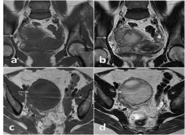
Figure 1a and figure 1b: Coronal sections from MRI pelvis, T1 and T2 fat sat sequences respectively, showing endometriotic deposits appearing as tiny nodular deposits over the peritoneal reflections as hypointense signals (white arrows). Figure 1c and figure1d: Axial sections from MRI pelvis, T1 and T2 fat sat sequences respectively, showing a large, multiloculated cyst appearing hyperintense on both sequences (white arrows)
Dear Editorial Team, Clinical Medical Reviews and Reports. My experience with the journal was highly positive. The peer-review process was rigorous, constructive, and completed in a timely manner. The reviewers provided valuable comments that helped improve the quality and clarity of our manuscript. The editorial office was professional, responsive, and supportive throughout all stages of the publication process. Communication was clear and efficient, and any questions were addressed promptly. Overall, I found the journal to maintain high scientific standards and an excellent publication workflow. I would be pleased to consider submitting future work to this journal. Best wishes from, Elena Popa.

It was my pleasure to submit my testimonial concerning the Reviewer Board of our Scientific Journal “Brain and Neurological Disorders”. The Reviewers focused on some modifications and their contribution was helpful. The ladies of our Editorial Office were also supported my efforts. It was my honor to have such a co-operation and I am looking forward for more collaboration.

Dear Grace Pierce, Editorial Coordinator of Journal of Clinical Research and Reports, Thank you for the speedy and efficient peer review process. I appreciate the fact that your peer reviewers do not take months to respond like with some other journals. I would also like to thank the editorial office for responding quickly to my questions. It is an excellent journal. I plan to submit more manuscripts in the future. Best wishes from, Robert W. McGee

Dear Grace Pierce, Editorial Coordinator of Journal of Clinical Research and Reports, Working with you and your team on our recent publication in JCRR has been a truly wonderful and enjoyable experience. The responses were prompt, and the reviewers were patient, constructive, and highly professional. One reviewer in particular gave me the feeling that a professor was carefully reading and commenting on my coursework, which was deeply touching. The entire process was straightforward and hassle‑free, with no tedious online forms to complete. I highly recommend this journal. Best wishes from, DR Aibing Rao, Head of R&D

I Appreciate the Opportunity to Share my Experience with the Journal of Clinical Research and Reports. The peer review process was timely and constructive, and the feedback provided helped improve the quality of our manuscript. The editorial office was professional, responsive, and supportive throughout the process, ensuring smooth communication and efficient handling of the submission. Overall, it was a positive experience collaborating with your team.

Dear Mercy Grace, Editorial Coordinator of Obstetrics Gynecology and Reproductive Sciences, We would like to express our gratitude for your help at all stages of publishing and editing the article. The editors of the magazine answer all the necessary questions and help at every stage. We will definitely continue to cooperate and publish other works in the Obstetrics Gynecology and Reproductive Sciences! Best wishes from, Alla Konstantinovna Politova,
