
Review Article | DOI: https://doi.org/10.31579/2690-1919/289
1 Infectious Diseases and Tropical Medicine Research Center (IDTMRC), Department of Aerospace and Subaquatic Medicine, AJA University of Medicinal Sciences, Tehran, Iran
2 Faculty of Pharmacy, Tehran Medical Sciences, Islamic Azad University, Tehran, Iran.
*Corresponding Author: Mohammad Darvishi. Infectious Diseases and Tropical Medicine Research Center (IDTMRC), Department of Aerospace and Subaquatic Medicine, AJA University of Medicinal Sciences, Tehran, Iran.
Citation: Mohammad Darvishi, Fatemeh Hajilou. (2023). Diagnostic Biomarkers of Diabetic Neuropathy, Journal of Clinical Research and Reports, 13(1) DOI:10.31579/2690-1919/289.
Copyright: © 2023 Mohammad Darvishi. This is an open-access article distributed under the terms of The Creative Commons Attribution License, which permits unrestricted use, distribution, and reproduction in any medium, provided the original author and source are credited.
Received: 10 December 2022 | Accepted: 22 December 2022 | Published: 09 January 2023
Keywords: biomarker; diabetic; neuropathy; diagnostic
Diabetes mellitus (DM) is a chronic disease that occurs due to inadequate production of insulin or decreased effect of available insulin. Considered one of the most critical life-threatening diseases of the 21st century, the number of patients has been increasing over the past few decades. These patients suffer many problems such as neuropathy, nephropathy and rethinopathy which the first can decrease their life expectations.
Diabetic peripheral neuropathy (DPN) is the most common complication in diabetic patients and more than one half of these patients develop nerve dysfunction through their life. The primary cause of diabetic foot disease is DPN which sleep disturbances, poor quality of life, depression and finally unemployment are the results of it.
In this article, we are studying different biomarkers of diabetic neuropathy using related papers in order to consider them in diabetic patients and prevent further complications in them.
Diabetes mellitus (DM) is a chronic disease that occurs due to inadequate production of insulin or decreased effect of available insulin. Considered one of the most critical life-threatening diseases of the 21st century, the number of patients has been increasing over the past few decades (World Health Organization 2019). One of the complications in these patients is diabetic neuropathy.
Diabetic peripheral neuropathy (DPN) is the most common complication in diabetic patients and more than one half of these patients develop nerve dysfunction through their life [1], [2]. One of the results of diabetes mellitus is diabetic foot which many factors affect recurrence of it [3]. The primary cause of diabetic foot disease is DPN [2], [4] which sleep disturbances, poor quality of life, depression and finally unemployment are its results [2] , [6], [7], [8], [9].
1-Nerve conduction studies (NCS)
NCS has been used as a diagnostic, staging, and prognostic biomarker for DPN. Historically, it has been gold standard for the diagnosis of DPN. This study includes supramaximal transcutaneous stimulation of defined upper and lower limb peripheral nerves. It has been done with surface electrodes noninvasively.
One of the prognostic features of DPN is slowing of motor nerve conduction velocity which can led to death [10],[2]. There are some limitations for NCS, too. Most of the time, neurophysiology departments have normal ranges but it is not the same for NCS and also there are not specified standards for it [11],[2].
2-Pain-related Evoked Potentials
It is involved recording of cortical activity through scalp electrodes in response to selective stimulation of nociceptive skin afferents. Distinct waveforms can be obtained for A-delta and C-fiber stimulation, but for the purposes of clinical evaluation only assessment of A-delta waveforms is currently believed to be reliable [12],[13],[2]. Nociceptor stimulation delivered by using a laser or a brief thermal stimulus.
The most reliable neurophysiological method to assess nociceptor pathways is Laser evoked potentials (LEPs) [12],[13],[2]. LEPs have a lot of advantages such as being noninvasive, almost rapid to perform and are an objective neurophysiological biomarker for SFN [14],[2] There are some downsides for LEP, too. This technique is expensive, not common and not available, and certain safety measures (eg, wearing of eye protection) are required during the investigation. Also, the waveform amplitudes are dependent on the participants’ attention [15],[2] and it cannot differentiate between central and peripheral nervous system pathology.
Mechanism of contact heat evoked potentials (CHEPs) is the same as LEPs but they are noninvasive and do not have safety issues as with LEPs. However, they require specialized equipment and are subject to significant habituation effects [15],[2].
Intra-epidermal electrical evoked potentials (IEEPs) using specialized electrodes that deliver a high current density to epidermal fibers. They do not need a specialized stimulator device such as a laser or thermode. Also, they are noninvasive and easy to perform but it is currently experimental, with few studies that address the use of IEEPs in DM and none that compare their use with other SFN biomarkers [16], [2]
3-Nerve Excitability Testing
One of noninvasive methods in which properties of human peripheral nerve axons can be assessed is using nerve excitability testing (NET) [17],[2]
NET is performed in a similar manner to NCS but uses submaximal as well as supra-maximal stimulation according to standardized excitability protocols (stimulus–response curve, strength–duration properties, threshold electrotonus and the current/threshold relationship, and the recovery cycle) to obtain a reflection of firing threshold [17],[18],[19],[2] The results provide a surrogate of ion channel and axonal membrane potential properties at the stimulation site. Although motor axons are more commonly investigated, because recordings are more stable and less affected by artifact, sensory nerves can also be assessed. NET has the advantage in that it assesses functional measures which may occur before pathologic axonal or demyelinating features and their NCS corollary become evident.
In diabetic patients, changes in excitability, including features compatible with sodium channel dysfunction, have been reported in association with subclinical neuropathy [20],[21],[2] These alterations progress with increasing neuropathy severity and are seen in patients with normal NCS, raising the possibility that NET could provide an opportunity to detect abnormalities before they become irreversible [21],[2]. The types of alteration are different between sensory and motor axons and occur at an earlier stage in sensory nerves [2].
NET is not widely available and requires specialist equipment and software. There are no clinically relevant normative ranges and, although alterations in NET can be shown despite normal NCS, it has yet to be validated as a biomarker for either DPN or pDPN. As with NCS, it does not provide information about the status of small nerve fibers. However, it is a useful, noninvasive method to explore disease pathophysiology and may prove a useful biomarker when assessing response to treatment for DPN or pDPN such as in trials of therapeutic agents that act on voltage-gated ion channels [2].
4-Microneurography
Microneurography involves inserting a fine tungsten electrode into a peripheral nerve. For evaluation of peripheral neuropathy, this typically involves recording from the peroneal or superficial peroneal nerves. The electrode is micromanipulated to enable recording of unmyelinated fibers. A typical paradigm involves stimulation of the cutaneous receptive field(s) of the unmyelinated fibers by using electrical stimuli. Using this method, several C-fibers can be recorded at one time. A raster plot is generated showing action potentials time-locked to the electrical stimulus with latencies appropriate for the slow conduction velocity of C fibers. This enables differentiation of nociceptor subtypes (eg, polymodalnociceptorsvs silent nociceptors [22],[2]. Normally, there is a stable baseline latency to low-frequency stimulation of the receptive field, but when a fiber is spontaneously active an irregular “saw-tooth” baseline is seen [22],[23],[24],[25],[26],[2] Also, assessment of “abnormal”sensitivity to cutaneous applied mechanical and thermal stimuli can be recorded [23],[28],[2]. The method can provide evidence of both spontaneous activity and sensitization. Patients with painful peripheral neuropathy have been shown to have abnormal hyperexcitability of nociceptor fibers [23], [26], [27], [28], [29],[2]. Microneurographic studies in DPN have shown variable findings. In patients with documented large fiber neuropathy, one study found an altered distribution of nociceptor subtypes with a higher proportion of mechanically insensitive to mechanically sensitive C-nociceptors, as well as evidence of loss of mechanical sensitivity in normally sensitive fibers [30],[2] Although a small proportion of nociceptor fibers were spontaneously active, this did not significantly differ between pDPN and DPN. Other studies have shown a higher proportion of hyperexcitablenociceptor fibers in patients with pDPN [26], [28], [2].
Although microneurography is an invaluable tool for investigating the presence of nociceptorhyperexcitability, its use as a biomarker is limited. The technique is invasive, albeit minimally and time consuming for both the patient and investigator. Furthermore, it requires specialist equipment and highly trained operators, and is performed only in a limited number of centers worldwide. These factors significantly affect the availability and cost effectiveness of microneurography. Also, age and potentially disease-specific definitions of normative and abnormal data are needed so that its sensitivity and predictive value can be determined, and it can be established as a clinical tool. Anotherdownside of it is that each session may yield only small numbers of nociceptor recordings that are suitable for analysis. For example, in one study, only 1 to 3 suitable nociceptor fibers were recorded per session [26],[2] This limits diagnostic applicability and capacity for monitoring interventions on an individual level. It is not known how microneurography compares with the indirect, but consider more widely available, QST techniques in identifying patients with hyperexcitablenociceptors. Furthermore, the relationship between abnormalities detected by microneurography and structural changes of small fibers in the skin is unknown. The method is potentially of use in clinical trials, especially those with a focus on underlying pain mechanisms, in which patients with and without evidence of hyperexcitability can be segregated [31],[26],[2]
5- Monocyte chemoattractant protein-1 (MCP-1)
MCP-1 protein also known as C–C motif ligand 2 (CCL2)/Monocyte Chemotactic and activation factor (MCAF), belongs to the chemokine family. MCP-1 is a monomeric polypeptide secreted by monocyte, macrophages, and dendritic cells at the site of infection, damage, and injury [32],[33]. MCP-1 secretion is induced by pro-inflammatory mediators like TNFa, interferon-gamma (INF-c), interleukin 1 beta (IL-1b), and platelet-derived growth factor (PDGF) during inflammation. According to scientific reports, MCP-1 aids mononuclear phagocytes relocation during hypoxia and inflammation. Investigators reported that serum, urinary levels of MCP-1 are significantly upregulated in early and late stages in type 2 diabetes. An increase in expression or upregulation of MCP-1 is mainly associated with monocyte binding. MCP-1 contributes to the treatment and management of diabetic neuropathic pain via C–C motif ligand receptor 2 (CCR2) receptors. MCP-1 acts by augmenting excitatory synaptic transmission. The literature showed that spinal astrocytes play a crucial role in neuropathic pain sensitisation by activation of c-Jun-N-terminal kinase (JNK). Some reports showed that TNF-a could activate JNK via TNF receptor 1. This activated TNF-a/JNK pathway transiently stimulates MCP-1. This upregulated levels of MCP-1 can be considered as a potential biomarker in the diagnosis of diabetic neuropathy pain during its early stage to avoid its diverse progression and damage of neurons. MCP-1 inhibitory activity is linked with the management of inflammation as well as the treatment of neuropathic pain [33].
6-Vascular endothelial growth factor (VEGF)
VEGF is an angiogenic factor secreted by various cells, including platelets, macrophages, renal mesangial cells, and keratinocytes. VEGF plays a critical role in cellular activities like haematopoiesis, bone formation, wound healing, and development [33],[34]. Hyperglycaemia modulates the VEGF pathway at protein and mRNA levels in Schwann cells and dorsal root ganglion. Various therapeutic approaches, like VEGF-A165b and VEGF gene transfer, are used to treat diabetic neuropathy. The VEGF-A165b initiates extravasation in dorsal root ganglia, plantar skin of hind paw, and saphenous nerve. This potentiates transient receptor potential cation channel, subfamily A, member1 (TRPA1) channel at the initial stages of diabetic neuropathy leading to altered neuronal stress and pain [33],[35]. A study by Veves et al. showed that VEGF gene transfer studies had reversed severe diabetic neuropathic conditions [33],[36]. DM also influences neuropeptide Y and VEGF during inflammation; both of them play a crucial role in the pathophysiology of diabetic neuropathy. VEGF exhibits polymorphism and halts the potential regulation of gene expression [33], [37] VEGF also reported for its association with atherosclerosis by increasing vascular permeability to low-density lipoproteins in the cardiovascular system, phosphorylation of Murine thymoma viral oncogene Akt and stimulation of neurons linked with neuroprotection in the central nervous system [33]. It is also reported that patients with diabetic peripheral neuropathy in the symptomatic stage have downregulated serum levels of VEGF [33],[38]. Studies also reported that Schwann cells, when cultured in the hyperglycaemic medium, resulted in impairment of neurite outgrowth. This impairment was due to alteration in VEGF receptors regulation in dorsal root ganglion. This causes decreased intracellular secreted protein levels without altering the mRNA level in Schwann cells. This leads to reduced expression of the VEGF both in Schwann cells and dorsal root ganglion [33].
7-Transient receptor potential vanilloid 1 or transient receptor potential cation channel subfamily V member (TRPV1)
TRPV1 is a transducer protein associated with noxious inflammatory and thermal stimuli. TRPV1 proteins are mainly found in nociceptive neurons of the peripheral nervous system and some tissues of the central nervous system. TRPV1 plays a crucial role in pain sensation. It is activated in the presence of capsaicin and acidic condition [33],[39]. It is also activated by ligands like phosphatidylinositol, tyrosine kinase, and proton influx, specifically in peripheral and nociceptive neurons [33],[40]. Diabetic peripheral neuropathy is mainly characterised by allodynia and hyperalgesia [33],[41] The severe pain in diabetic neuropathy is primarily associated with the upregulation of TRPV1 in peripheral nerves [33],[42]. The elevated levels of TRPV1 were reported due to enhanced reflex arc functioning in diabetics [33], [43] It is also said that in peripheral sensory neurons, the inhibitory activity of TRPV1 is due to the activation of the m-opioid receptor. This inhibition of TRPV1 is due to impaired function in the dorsal root of the ganglion and TRPV1 activity [33], [44] This impairment of TRPV1 was confirmed in human embryonic kidney cells. These cells heterologously expressed TRPV1 by stimulating PKA and PKC mediated phosphorylation and resulted in the modulation of TRPV1. Scientific reports also showed that the TRPV1 activation is mainly associated with increased stress in dorsal root ganglion.TRPV1 also has a potential role in pain and post-traumatic neuropathy [33],[45]
The involvement of C fibres was assessed through microneurography. The mechanism of transduction channels TRPA1 and TRPV1 in methylglyoxal-induced pain sensation was investigated by using specific ion channel blockers. The study showed that the selective pharmacological blockade of TRPV1 showed that TRPV1 is crucially involved in methylglyoxal- induced chemical pain sensation and heat hyperalgesia.
The study concluded that methylglyoxal could be a mediator of diabetes-induced neuropathic pain through TRPV1 activation and sensitisation of the voltage-gated sodium channel subtype 1.8 [33],[46]
Cellular oxidative stress plays a key role in the post-translational modification of proteins via SUMOylation. The effect of SUMOylation on the TRPV1 ion channel was carried out by Agarwal and group in diabetic mice and patients. In this study, they have identified the novel molecular targets, that is, key enzymes involved in the regulation of metabolic pathways and ion channels of SUMOylation. In the study sensory neurons of diabetic mice and diabetic patients showed significant changes in the SUMOylation status of metabolic enzymes and ion channels in western blot analysis. These changes lead to metabolic dysfunction, sensory loss, and accelerated neuropathology in diabetic gene-targeted mice selectively lacking the ability to SUMOylate proteins in peripheral sensory neurons. The study showed that diabetes induced de-SUMOylation can impair the functions of TRPV1. The diabetes-induced and metabolic imbalance is caused by de-SUMOylation of various metabolic enzymes that facilitate diabetic sensory loss. The study concluded that endogenous post-translational mechanism regulates TRPV1 function in diabetic neuropathy [33], [47]
It is documented that a-lipoic acid has the ability to regulate TRPV1 in dorsal root ganglion neurons of rats with diabetes. Alpha-lipoic acid acts via alleviating the neuropathic pain in diabetes by regulating TRPV1 expression [33],[48]. All scientific reports insist that the regulation of TRPV1 will provide the new strategy for the management of painful diabetic neuropathy.
Nuclear factor kappa-light-chain-enhancer of activated B cells (NF-jB) NF-jB is a pro-inflammatory mediator predominantly involved in adaptive immunity, stress responses, lymphoid organogenesis, and B-cell development. The pro-inflammatory mediators, namely TNF-a and IL-6, are responsible for the activation of NF-jB during cellular damage and inflammation [33], [49]. NF-jB is involved in pro-inflammatory cytokine production during immunological responses. Hyperglycaemia causes activation of NF-jB. This activated NF-jB further triggers expressions of inflammatory mediators. Literature showed that NF-jB is associated with activation, survival, and differentiation of inflammatory T cells and innate immune cells. NF-jB is mainly expressed in endothelialcells linked with its dysfunction and progressing of diabetic neuropathy. The overexpression of NF-jB triggers damage to endothelial cells [33],[50 ]
8-Oxidative biomarkers Nuclear factor (erythroid-derived 2) (NFE2L2)
Nrf2 or NFE2L2 is a dynamic controller of antioxidant responses and cellular damage. Oxidative stress is a factor involved in many metabolic disorders, including diabetes and its complications. Oxidative stress is linked to insulin resistance [33],[51]. The activation of “NFE2L2” plays an important role in maintaining blood glucose level, vascular complications, and endothelial dysfunction by reducing oxidative stress. Hyperglycaemia induced oxidative stress leads to disturbance in the normal physiology of nervous tissue. Increased oxidative stress is one of the early events in the development of insulin resistance, inflammation, and alteration of the aldose-reductase pathway [33] The endogenous disturbance body stimulates major reactive oxygen species defense machinery, NFE2L2. Itscavenges free radicales and increases insulin sensitivity and lipid metabolism. The excess oxidative stress during prolonged hyperglycaemia cannot be controlled by NFE2L2. This leads to the downregulation of NFE2L2 in the sciatic nerve of diabetic animals [33], [52]
9-Adiponectin
Adiponectin hormone is made of 147 amino acids with 30 kDamolecular weight. Adiponectin involved in homeostatic control of blood glucose circulation, lipid oxidation, insulin sensitivity, coronary heart diseases and atherosclerosis [33],[53]. Adiponectin secreted by adipocytes and white adipose tissue is associated with the progression of diabetic neuropathy. Adiponectin gene polymorphism and glypican secretions are significantly involved in diabetic peripheral neuropathy. The upregulation of serum adiponectin levels is positively correlated with aching diabetic neuropathy [33]. Increased levels of adiponectin were associated with a decrease in heart rate and an increase in heart rate velocity leading to cardiovascular autonomic neuropathy. Type 1 diabetics are more prone to cardiovascular autonomic neuropathy, but these findings were found to be limited in type 2 diabetics.Researches have been shown that the adiponectin polymorphism at G276T and T45G is associated with a significantly increased risk of diabetic neuropathy in Type 2 diabetic patients. There is a strong connection between adiponectin and decreased nerve conduction velocity in inflammation and progression of diabetic sensorimotor neuropathy [33],[54]. Recently, ketogenic diet is reported to control upregulatedadiponectin levels in diabetic neuropathy. Ketogenic diet (High-fat, adequate-protein, and low carbohydrate) strategy is usually used to control several diseases such as diabetes, neurological disorders, and cancer. Ketogenic diet replaces plasma glucose with ketone bodies.
The ketogenic diet gives the body the capacity to fight against several diseases such as metabolic disorders, epileptic seizures, neurodegeneration to skeletal muscle atrophy, autosomal dominant polycystic kidney disease, and peripheral neuropathy. A ketogenic diet control the diabetic neuropathy via reducing oxidative stress, enhancing mitochondrial efficiency via regulating the phospho-AMP-activated protein kinase, Naþ/ Kþpump, ketogenesis, gamma-aminobutyric acid-glutamate, ghrelin and leptin levels, lipolysis, lipogenesis, and gluconeogenesis. Anti-inflammatory and antioxidant activities of the ketogenic diet reduce metabolic syndromes-associated allodynia and stimulate peripheral sensory and axonal regeneration [33],[55]. Further studies should be planned to study the detailed molecular mechanism of adiponectin in diabetic neuropathy.
10-Nicotinamide adenine dinucleotide phosphate oxidase (NOX1)
A multicomponent enzyme, NOX, mediates electron transfer from NADPH to molecular oxygen.Mostly they are present in the phagocytic cells and primary cells in the brain, such as vascular endothelial cells, microglia neurons and astrocytes. NOXs mediate neuropathic painby production of reactive oxygen species like superoxide in phagocytic cells. The prolonged hyperglycaemic condition develops upregulation of NOX 4 via tyrosine kinase and MAPK pathway. Reactive oxygen species (ROS) is a triggering factor for activation of NOX 4 via angiotensin II (Ang II). NOX is involved in the generation of peroxynitrite (powerful oxidant), which acts via Ang II in endothelial nitric oxide synthase uncoupling in nerve tissue. Hyperglycaemia-induced oxidative stress plays a crucial role in the progression of diabetic complications. NOX-ROS significantly contributes to the activation of oxidative stress-associated inflammatory pathways that lead to vascular tissue damage including nerves [33],[34] These pathways are basically associated with hypertrophy, angiogenesis, and inflammation in nerve cells. These pathways are responsible for the upregulation of the NOX in diabetic neuropathy [33]. Recently, an in vivo study was carried out by Oghbaei and group to observe the effect of a low dose of sodium nitrate on the diabetic peripheral neuropathy in male Wistar rats.
Diabetes was caused using STZ (60 mg/kg i.p.). Diabetic animals were treated with 100 mg/L sodium nitrate solution (SC) administered for 60 days. Behavioural studies, mechanical allodynia like von Frey filament test, thermal algesia like tail withdrawal test, hot plate test were carried out. Blood samples were analysed for serum insulin and NOX levels at the end of the study. The NOX levels were upregulated in animals with diabetic neuropathy. The study revealed that sodium nitrate has a protective effect on diabetic neuropathy [33].
11-Ceruloplasmin
Copper is an essential factor necessary for homeostasis. Ceruloplasmin, , also known as a 2- glycoprotein, is a serum ferroxidase produced by liver cells and contains almost 95% of copper found in plasma [33], [56]. It plays a key role in copper transportation and metabolism within the plasma. Ceruloplasmin also monitors iron efflux from cells with the enrollment of iron [33], [57]. Recently, scientific data reported that ceruloplasmin gene abnormality is associated with various neuronal diseases, including neuropathy and neurodegenerative disorders [33]. It is also known as “moonlighting protein” for its versatile pro-oxidant and antioxidant activities. The pro-oxidant activity is linked with amine oxidase and antioxidant activity linked with glutathione peroxidase activity. The upregulation of ceruloplasmin is linked with the acute phase of trauma, inflammation, diabetes, and its complications [33]. The damage to ceruloplasmin is coupled with extrapyramidal diseases, progressive dementia, cerebellar ataxia, and diabetes mellitus [33]. Scientists also expected significantly increased levels of ceruloplasmin in diabetic neuropathy. More studies are required to understand the mechanism of action of ceruloplasmin in diabetic neuropathy [33].
Haem oxygenase-1 (HO-1)
Haem oxygenase-1, is associated with stress response and is widely distributed in systemic tissues. HO-1 is mainly regulated by the Nrf2 gene and induced by haem, inflammatory cytokines, prostaglandins, heat shock protein, endotoxins, and heavy metals. HO-1 helps in the regulation of haem metabolism, cellular homeostasis, and vascular inflammation [33],[58]
HO-1 is associated with the rate-limiting step involved in oxidative cleavage of haem degradation, which generates biliverdin, carbon monoxide, and free iron. HO-1 is reported for different activities, namely anti-inflammatory, antioxidant, immunomodulatory, antiapoptotic, and antiproliferative activities. The upregulation ofHO-1 is associated with the activation of multiple signaling pathways like PI3K/Akt, p38 MAPK, and JAK-STAT pathway. HO-1 is implicated in oxidative stress and cellular defense, specifically in neuro-inflammation and also in the progression of diabetic neuropathy
The function of HO-1 in diabetic neuropathy was studied by Leng and group. Diabetes was induced in male C57 mice with a high-fat diet (8 weeks) and STZ (100 mg/kg, i.p. for 2 successive days). Diabetic animals were treated with diosgenin (50 & 100 mg/kg) for 14 days (after 6 weeks of induction). The study showed that diosgenin significantly increased tail withdrawal latency and mechanical hyperalgesia. Diabetic animals showed down-regulation of HO-1 compared to normal control in western blot analysis. Diosgenin treatment upregulated the HO-1 expression as compared to diabetic animals. The study concluded that diosgenin has a neuroprotective effect in diabetic peripheral neuropathy via reducing oxidative stress in Nrf2/HO-1 pathway [33[,[59]. Similarly in vitro studies were carried out in dorsal root ganglionic cells. The cells exposed to high glucose displayed downregulation of HO-1 in diabetic peripheral neuropathy. The cells treated with hirudin protected dorsal root ganglion neuronal cells from apoptosis by scavenging ROS, upregulating Nrf-2/HO-1 expressions, inhibiting NF-j B, and Caspase-3 pathway [33],[52]
12-Dipeptidyl peptidase-4 (DPP4)
DPP4 is an immune regulated enzyme involved in signal transduction in glucose metabolism. It also helps in the degradation of glucose like peptide-1 (GLP-1), cytokines, and growth factors. DPP4 inhibitors improve levels of incretin hormones and insulin sensitivity, which regulate glucose level. The higher levels of serum DPP4 have been reported during a condition called metabolic syndrome which obesity, non-alcoholic fatty liver disease, and diabetes are its signs .
Alterations in regulations of DPP4 are linked with neuroinflammation in hyperthermia [33],[60] The mechanism of action of DPP4 is still not understood clearly.
Poly ADP ribose polymerase a (PARP a) Eukaryotic cells manage various endogenous and environmental genotoxic agents, reactive nitrogen species (nitrosative) and oxidative stress through PARP a. The PARP repair DNA by the removal of base and cell proliferation mechanism [33],[61]. PARP activation is an essential downstream effector in metabolic changes in the diabetic neuropathy. Enzyme PARP can cleave nicotinamide adenine dinucleotide and give rise to poly (ADP-ribose) polymer and nicotinamide. These end products have a considerable role in neurodegenerative diseases. It is well known that the relation between PARP activation and oxidative-nitrosative pressure is unidirectional, and it is just the outcome of scavenging free radicles and oxidation [33],[62]. The mechanism of PARP involves the reduction of NADþ and moderates ATP generation, electron transport, and glycolysis. Overall results revealed in acute endothelial dysfunction in blood vessels during diabetic neuropathy. It is discovered that PARP activation leads to NADþdepletion, which further leads to alteration in transcriptional regulation, impaired signal transduction, gene expression, and damage to neurons and neuroglial cells.
In diabetic neuropathy, PARP activation was basically found in peripheral nerves, dorsal root ganglia and spinal cord. This activated PARP causes up-regulation of inducible nitric oxide synthase (iNOS) as a pro-inflammatory response. This nitrosative stress may cause damage to nerves and nerve fibres, which leads to diabetic neuropathy [33],[63]
The effect of Lipoic acid was studied on PARP expression in type 2 diabetic Sprague Dawley rats by Chen and group. Type 2 diabetes was induced using a high-fat diet followed by STZ (35 mg/kg, i.p.). Diabetic animals were treated with100 mg/kg i.p. lipoic acid for 8 weeks. The results showed that diabetic animals showed demyelinating changes to sciatic nerve fibres.PARP expression and the apoptosis index of sciatic nerve cells were significantly higher than normal animals in immunohistochemistry. Lipoic acid treatment showed significant improvement in the symptoms of diabetic neuropathy by reducing PARP activity and inhibiting apoptosis [33],[63]
13-Sirtuin 1 (SIRT1)
SIRT, also known as metabolic sensor, belongs to the family of Nicotinamide adenine dinucleotide (NAD)-dependant histone deacetylases. It plays a critical role in the assessment of the energy status of the body by regulation of mitochondrial function, oxidative stress, and inflammation. The imbalance between the energy requirement and supply may lead to the progression of various forms of diabetic neuropathy [32],[64]. This epigenetic enzyme act by deacetylation of multiple factors. Inactivation of SIRT1 is one of the underlying causes of pathogenesis in hyperglycaemia associated with vascular complications and insulin resistance. Studies have been shown that the inhibitory activity of SIRT1 is due to the interaction between pro-inflammatory mediators like IL-6, TNF-a, and NF-jB [32]. The downregulation of SIRT1 has been majorly documented in type 2 diabetic neuropathy. Downregulation of SIRT1 may lead to hypoxia, oxidative, and metabolic stress which leads to the progression of diabetic neuropathy. SIRT1 associated antioxidant signalling in experimental diabetic neuropathy works by reducing oxidative stress and improving mitochondrial impairment [32],[65] . SIRT also regulates the synaptic plasticity in the brain and spinal dorsal horn neurons. The role of SIRT in synaptic plasticity is still unclear. But literature showed that the upregulation of SIRT is linked with the neuroprotective activity of diabetic neuropathy.
14-Metastasis associated lung adenocarcinoma transcript 1 (MALAT1)
MALAT1 which is the first identified non-protein-coding long noncoding RNA (lncRNA), has a highly conserved gene [32]. MALAT1 is located in various tissues like kidney, liver, lung, brain, and heart. The p53 gene regulates the MALAT1 expression during haematopoiesis. MALAT1 helps in retaining the proliferation of haematopoietic cells [33],[66]. Glucose imbalance leads to activation of MALAT1 by inflammatory mediators and may increase serum amyloid antigen 3 significantly [33]. MALAT1 overexpression occured during the progression of diabetic neuropathy. Scientific data showed that the MALAT1 was also associated with an anti-inflammatory role during nerve damages. The study revealed that MALAT1 can be the potential biomarker for diabetic neuropathy [33].
15-microRNA (miRNA)
MiRNAs whichare small (19–25 nucleotides) single-stranded, noncoding RNA transcripts, are present in tissue and plasma samples. They act by inducing messenger RNA (mRNA) degradation or blocking protein translation during post-translation modifications of genes.While the mechanism of miRNA in the progression of diabetic neuropathy is still unclear [33],[67] it has been shown that the degree of miRNA aberration is directly associated with painful diabetic neuropathy. For instance, miRNAs -146, -199a-3p and -499a can be used as circulating biomarkers for the detection of diabetic neuropathy. Some miRNAs -25, -190a-50, -23a, -9 and -29c have a potential role as therapeutic targets in diabetic neuropathy.
Recently, the literature showed that miRNA plays a key role in the progression of diabetes-associated with neuropathic pain. But the mechanism ofmiRNA accumulation and diabetic neuropathy is unclear. The role of miR-155 in diabetic peripheral neuropathy in vivo and in vitro was studied by Chen and group. The study was done in Schwann cells. Hyperglycaemia was caused in Schwann cells by exposing the cells to 5.5mM glucose. Functional studies were carried out to determine the effect of miR-155 on cellular function, reactive oxygen species, Nrf2 and inflammation.In an in vivo study in type 1 diabetic rats,diabetes was induced by using STZ (60 mg/kg, i.p.). Diabetic rats were treated with miR-155 antagomir or agomirin order to study the role of miR-155 on nerve conduction velocities, angiogenesis, and inflammatory response. In vitro silencing of miR-155 was carried out in Schwann cells. The results of the study showed the inhibition of apoptosis and alleviated inflammation. In vitro and in vivo treatment of miR-155 antagomir-induced inhibition enhanced nerve conduction velocities, strengthened angiogenesis, and alleviated inflammation. The study showed that miR-155 was involved in the regulation of Nrf2 in diabetic peripheral neuropathy. The study concluded that the silencing of miR-155 alleviate sciatic nerve injury in diabetic peripheral neuropathy. Hence miR-155 would be considered the potential therapeutic target for the management of diabetic peripheral neuropathy [33]. Further studies should be done to confirm the mechanism of miRNA in the progression of diabetic neuropathy.

Image1-Summery of some of the biomarkers
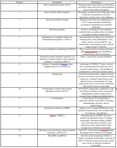
Table 1-Summery of biomarkers of DPN
Dear Editorial Team, Clinical Medical Reviews and Reports. My experience with the journal was highly positive. The peer-review process was rigorous, constructive, and completed in a timely manner. The reviewers provided valuable comments that helped improve the quality and clarity of our manuscript. The editorial office was professional, responsive, and supportive throughout all stages of the publication process. Communication was clear and efficient, and any questions were addressed promptly. Overall, I found the journal to maintain high scientific standards and an excellent publication workflow. I would be pleased to consider submitting future work to this journal. Best wishes from, Elena Popa.

It was my pleasure to submit my testimonial concerning the Reviewer Board of our Scientific Journal “Brain and Neurological Disorders”. The Reviewers focused on some modifications and their contribution was helpful. The ladies of our Editorial Office were also supported my efforts. It was my honor to have such a co-operation and I am looking forward for more collaboration.

Dear Grace Pierce, Editorial Coordinator of Journal of Clinical Research and Reports, Thank you for the speedy and efficient peer review process. I appreciate the fact that your peer reviewers do not take months to respond like with some other journals. I would also like to thank the editorial office for responding quickly to my questions. It is an excellent journal. I plan to submit more manuscripts in the future. Best wishes from, Robert W. McGee

Dear Grace Pierce, Editorial Coordinator of Journal of Clinical Research and Reports, Working with you and your team on our recent publication in JCRR has been a truly wonderful and enjoyable experience. The responses were prompt, and the reviewers were patient, constructive, and highly professional. One reviewer in particular gave me the feeling that a professor was carefully reading and commenting on my coursework, which was deeply touching. The entire process was straightforward and hassle‑free, with no tedious online forms to complete. I highly recommend this journal. Best wishes from, DR Aibing Rao, Head of R&D

I Appreciate the Opportunity to Share my Experience with the Journal of Clinical Research and Reports. The peer review process was timely and constructive, and the feedback provided helped improve the quality of our manuscript. The editorial office was professional, responsive, and supportive throughout the process, ensuring smooth communication and efficient handling of the submission. Overall, it was a positive experience collaborating with your team.

Dear Mercy Grace, Editorial Coordinator of Obstetrics Gynecology and Reproductive Sciences, We would like to express our gratitude for your help at all stages of publishing and editing the article. The editors of the magazine answer all the necessary questions and help at every stage. We will definitely continue to cooperate and publish other works in the Obstetrics Gynecology and Reproductive Sciences! Best wishes from, Alla Konstantinovna Politova,
