
Research Article | DOI: https://doi.org/10.31579/2690-1897/222
1 Department of Biological Sciences, Federal University Gusau.
2 Department of Microbiology, Federal University Gusau.
3 Department of Pharmaceutical Technology, Federal Polytechnic Kabo.
*Corresponding Author: Ali M, Department of Microbiology, Federal University Gusau.
Citation: Lurwan Mu’azu., Ali M., Idris I., Zage A.U, (2024), Determination of Toxicity Level of Sclerocarya birrea Stem bark Ethanol Extract against Wistar Rats, J, Surgical Case Reports and Images, 7(9); DOI:10.31579/2690-1897/222
Copyright: © 2024, Ali M. This is an open access article distributed under the Creative Commons Attribution License, which permits unrestricted use, distribution, and reproduction in any medium, provided the original work is properly cited.
Received: 23 October 2024 | Accepted: 31 October 2024 | Published: 08 November 2024
Keywords: toxicity level; sclerocarya birrea; stem bark; wistar rat
Many plants are highly poisonous when ingested. The study was aimed to study the toxicity effects of ethanol extract Sclerocarya birrea stem bark extracts against liver and kidney of wistar rats. The stem bark of S. birrea was extracted using ethanol by Soxhlet extraction method. Twenty-one (21) adult wistar rats were used in the study. Lorkeʼs method was used to test the acute toxicity of the extracts. The animals were divided into seven groups with three rats in each group. Animals in groups were orally administered with the extract of 62.5, 125, 250, 500, 1000 and 2000 mg/Kg body weight once daily for 10 days respectively. The result showed that here is no mortality in wistar rat after oral administration of the extract at a dose up to 2000 mg/Kg. Other clinical sign such as salivation, loss of hair, decrease in respiration rate (pulsating) and diarrhea were absent. However, the animals experienced restlessness at a concentration of 1000 and 2000 mg/Kg which disappeared within 24 hours. The histopathological analysis of the liver and kidney following administration of different concentration of the extract showed little loss of hepatocellular boundaries and vacuolation in the hepatocytes at 1000 and 2,000 mg/Kg in the liver and mild tissue degeneration and glomerulus shrinkage at 1000 and 2,000 mg/Kg in the kidney, but there is no visible degeneration at lower concentration. It is concluded that the ethanol extract of S. birrea may be safe for human consumption when use at low or moderate concentrations.
Since many years, human population across the world utilized elements of their environment, in particular plants, to treat themselves. Most pharmaceutical products depend on plants as the main source of material for their production. Plant derived medicines can serve as a basis for the manufacture of different more active drugs [1]. Most plants are made up of bioactive compounds such as lipids, phytochemicals, pharmaceutics, flavons, fragrances and pigments. To date, even with the spectacular progress accomplished in the field of science, an estimate of 66% to 85% of the world’s population, especially from developing countries, depend directly on plants as medicines in treating all sorts of diseases [2,3]. Various studies have reported that medicinal plants contain numerous biologically active compounds such as flavonoids, terpenoids, carotenoids, steroids, simple phenolic, glycosides, tannins, saponins, polyphenols, to mention a few, which have shown medicinal activities [4]. Most of these phytochemicals, commonly referred as secondary metabolites, were reported to act as antimicrobials [5]. According to World Health Organization, even developed countries are beginning to turn to plants for their medicine source due to the increased resistance of most existing antimicrobial drugs [6]. Sclerocarya birrea (commonly called marula) is a savannah tree, belongs to the family Anacardiaceae, with a plum-like pale yellow fruit of 3-4 cm in diameter with a juicy mucilaginous flesh. Sclerocarya birrea is deciduous and mainly dioecious, although there have been reports of monoecious trees [7]. It is a medium sized tree reaching heights of between 7 to 17 m, with grey fissured bark, stout branch lets and pale foliage. The leaves are compound, pinnate and the flowers greenish-white or reddish. The fruits are yellow, resembles a mango. Rough stems-bark is flaky, with a mottled appearance due to contrasting grey and pale brown patches [8]. The leaves are divided into 10 or more pairs of leaflets, each about 60 mm long, dark-green above, and sharp point. The flowers are borne in small, oblong clusters [7]. In some African countries, the stems-bark, roots and leaves of Sclerocarya birrea are used for an array of human ailments, including: malaria and fevers, diarrhea and dysentery, stomach ailments, headaches, sore eyes, toothache, backache and body pains, infertility, schistosomiasis, constipation, abdominal cramps and some other unspecified gastro-intestinal problems, toothaches and swollen or infected gums, cough, hypertension, arthritis, proctitis, epilepsy, diabetes mellitus, sores, boils, carbuncles, abscesses and certain other bacterial infections [9]. Traditionally the stem bark is used for the treatment of various gastrointestinal disorder especially dysentery/diarrhea, hemorrhoid, stomach ulcers and pain, sore throat/mouth and toothache [10]. Both the ethanolic and aqueous extract of this plant was found to be anti-inflammatory in rats paw induced edema, and with some antioxidant activity [11]. Despite the medicinal uses of plants extracts, the plants can be toxic when used at certain concentrations. Hence, the study was aimed to study the toxicity effects of Sclerocarya birrea stem bark ethanol extracts against the liver and kidney of wistar rats.
Collection and Identification of Plant Materials
The stem bark Sclerocarya birrea was collected at Kibiya town in Kibiya Local Government Kano. The Identification and authentication of the plant materials was conducted at Herbarium unit in the Department of Plant Science Bayero University Kano and Department of Biological Science, Kano University of Science and Technology, Wudil and the following voucher number was assigned BUKHAN 0435. Voucher specimen was deposited there for future reference. The samples were washed with water to remove dust and rinsed with distilled water, air dried for two-weeks and pulverized into powder form using sterile mortar and pestle in the laboratory as described by Ali et al. [12]. The powdered sample was bagged in a black polythene bag and store in air tight container for further use.
Extraction of Plant Materials
Ethanol was used as solvent in the extraction process. One hundred grams (100g) of the powdered leaves was weighed out and mixed with 500 ml of the solvent in a sterile conical flask and extracted using soxhlet extraction method. The filtrate was evaporated and dried at 40 °C under reduced pressure using rotary vacuum evaporator. The extract yields was weighed, stored in dark air tight container at 4°C [13].
Preparation of Extracts Concentrations
The solid residues (extracts) obtained was weighed and dissolved in 30% DMSO at a stock concentration of 2000mg/mL by dissolving 20 g in 10 mL. Various concentrations (1000, 500, 250, 125 and 62.5mg/mL) were made from the stock solution and stored at 40C until use [12].
Experimental Animals
Twenty one (21) adult wistar rats were obtained from Department of Pharmaceutical Science, College of Natural and Pharmaceutical Sciences, Bayero University Kano, Nigeria. The rats were kept in cage and maintained under laboratory condition of light and temperature with free access to food and water. The experimental animals were left for 10 days to adapt to the environment (acclimatization) and the animals were divided into 7 groups (A, B, C, D, E, F and G) of three animals each. The experimental protocol for the experiment was in accordance with OECD guidelines for the testing of chemicals.
Acute Toxicity Testing
Lorkeʼs method was used to test the acute toxicity of the extracts [14]. The animals were divided into seven groups with three rats in each group. Animals in Groups A, B, C, D, E, F and G were orally given extract of 62.5, 125, 250, 500, 1000 an 2000mg/kg body weight once daily for 10 days respectively. Group A was the control group and received only distilled water by the same route. Soon after dosing the rats were observed in an effort to note any changes in behavior. These included noting any changes in behaviour and structure such as salivation, restlessness, pulsating, loss of hair, diarrhea and death.
Histopathological Examination
The liver and one of the kidneys for each rat were fixed and preserved in 10% formaldehyde before subjection to tissue processing procedures for the preparation of permanent mount of each tissue as described by Baker and Silverton [15]. The tissues were dehydrated through various grades of alcohol 30%, 50%, and 70
Acute Toxicity and Lethality Tests
Toxicity and Lethality Profile of S. birrea Stem Bark Extract
The toxicity test result of different concentration of S. birrea stem bark extract is presented in Table 1 below. The result showed that here is no mortality in wistar rat after oral administration of the extract at dose as high as 2000mg/Kg. Other clinical sign such as salivation, loss of hair, decrease in respiration rate (pulsating) and diarrhea were absent. However, the animals experienced restlessness at a concentration of 1000 and 2000mg/Kg which disappeared within 24 hours.
| Dose (mg/Kg) | Salivation | Restlessness | Loss of hair | Pulsating | Diarrhea | Death |
| Group A (control) | - | - | - | Normal | - | 0/3 |
| Group B (62.5) | - | - | - | Normal | - | 0/3 |
| Group C (125) | - | - | - | Normal | - | 0/3 |
| Group D (250) | - | - | - | Normal | - | 0/3 |
| Group E (500) | - | - | - | Normal | - | 0/3 |
| Group F (1000) | - | + | - | Normal | - | 0/3 |
| Group G (2000) | - | + | - | Normal | - | 0/3 |
Table 1: Toxicity and Lethality Profile of S. birrea Stem Bark Extract against wistar rat
Histopathological Examination of Liver
The result of histopathological examination of liver of the experimenting animals is presented in figure 1–5 below. The result showed that there were no significant histopathological changes in the liver of the animal in group C (125mg/kg) and E (500mg/kg) when compared to control group. However, there some histopathological changes observed on animals in group F (1000mg/kg) and G (2000mg/kg). These changes include: the presence of congested blood vessels, peritoneal inflammation, presence of fatty cell vacuoles, and cell necrosis.
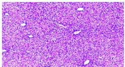
Figure 1: Section of Liver of Control group showing normal Liver tissue/hepatocytes
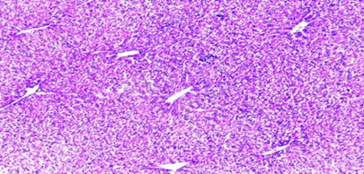
Figure 2: Section of Liver of group (125mg/kg) animal showing normal Liver tissue/hepatocytes
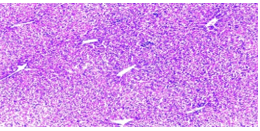
Figure 3: Section of Liver of group E (500mg/kg) animal showing normal Liver tissue/hepatocytes
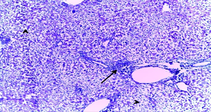
Figure 4: Section of Liver of Group F animal trated with 1000 mg/kg of the extract showing the presence of congested blood vessels, fatty cell vacuoles, and cell necrosis.
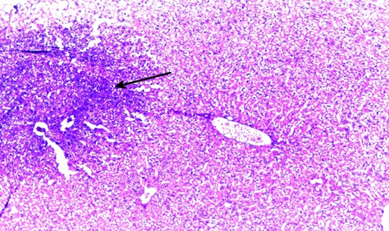
Figure 5: Section of Liver of Group G animal trated with 2000 mg/kg of the extract showing the presence of congested blood vessels, peritoneal inflammation, presence of fatty cell vacuoles, and cell necrosis.
Histopathological Examination of Kidney
The result of histopathological examination of kidney of the experimenting animals is presented in figure 6 – 10 below. The result showed no significant histopathological changes in the liver of the animal
in group B (62.5mg/kg) and E (500mg/kg) when compared to control (Group A). However, there some histopathological changes observed on animals in F (1000mg/kg) and G (2000mg/kg) showing areas of mild tissue degeneration, Glomerulus shrinkage and disintegration.
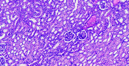
Figure 6: Section of Kidney of Control group showing normal nephron (Bowman capsule, glomerulus and renal tubule)
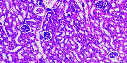
Figure 7: Section of Kidney of Group B animal treated with 62.5 mg/kg of the extract showing normal nephron
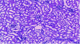
Figure 8: Section of Liver of Group B animal treated with 500 mg/kg of the extract showing normal nephron
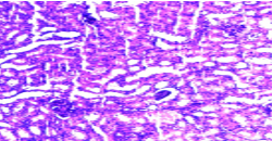
Figure 9: Section of Liver of Group F animal treated with 1000 mg/kg of the extract showing the presence of tissue degeneration and Glomerulus shrinkage
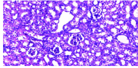
Figure 10: Section of Liver of Group G animal treated with 2000 mg/kg of the extract showing the presence of tissue degeneration, Glomerulus shrinkage and disintegration
Toxicity is the degree to which a substance can harm animals [16]. Toxicity can refer to the effect on a whole organism, such as an animal, bacterium, or plant, as well as the effect on a substructure of the organism, such as a cell (cytotoxicity) or an organ (organ toxicity), such as the liver (hepatotoxicity) [17]. A poisonous substance is any substance that produces disease conditions, tissue injury, or otherwise interrupts natural life processes when in contact with or absorbed into the body [18]. Many plants are highly poisonous when ingested. The study was aimed to determine the toxicity level of different concentration of S. birrea ethanol extract on wistar rat. In the present study, no mortality was observed in the acute toxicity study at all the concentrations (62.5 – 2000mg/kg) dosages indicating that the LD50 of the ethanol extract of S. birrea stem bark is greater than 2,000 mg/kg. Similarly, other clinical sign such as salivation, loss of hair, decrease in respiration rate (pulsating) and diarrhea were absent in all the concentrations. However, the animals experienced restlessness at a concentration of 1000 and 2000mg/kg which disappeared within 24 hours. Finding of this study was in conformity with that of and Thakur and Mengi [19] and Balogun et al. [20] who both reported report that the locomotory activity is considered to be an index of alertness, and any decrease in locomotion can indicate sedation. Acute toxicity study results suggest that S. birrea might be alerting the animal when high doses (1000 and 2000 mg/kg) are used.
Results obtained from the histopathological analysis of the liver and kidney following administration of different concentration of ethanol extract of S. birrea stem bark showed little loss of hepatocellular boundaries and vacuolation in the hepatocytes at 1000 and 2,000 mg/kg respectively indicating mild hepatotoxicity as compared to the 500, 250 and 125 mg/kg dosage groups which showed normal hepatocytes similar to control group. On the other hand, the sections obtained from the kidney showed areas of mild tissue degeneration; glomerulus shrinkage and disintegration at concentration of 1000 and 2,000 mg/kg respectively, but no visible degeneration were observed at lower concentration. Finding of this study supported the finding of Mawoza et al. [21] who reported mild degeneration of kidney when administered with 2000 and 5000 mg/kg respectively. Finding of this study showed that S. birrea stem bark ethanol extract is considered safe for human use when use at low or moderate concentrations. S. birrea is a medicinal plant having vast nutritional and therapeutic potential [22].
Based on the finding of this study, it is concluded that the lethal dose of ethanol extract of S. birrea stem bark is higher than 2,000 mg/kg as there is no mortality in the experimental animal. Similarly, clinical sign such as salivation, loss of hair, decrease in respiration rate (pulsating) and diarrhea were absent. However, the animals experienced restlessness at a concentration of 1000 and 2000 mg/Kg which disappeared within 24 hours which shows that the extract is safe for human use when use at low or moderate concentrations. It is recommended that the extracts from plant should be used at lower concentration.
The authors wish to acknowledge the technical staff of the Department of Pharmaceutical Sciences, College of Natural and Pharmaceutical Sciences, Bayero University Kano, Nigeria for provision and rearing of the experimental animals. Thanks to staff of Specialist laboratories and materials Kano for provision of materials and use of laboratory facilities.
Dear Editorial Team, Clinical Medical Reviews and Reports. My experience with the journal was highly positive. The peer-review process was rigorous, constructive, and completed in a timely manner. The reviewers provided valuable comments that helped improve the quality and clarity of our manuscript. The editorial office was professional, responsive, and supportive throughout all stages of the publication process. Communication was clear and efficient, and any questions were addressed promptly. Overall, I found the journal to maintain high scientific standards and an excellent publication workflow. I would be pleased to consider submitting future work to this journal. Best wishes from, Elena Popa.

It was my pleasure to submit my testimonial concerning the Reviewer Board of our Scientific Journal “Brain and Neurological Disorders”. The Reviewers focused on some modifications and their contribution was helpful. The ladies of our Editorial Office were also supported my efforts. It was my honor to have such a co-operation and I am looking forward for more collaboration.

Dear Grace Pierce, Editorial Coordinator of Journal of Clinical Research and Reports, Thank you for the speedy and efficient peer review process. I appreciate the fact that your peer reviewers do not take months to respond like with some other journals. I would also like to thank the editorial office for responding quickly to my questions. It is an excellent journal. I plan to submit more manuscripts in the future. Best wishes from, Robert W. McGee

Dear Grace Pierce, Editorial Coordinator of Journal of Clinical Research and Reports, Working with you and your team on our recent publication in JCRR has been a truly wonderful and enjoyable experience. The responses were prompt, and the reviewers were patient, constructive, and highly professional. One reviewer in particular gave me the feeling that a professor was carefully reading and commenting on my coursework, which was deeply touching. The entire process was straightforward and hassle‑free, with no tedious online forms to complete. I highly recommend this journal. Best wishes from, DR Aibing Rao, Head of R&D

I Appreciate the Opportunity to Share my Experience with the Journal of Clinical Research and Reports. The peer review process was timely and constructive, and the feedback provided helped improve the quality of our manuscript. The editorial office was professional, responsive, and supportive throughout the process, ensuring smooth communication and efficient handling of the submission. Overall, it was a positive experience collaborating with your team.

Dear Mercy Grace, Editorial Coordinator of Obstetrics Gynecology and Reproductive Sciences, We would like to express our gratitude for your help at all stages of publishing and editing the article. The editors of the magazine answer all the necessary questions and help at every stage. We will definitely continue to cooperate and publish other works in the Obstetrics Gynecology and Reproductive Sciences! Best wishes from, Alla Konstantinovna Politova,
