
Case report | DOI: https://doi.org/10.31579/2578-8949/108
Department of Dermatology and Venereology, CHU Ibn Sina, Mohammed V University, Rabat
*Corresponding Author: Najoua Ammar, Department of Dermatology and Venereology, CHU Ibn Sina, Mohammed V University, Rabat
Citation: Najoua Ammar, Mariame Meziane, Laila Benzekri, Nadia Ismaili, Karima Senouci (2023), Dermatofibrosarcoa of Darier-Ferrand with an Atypical Location. Dermatology and Dermatitis. 8(1); DOI:10.31579/2578-8949/108
Copyright: ©2023 Najoua Ammar, This is an open-access article distributed under the terms of The Creative Commons. Attribution License, which permits unrestricted use, distribution, and reproduction in any medium, provided the original author and source are credited.
Received: 29 November 2022 | Accepted: 17 December 2022 | Published: 20 January 2023
Keywords: darier–ferrand dermatofibrosarcoma; dermatofibrosarcoma protuberans
Dermatofibrosarcome protuberans (DFSP) is a rare malignant soft tissue tumor characterized by aggressive local béhavioral, high frequency of local recurrence with low metastatic potential. It occurs most often in patients between 20 and 50 years of age, and is preferentially located on the trunk. The standard treatment remains complete surgical excision with wide margins (>3 cm) or Mohs micrographic surgery. We report here a case of DFS unusual by its location on the leg
Darier-Ferrand dermatofibrosarcoma or dermato fibrosarcome protuberans (DFSP) is a rare cutaneous fibrous tumor, characterized by aggressive local behavior and low metastatic potential.
It was first described in 1924 by Darier and Ferrand. It represents 0.1% of all cancers and 1.8% of all soft tissue sarcomas [1].
The most frequent tumor location is the trunk but rarely the extremities. Herein we are reporting a case of a Darier-Ferrand dermatofibrosarcoma of the leg
We present the observation of a 45-year-old patient, with no previous medical history, who was referred to us for the management of a protruding mass in the leg that had been evolving for at least 8 years and that had rapidly increased in size and become painful over the past year. No other systemic or local symptoms were reported. Physical examination revealed a 10-cm, firm, tender mass on the left leg (Figure. 1). There was no evidence of ulceration or bleeding. No adenopathy was found. The lesion was fixed to the overlying skin but mobile in the deeper tissues.
Standard radiographs showed a softs tissus mass without bony involvement.
A punch biopsy was performed, revealing a malignant mesenchymal proliferation, composed of spindle cells with little atypical and low mitotic activity arranged in storiform fascicles. Immuno histo chemistry showed a high expression of CD34 concluding to a dermatofibrosarcoma.
The patient underwent a wide local excision of the mass.
Dermatofibrosarcoma protuberans (DFSP) was first described by Taylor en 1890, followed by Darier and Ferrand en 1924 as dermatofibroma or recurrent progressive fibrosarcoma of the skin, and finally by Hoffmann en 1925 as dermatofibrosarcoma protuberans [2].
It is a rare mesenchymal tumor that belongs to the sarcomas of low grade of malignancy with an aggressive local behavior and has a low metastatic potential. It represents 1.8% of all soft tissue sarcomas and only 0.1% of all cancers with a higher incidence in women and in the black population. It is most often seen in patients between 20 and 50 years of age and localizes preferentially on the trunk and rarely on the extremities [3].
Clinically, it begins as a simple brownish or purplish infiltrated plaque or as a small nipple nodule, usually asymptomatic. It evolves slowly over several years to a large multinodular mass with progressive evolution. There are no known predisposing risk factors for the development of DFSP, but a history of trauma has been noted [4,5].
Histologically, the cells are monomorphic, spindle-shaped, cartwheel-shaped, with few atypia and low mitotic activity. The neoplastic cells often infiltrate the surrounding adipose tissue in a honeycomb pattern. Immunohistochemistry shows diffuse and strong expression of CD34, and loss of expression of other biomarkers such as S100 protein, factor XIIIa, smooth muscle alpha actin and melanin [6].
Differential diagnoses are mainly neurofibroma, dermofibroma, hemangioma, giant cell fibroblastoma, pilomatrixoma and malignant melanoma.
The gold standard treatment for DFSP is wide local excision (WLE), with safety margins ranging from 3 to 5 cm Re-excision is recommended if the surgical margin is positive. Mohs micrographic surgery (MMS) is an alternative to wide local excision [6].
Darier-Ferrand dermatofibrosarcoma is an uncommon recurrent tumor that can occur in sites other than the trunk.
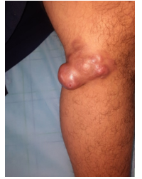
Figure 1: Dermatofibrosarcoma of Darier and Ferrand presented as a mass on the left leg
None of the authors has a conflict of interest to declare.
Dear Editorial Team, Clinical Medical Reviews and Reports. My experience with the journal was highly positive. The peer-review process was rigorous, constructive, and completed in a timely manner. The reviewers provided valuable comments that helped improve the quality and clarity of our manuscript. The editorial office was professional, responsive, and supportive throughout all stages of the publication process. Communication was clear and efficient, and any questions were addressed promptly. Overall, I found the journal to maintain high scientific standards and an excellent publication workflow. I would be pleased to consider submitting future work to this journal. Best wishes from, Elena Popa.

It was my pleasure to submit my testimonial concerning the Reviewer Board of our Scientific Journal “Brain and Neurological Disorders”. The Reviewers focused on some modifications and their contribution was helpful. The ladies of our Editorial Office were also supported my efforts. It was my honor to have such a co-operation and I am looking forward for more collaboration.

Dear Grace Pierce, Editorial Coordinator of Journal of Clinical Research and Reports, Thank you for the speedy and efficient peer review process. I appreciate the fact that your peer reviewers do not take months to respond like with some other journals. I would also like to thank the editorial office for responding quickly to my questions. It is an excellent journal. I plan to submit more manuscripts in the future. Best wishes from, Robert W. McGee

Dear Grace Pierce, Editorial Coordinator of Journal of Clinical Research and Reports, Working with you and your team on our recent publication in JCRR has been a truly wonderful and enjoyable experience. The responses were prompt, and the reviewers were patient, constructive, and highly professional. One reviewer in particular gave me the feeling that a professor was carefully reading and commenting on my coursework, which was deeply touching. The entire process was straightforward and hassle‑free, with no tedious online forms to complete. I highly recommend this journal. Best wishes from, DR Aibing Rao, Head of R&D

I Appreciate the Opportunity to Share my Experience with the Journal of Clinical Research and Reports. The peer review process was timely and constructive, and the feedback provided helped improve the quality of our manuscript. The editorial office was professional, responsive, and supportive throughout the process, ensuring smooth communication and efficient handling of the submission. Overall, it was a positive experience collaborating with your team.

Dear Mercy Grace, Editorial Coordinator of Obstetrics Gynecology and Reproductive Sciences, We would like to express our gratitude for your help at all stages of publishing and editing the article. The editors of the magazine answer all the necessary questions and help at every stage. We will definitely continue to cooperate and publish other works in the Obstetrics Gynecology and Reproductive Sciences! Best wishes from, Alla Konstantinovna Politova,
