
Case Report | DOI: https://doi.org/10.31579/ 2690-8816/153
*Corresponding Author: Dr Sule Muhammad Baba. Radiology Department, Usmanu Danfodiyo University, Sokoto.
Citation: Shamaki AMB, Sule MB, Gele IH, Aminu UU, (2025), DENTIGEROUS CYST PRESENTING AS LOWER JAW SWELLING IN AN ADULT MALE: RADIOLOGICAL FEATURES AND A CASE REPORT, J Clinical Research Notes, 6(1); DOI:10.31579/ 2690-8816/153
Copyright: © 2025, Sule Muhammad Baba. This is an open access article distributed under the Creative Commons Attribution License, which permits unrestricted use, distribution, and reproduction in any medium, provided the original work is properly cited.
Received: 06 January 2025 | Accepted: 13 January 2025 | Published: 21 January 2025
Keywords: dentigerous; cyst; jaw; radiographic
Dentigerous cyst (DC) is one among the jaw swellings that appear as a well circumscribed radiolucency surrounding the crown of an unerupted tooth and most commonly diagnosed by their radiographic appearance. Most often DC appear as unilocular well-defined pericoronal radiolucency centered on an impacted tooth within the mandible. The incidence is reported as 1.44 in every 100 unerupted teeth, and more prevalent in males especially in the 2nd and 3rd decades of life.
This is a forty-year-old man who presented from a peripheral health care center for plain radiography of the jaw on account of a painless left lower jaw swelling. The plain radiograph of the jaw demonstrated a lucent well circumscribed swelling with sclerotic margin over the crown of an unerupted left lower mandible most likely the third molar. No dental anarchy or loss of the lamina dura was noted on the jaw radiograph. The patient had surgical excision of the jaw mass and unerupted/impacted tooth with histopatholgical assessment of the sample that confirmed the epithelial lining likened to dentigerous cyst.
We present a case of dentigerous cyst presenting in an adult male as a lower jaw swelling because of its peculiar radiographic appearance and to further review the literature.
Dentigerous cyst is a non-inflammatory odontogenic cyst, otherwise known as follicular cyst, develops from the pericoronal tissue which could either be dental sac or dental follicle of an impacted tooth be it permanent or deciduous or supernumerary1-3.
Dentigerous cysts seen around the crown of an unerupted tooth happens to be the second most common cyst of dental origin when compared to cysts related to the roots of the teeth such as periapical cysts and also accounts for about 14-24% of all jaw cysts4,5.
Follicular cysts are most often painless, and discovered accidentally following clinical and radiographic examination, may be large enough resulting into a palpable mass, such growth may also lead to displacement of adjacent teeth4,6. Dentigerous cysts are often asymptomatic but occasionally may present with pain most times caused by infection or paresthesia following compressive effect on a nerve1,7.
Dentigerous cysts are mainly imaged radiologically, this could either be following a panoramic dental X-ray (OPG), plain radiography of the jaw, computed tomography and magnetic resonance imaging with the OPG being the most frequent diagnostic imaging1,7.
Follicular cysts do have a peculiar radiologic appearance, they appear as well-defined oval unit-locular homogenous radiolucent lesion adherent to the cemento enamel junction (crown) of an unerupted/impacted tooth, usually the third molars and predominantly in the mandible, and present in three main radiographic forms; the central, lateral, and circumferential forms1,7-9.
Dentigerous cyst has varying treating modalities, ranging from enucleation to marsupialization, this may be influenced by the age of the affected individual, severity of impaction, and root form of the associated tooth/teeth10.
This is a forty-year-old man who presented from a peripheral health care center for plain radiography of the jaw on account of a painless left lower jaw swelling.
On examination, the patient presented with a non-tender swelling on the left lower jaw region, the patient is stable, not pale, not dehydrated, not jaundiced and not in any form of obvious distress.
The plain radiographs of the jaw (Anterior-Posterior and left lateral views) demonstrated a lucent well circumscribed swelling with sclerotic margin over the crown and root of an unerupted tooth on the left lower hemi-mandible most likely the third molar; this is most likely the circumferential form of the dentigerous cyst (Figures 1 and 2). No dental anarchy or loss of the lamina dura was noted on the jaw radiograph. The patient had surgical excision of the jaw mass and unerupted/impacted tooth with histopatholgical assessment of the sample that confirmed the epithelial lining likened to dentigerous cyst.
We present a case of dentigerous cyst presenting in an adult male as a lower jaw swelling because of its peculiar radiographic appearance and to further review the literature.
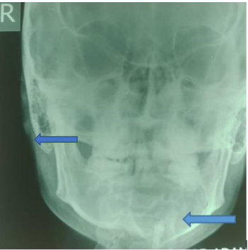
Figure 1: An anterior-posterior view of the jaw demonstrating a well circumscribed oval lucency with sclerotic margin over the crown and root of an unerupted tooth in the region of the body of the left hemi-mandible in keeping with the classical appearance of a circumferential form of dentigerous cyst (Left blue arrow).
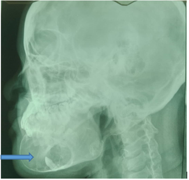
Figure 2: A left lateral view of the jaw demonstrating a well circumscribed oval lucency with sclerotic margin over the crown and root of an unerupted tooth in the region of the body of the left hemi-mandible in keeping with the classical appearance of a circumferential form of dentigerous cyst (right blue arrow).
Dentigerous or follicular cyst is most prevalent in younger patients, most often in the second and third decades of life, with a high prevalence in males and commonly associated with an impacted third molars of the mandible. The index case is however forty years of age, but a male and the cyst were associated with an unerupted tooth on the mandible.
Follicular cyst is most often painless, and discovered accidentally following clinical and radiographic examination, may be large enough resulting into a palpable mass, such growth may also lead to displacement of adjacent teeth4,6. The case under review was also diagnosed following clinical and radiographic evaluations, presenting with a left lower jaw palpable mass surrounding an unerupted tooth, thereby conforming to these literatures.
Dentigerous cysts are mainly imaged radiologically, this could either be following a panoramic dental X-ray, plain radiography of the jaw, computed tomography (CT) and magnetic resonance imaging (MRI) with the OPG being the most frequent diagnostic imaging1,7. The index case was also diagnosed following radiographic examination; plain radiographs of the jaw, thereby conforming to these literatures. The case under study however did not undergo panoramic radiography, CT and MRI.
Follicular cysts do have a peculiar radiologic appearance, they appear as well-defined oval uni-locular homogenous radiolucent lesion adherent to the cemento-enamel junction (crown) of an unerupted/impacted tooth, usually the third molars and predominantly in the mandible, and present in three main radiographic forms; the central, lateral, and circumferential forms1,7-9. The case under review also appeared as a well circumscribed oval lucency over the crown and root of an unerupted tooth on the left hemi-mandible, thereby agreeing with these literatures.
Dentigerous cyst has varying treating modalities, ranging from enucleation to marsupialization, this may be influenced by the age of the affected individual, severity of impaction, and root form of the associated tooth/teeth10. The case under review had enucleation of the cyst with the unerupted tooth, thereby conforming to this literature.
Palpable jaw mass could be Dentigerous cyst, this can be diagnosed following basic plain radiographs of the jaw due to its characteristic appearance radiographically thereby aiding in immediate institution of the required management.
Dear Editorial Team, Clinical Medical Reviews and Reports. My experience with the journal was highly positive. The peer-review process was rigorous, constructive, and completed in a timely manner. The reviewers provided valuable comments that helped improve the quality and clarity of our manuscript. The editorial office was professional, responsive, and supportive throughout all stages of the publication process. Communication was clear and efficient, and any questions were addressed promptly. Overall, I found the journal to maintain high scientific standards and an excellent publication workflow. I would be pleased to consider submitting future work to this journal. Best wishes from, Elena Popa.

It was my pleasure to submit my testimonial concerning the Reviewer Board of our Scientific Journal “Brain and Neurological Disorders”. The Reviewers focused on some modifications and their contribution was helpful. The ladies of our Editorial Office were also supported my efforts. It was my honor to have such a co-operation and I am looking forward for more collaboration.

Dear Grace Pierce, Editorial Coordinator of Journal of Clinical Research and Reports, Thank you for the speedy and efficient peer review process. I appreciate the fact that your peer reviewers do not take months to respond like with some other journals. I would also like to thank the editorial office for responding quickly to my questions. It is an excellent journal. I plan to submit more manuscripts in the future. Best wishes from, Robert W. McGee

Dear Grace Pierce, Editorial Coordinator of Journal of Clinical Research and Reports, Working with you and your team on our recent publication in JCRR has been a truly wonderful and enjoyable experience. The responses were prompt, and the reviewers were patient, constructive, and highly professional. One reviewer in particular gave me the feeling that a professor was carefully reading and commenting on my coursework, which was deeply touching. The entire process was straightforward and hassle‑free, with no tedious online forms to complete. I highly recommend this journal. Best wishes from, DR Aibing Rao, Head of R&D

I Appreciate the Opportunity to Share my Experience with the Journal of Clinical Research and Reports. The peer review process was timely and constructive, and the feedback provided helped improve the quality of our manuscript. The editorial office was professional, responsive, and supportive throughout the process, ensuring smooth communication and efficient handling of the submission. Overall, it was a positive experience collaborating with your team.

Dear Mercy Grace, Editorial Coordinator of Obstetrics Gynecology and Reproductive Sciences, We would like to express our gratitude for your help at all stages of publishing and editing the article. The editors of the magazine answer all the necessary questions and help at every stage. We will definitely continue to cooperate and publish other works in the Obstetrics Gynecology and Reproductive Sciences! Best wishes from, Alla Konstantinovna Politova,
