AUCTORES
Globalize your Research
Case Report | DOI: https://doi.org/10.31579/2690-4861/590
1 Gynecological Oncology department, Hospital Universitario de Basurto, Bilbao, Spain.
2 Pathological Anatomy department, Hospital Universitario de Basurto, Bilbao, Spain.
*Corresponding Author: Cristina Berriozabal, Gynecological Oncology department, Hospital Universitario de Basurto, Bilbao, Spain.
Citation: Cristina Berriozabal, Eva Beiro, Tamara Dehesa, Carlos D. Pérez, Jesús Hilario De La Rosa, et al, (2024), Delayed Vulvar Metastases from Rectal Carcinoma: A Case Report, International Journal of Clinical Case Reports and Reviews, 19(4); DOI:10.31579/2690-4861/590
Copyright: © 2024, Cristina Berriozabal. This is an open-access article distributed under the terms of the Creative Commons Attribution License, which permits unrestricted use, distribution, and reproduction in any medium, provided the original author and source are credited.
Received: 23 October 2024 | Accepted: 01 November 2024 | Published: 11 November 2024
Keywords: vulvar metastases; gynecological metastases; colorrectal adenocarcinoma
Primary tumors of the vulva are a rare entity, accounting for only 3-5% of all gynecologic malignancies. However, metastatic tumors of the vulva are even rarer, constituting 5-8% of all vulvar tumors. These metastases can be the first manifestation of another primary tumor or may appear years after initial treatment. This case presents a 78-year-old woman treated for rectal adenocarcinoma who developed vulvar metastases a year and a half after completing treatment. Previously, she had a liver metastasis that was treated with laparoscopic resection. On physical examination, two firm vulvar masses were detected, and the histopathological analysis revealed adenocarcinoma infiltration, likely of rectal origin. After a negative extension study via CT scan, a local resection of the vulvar metastases was performed. Two new masses were detected a month later, leading to a second local resection.
This case underscores the importance of an accurate diagnosis of vulvar lesions, particularly in women with a history of another primary tumor. Vulvar metastases can easily be misdiagnosed as a primary vulvar tumor or even as a benign condition. Despite their low incidence, they should be considered, as an accurate diagnosis is crucial for appropriate management and prognosis. Immunohistochemical techniques play a key role in identifying the origin of the primary tumor. Due to the limited number of cases, the optimal management of metastatic tumors to the vulva remains uncertain, this is the reason why multidisciplinary discussion is essential to determine the most appropriate treatment. Finally, the prognosis remains poor due to distant metastatic spread.
Primary cancer of the vulva is a rare entity, accounting for only 3-5% of all gynecologic malignancies and less than 1% of all cancers in women [1]. Most primary malignancies of the vulva are squamous cell carcinomas, with occasional cases of basal cell carcinomas and melanomas [2]. Metastatic tumors of the vulva are even rarer, constituting 5-8% of all vulvar tumors. These lesions occur most frequently in postmenopausal women and their diagnosis can be challenging, especially in the absence of a known primary malignancy. Clinicians should therefore consider biopsy of these lesions to enable a thorough histopathological analysis. These metastases are often associated with widespread disease and frequently represent a preterminal event [1,2]. Differentiating a metastatic lesion from a primary tumor is crucial for both: management and prognosis [3,4].
Metastases to the female genital tract originating from gynecological tumors are relatively common; however, metastases from extra-genital tumors are unusual. The most frequent extra-genital primary tumors that metastasize to the genital tract are colorectal and breast cancers, which typically involve the ovaries and vagina [4,5,6]. Vulvar metastases are particularly uncommon, accounting for only 2% of cases [6]. Here, we present an unusual case of recurrent rectal carcinoma manifesting as vulvar metastasis, initially misdiagnosed as vulvar folliculitis.
We present the case of a 76-year-old patient referred to the gynecology department by her primary care physician with suspected vulvar folliculitis. For this case, ethical approval and informed patient consent were obtained. The most relevant aspect of her medical history was the diagnosis of rectal carcinoma a year and a half prior. She underwent neoadjuvant chemoradiotherapy followed by surgery. A laparoscopic abdominoperineal oncologic resection, assisted by the Da Vinci system, was performed with the placement of a terminal colostomy. The histopathological report revealed low-grade residual adenocarcinoma infiltrating the submucosal layer, with free surgical margins: G2 ypT1 pN1c pMx L0 V0 Pn0 GRT-2. After surgery, she received six additional cycles of chemotherapy with capecitabine. One year later, a follow-up Computerized Tomography (CT) scan revealed a solitary liver lesion in segment IVA, which was treated via laparoscopic resection. The histopathological examination confirmed metastasis of rectal adenocarcinoma. As the recurrence occurred within the first six months after chemotherapy, no further cycles were administered, and the patient remained under regular follow-up.
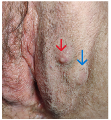
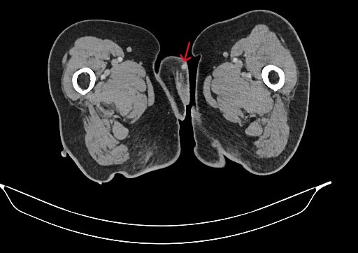
Three months after the liver resection, vulvar lesions appeared, and the patient was referred to the gynecology service. On physical examination, two firm masses were detected: one measuring 7 mm on the left labium majus and another measuring 15 mm near the left inguinal fold (Figure 1). The rest of the physical examination was unremarkable, and no inguinal lymph nodes were palpable. The histopathological results revealed infiltration by adenocarcinoma, likely of rectal origin, with an immunohistochemical profile of CK20+/CDX2+/CK7- (Figure 2). The extension study with a CT scan ruled out distant metastasis, identifying only the vulvar lesions (Figure 3). Consequently, the multidisciplinary tumor board recommended local resection of the tumor.

Figure 1: Physical examination of initial vulvar metastases: one located in the left labium majus mimicking vulvar folliculitis (red arrow) and another near the left inguinal fold (blue arrow).

Figure 2: Immunohistochemical profile of the vulvar metastase (CK20+/CDX2+).
A: Intense and diffuse expression of CK-20 (2x). The area of stained cells is marked with arrows.
B: Intense and diffuse expression of CK-20 (10x).
C: Heterogeneous expression of CDX-2 (2x). The area of stained cells is marked with arrows.
D: Heterogeneous expression of CDX-2 (20x).

Figure 3: Initial vulvar metastasis in the left labium majus as seen on CT scan.
A month after surgery, during follow-up, two new masses were detected (Figures 4 and 5). Histopathological analysis confirmed that they were again metastases of rectal adenocarcinoma with the same immunohistochemical profile. Another extension study was performed, and the CT scan again showed only the vulvar masses, with no evidence of distant spread (Figure 6), so another local resection was performed.


Figures 4 and 5: Second vulvar metastases.

Figure 6. Second vulvar metastasis at the top of the left labium majus on CT scan.
After the second vulvar surgery, the multidisciplinary tumor board decided to administer chemotherapy. No further recurrences were observed during treatment, and she is currently undergoing regular follow-up with the oncology department.
Metastases to the female genital tract originating from gynecological tumors are relatively common. However, metastases from extra-genital tumors are a very rare entity. The most common extra-genital tumors that metastasize to the genital tract are colorectal (37%) and breast cancers (34%) [4,5,6]. The ovaries and vagina are the most frequent metastatic sites for both extra-genital and genital primaries [5]. Other sites, such as the vulva, account for only 2% of genital tract metastases [6]. Within this rare group, we include our patient's unusual case.Given the low incidence of vulvar metastases, very few cases or case series have been reported in the literature. We will now analyze some of these reports.
Dehner was the first to publish a retrospective analysis defining the clinical and morphological features of vulvar metastases. In his study, 262 vulvar tumors were analyzed, of which 22 were metastatic (8.3%). The results showed that 81.8% of vulvar metastases originated from primary genital carcinomas, with the cervix being the most frequent site (10 cases: 45.4%), followed by endometrial, urethral, vaginal, and ovarian carcinomas (total of 8 cases: 36.4%). Primary extra-genital carcinomas accounted for only 4 cases (18.2%), including metastases from breast, kidney, and lymph nodes [7].Mazur et al. analyzed 325 cases of metastatic tumors affecting the female genital tract. Of these, 176 cases (54.15%) were metastases originating from primary tumors of the genital tract, while the remaining 149 cases (45.85%) were metastases from extragenital sites. The ovaries and vagina were the most common sites of metastasis, regardless of the location of the primary tumor. No vulvar metastases were reported. The gastrointestinal tract, particularly the stomach and colorectal regions, was the most frequent source of extragenital tumors metastasizing to the female genital tract, predominantly involving the ovaries [5].
Karpathiou et al. conducted a retrospective study of 196 secondary lesions involving the gynecologic tract. The organs affected were the ovaries (50%), vagina (22%), myometrium (10.7%), cervix (10.2%), endometrium (3.6%), vulva (2%), and Fallopian tubes (1.5%). The most common primary tumors were colorectal (39.8%), endometrial (15.3%), breast (12.7%), ovarian (10.7%), and gastric (5.6%) [6].Neto et al. conducted a study of 66 cases of metastatic tumors of the vulva at the University of Texas M.D. Anderson Cancer Center over 57 years. In 46.9% of the cases, the primary tumor was of gynecologic origin, including cervical (22.7%), ovarian (9%), endometrial (3%), and vaginal (3%) cancers. In 43.9% of cases, the primary tumor originated from non-gynecologic sites, including the gastrointestinal tract (18.2%), breast (6%), cutaneous melanoma (6%), lung (4.5%), lymphoma (4.5%), genitourinary system (3%), and pancreas (1.5%). In the remaining 9.1% of cases, the primary tumor could not be identified. Metastases were detected between 1 and 264 months (mean: 35.6 months) after diagnosing the primary tumor. The prognosis in these cases was poor, with a median survival of 7.5 months [1].
Chao et al. also reported 78 cases of metastatic tumors of the vulva with similar findings. In their study, the most common primary tumor was cervical cancer (61 cases), followed by urethral (5 cases), vaginal (4 cases), endometrial (3 cases), breast (2 cases), ovarian (1 case), rectal (1 case), and malignant lymphoma (1 case). The prognosis of these metastases was similarly poor [8].As reported by Dehner, Neto, and Chao, cervical, ovarian, and endometrial cancers (in that order) are the most common gynecologic malignancies that metastasize to the vulva. However, the gastrointestinal system remains the most frequent extragenital site of origin for vulvar metastases [1,2,4,7].As reported by Mazur and Karpathiou, the ovaries and vagina are the most common sites of metastasis in both the genital and extragenital tracts. The gastrointestinal tract, particularly the colorectal region and stomach, is the most frequent extragenital source of tumors that metastasize to the female genital tract [5,6].
Colorectal cancer is the third most commonly diagnosed malignancy worldwide and the second leading cause of cancer-related deaths [9]. It is also one of the malignancies most prone to metastasize, accounting for nearly 32.2% of diagnosed cases. Colorectal carcinoma typically metastasizes to the liver and lungs [10]. These recurrences are associated with poor prognoses and generally occur within the first five years after treatment [4]. The case of our patient fits within this category, with metastatic spread to the vulva one year after treatment; however, it represents an extremely rare site of metastasis.According to the study by Neto et al., the most frequently involved vulvar site in metastatic spread was the labium majus (44 out of 66 cases). Other locations were less common: clitoris (4 cases), and the labium minus, mons pubis, and introitus (3 cases each) [1].
In many cases, vulvar metastasis may be the first manifestation of the disease [1, 5]. The clinical presentation of vulvar metastases can be challenging, as they may mimic primary tumors or, as in our case, even resemble benign conditions. Therefore, a thorough clinical history and appropriate complementary studies (such as pathology and imaging) are crucial for accurate diagnosis [3,4,9-12].
The studies mentioned above provide important epidemiological data but do not explore the factors that may influence the development of these metastases. The pathogenesis of secondary gynecological tumors remains unclear, with lymphatic, hematogenous, and transcoelomic pathways being proposed [6]. Most metastases from the colon and rectum involve the ovaries, likely due to their extensive lymphatic network and anatomical location within the abdominal cavity. Malignant cells can also spread directly to adjacent structures, such as the cervix, uterus, and vulva. Typically, metastases to the upper third of the vagina originate from the genital tract, whereas those in the lower third and vulva may arise from the gastrointestinal tract [10]. The hematogenous route is also significant. When metastases develop within the first few years after surgery, this may be attributed to exfoliated tumor cells spreading via the hematogenous route [13].
Accurate and early diagnosis is critical for these patients. Immunohistochemical techniques play a key role in identifying the origin of the primary tumor. In the literature, some cases of primary vulvar or vaginal adenocarcinoma of intestinal type are described, and differentiating these from metastatic disease is essential for appropriate management [14-16]. The combined detection of CK20, and CK7 can assist in this distinction [10]. CK20 is typically expressed in the gastrointestinal tract and urothelium, whereas CK7 is present in most transitional and glandular epithelia. It is expressed in lung adenocarcinomas, breast cancer, and some subtypes of ovarian cancer, but not in colorectal tumors [4,10]. A CK20+/CK7- immunoprofile strongly suggests colorectal carcinoma [2-4,9-11]. CDX2, which indicates enteric differentiation, is more specific than CK20 for this diagnosis [10].
Primary vulvar or vaginal adenocarcinomas of intestinal type generally exhibit a CK20+/CK7+ pattern [14]. In our patient, the biopsy showed a CK20+/CDX2+/CK7- immunoprofile, pointing towards a colorectal metastasis and ruling out a primary vulvar tumor of intestinal origin.
Due to the limited number of cases, the optimal management of metastatic tumors to the vulva remains uncertain. The literature suggests a multimodal approach that includes surgery, chemotherapy, and radiotherapy [3-4,10-12,17]. Treatment decisions depend on the primary neoplasm as well as the extent and localization of metastases. For patients with widespread disease, surgical intervention is typically recommended to achieve local control and relieve symptoms [10]. In addition to surgery, case reports reviewed in this article describe the use of the FOLFOX chemotherapy regimen (leucovorin calcium, fluorouracil, and oxaliplatin) which is standard I colorectal carcinoma treatment [3-4,10]. This regime may be combined with Cetuximab, however, emerging studies in molecular analysis suggest that KRAS mutations can predict prognosis and response to chemotherapy. For instance, the KRAS p.G13D mutation has been linked to resistance to cetuximab [17]. Radiotherapy is also reported as a means of achieving local control [10-11], though dosage recommendations are not well established, and treatment may sometimes need to be discontinued due to severe adverse effects [4].
The prognosis remains poor due to distant metastatic spread [1,5,7,8]. Neto et al. reported 52 deaths out of 66 patients within 1 to 81 months of diagnosis [1]. Similarly, Dehner reported 20 deaths out of 22 patients within the first year following the diagnosis of vulvar metastases [7]. Nevertheless, since the largest and latest series were published years ago, new chemotherapy regimes have lengthened life expectancy of these patients [3].
Vulvar metastases can easily be misdiagnosed as a primary vulvar tumor or, as in our case, as a benign condition. They may also obscure the primary tumor and, in some cases, be diagnosed before it. Despite their low incidence, accurate diagnosis is crucial, as the management and prognosis differ significantly between metastatic disease and primary tumors. Multidisciplinary discussions are essential in determining the appropriate treatment plan. Given the rarity of these cases, there is no standardized treatment, though a combination of surgery and chemotherapy may be considered.
In conclusion, this rare entity should be considered in patients presenting with vulvar lesions and a history of another primary tumor, particularly of gynecologic or colorectal origin.
This case report has inherent limitations due to its nature, as it describes a single case, which restricts the generalizability of the findings. The few cases of vulvar metastases from colorectal carcinoma described in the literature limit direct comparison of findings with similar studies and the development of standardized management protocols. In this report, we present the clinical findings, diagnosis, and management of our case; however, further studies with a larger number of cases are needed to establish more defined, evidence-based clinical guidelines.
Author Controbutions
Cristina Berriozabal: literature review, manuscript writing, submission; Eva Beiro: literature review, manuscript writing; Tamara Dehesa: literature review, manuscript writing; Carlos David Pérez: manuscript review, images and editing, Jesús Hilario De La Rosa: leading surgeon and supervision; Ane Gartzia: images and review.
All authors (Cristina Berriozabal, Eva Beiro, Tamara Dehesa, Carlos David Pérez, Jesús Hilario De La Rosa and Ane Gartzia) meet the ICMJE authorship criteria.