
Research Article | DOI: https://doi.org/10.31579/2690-8794/204
+ These authors contributed equally to this work.
1 Department of Dermatology, Huashan Hospital, Fudan University, Shanghai, China.
2 UNISKIN Research Institute on Skin Aging, Inertia Shanghai Biotechnology Co., Ltd., Shanghai, China.
3 Derma Health Shanghai Biotechnology Co., Ltd., Shanghai, China.
*Corresponding Author: Fan Hu, UNISKIN Research Institute on Skin Aging, Inertia Shanghai Biotechnology Co., Ltd., Shanghai, China.
Citation: Qianqian Wang, Nihong Li, Mingyu Wang, Rui Ye, Le Du, et al., (2024), Deciphering the Dynamics of Facial Skin Aging and the Efficacy of Anti-Aging Eye Cream Through In Vivo Two-Photon Microscopy, Clinical Medical Reviews and Reports, 6(2); DOI:10.31579/2690-8794/204
Copyright: © 2024, Fan Hu. This is an open access article distributed under the Creative Commons Attribution License, which permits unrestricted use, distribution, and reproduction in any medium, provided the original work is properly cited.
Received: 08 March 2024 | Accepted: 18 March 2024 | Published: 29 March 2024
Keywords: DEJ; periocular; facial skin aging; eye cream; two-photon microscopy
Background: Skin aging is a multifactorial process influenced by genetic and environmental factors, manifesting prominently on the face. Variations in aging effects across facial areas necessitate advanced imaging for detailed assessment.
Materials and Methods: We utilized a portable handheld two-photon microscope (TPM) for in vivo exploration of skin aging alterations in the periocular and cheek areas of a Chinese female population. This study included 107 healthy volunteers, aged between 18 to 60 years, categorized into four age intervals. Our research extended to a clinical trial to evaluate the efficacy of a novel anti-aging eye cream rich in plant extracts, peptides, and antioxidants. This formulation was assessed for its potential in enhancing skin hydration, elasticity, and reducing wrinkles. Participants applied the eye cream twice daily for 8 weeks, with skin parameters measured at baseline and at intervals throughout the study period.
Results: Our findings reveal significant variations in epidermal thickness (ET) and dermal-epidermal junction (DEJ) indices with age, particularly noting a decrease in ET and flattening of the DEJ in older age groups. The anti-aging eye cream demonstrated marked improvements in skin hydration levels, elasticity, and a reduction in under-eye wrinkles and crow's feet, underscoring the formula's anti-aging benefits.
Conclusion: This study highlights the value of two-photon microscopy in providing detailed insights into the structural changes associated with skin aging, particularly in facial regions. The clinical evaluation of the anti-aging eye cream further confirms its efficacy in addressing key signs of aging, paving the way for personalized skincare treatments. Our findings underscore the therapeutic promise of targeted anti-aging formulations in mitigating the visible effects of skin aging.
The aging of human skin is a multifaceted process influenced by an intricate interplay of intrinsic and extrinsic factors. Intrinsic aging is governed by genetics and the natural chronological progression, whereas extrinsic aging is accelerated by environmental exposures such as ultraviolet radiation, pollution, and lifestyle habits [1]. These factors collectively precipitate a spectrum of cutaneous changes, notably in facial skin due to its constant exposure and expressive functionality [2, 3]. These changes are characterized by alterations in skin structure, including epidermal thinning, collagen degradation, reduction in elastin quality, and diminished hydration levels, leading to the visible signs of aging such as wrinkles, sagging, and texture irregularities [4, 5].
Despite the advances in dermatological science, the non-invasive quantification and characterization of skin aging, particularly on the human face, present considerable challenges. These challenges are attributed to the inherent heterogeneity of skin properties across different individuals and facial regions [6], as well as the technical limitations associated with current non-invasive diagnostic tools [7]. The advent of advanced dermatological imaging technologies, particularly two-photon microscopy (TPM), has revolutionized our understanding of skin aging. TPM's unmatched resolution and depth penetration allow for a detailed exploration of the skin's epidermal and dermal layers [8], providing insights into the microstructural changes associated with aging. This technique offers a powerful tool for the non-invasive examination of skin, revealing the complex dynamics of aging processes across different facial regions, especially in sensitive areas like the periocular region and cheeks.
In response to the nuanced understanding of skin aging mechanisms, the cosmetic and dermatological fields have seen a surge in the development of targeted anti-aging treatments. These include formulations enriched with peptides [9], antioxidants [10], and botanical extracts [11], designed to specifically counteract the molecular pathways of aging. The evaluation of such treatments through clinical trials, utilizing TPM for in-depth skin analysis, underscores their potential effectiveness. These trials not only provide empirical evidence of the treatments' benefits on skin hydration, elasticity, and appearance but also pave the way for personalized skincare regimens. Tailored treatments promise to meet the individualized needs and preferences of users, offering a proactive approach to mitigating the visible signs of aging.
This study leverages TPM's capabilities to delve into the aging dynamics of facial skin, particularly focusing on the periocular and cheek regions, within a homogeneous population sample. Additionally, we assess the impact of a novel anti-aging eye cream, rich in a synergistic blend of active ingredients, on identified aging markers. Through this approach, we aim to enhance the understanding of facial skin aging and validate the efficacy of targeted interventions. Our findings contribute to the broader narrative of dermatological research, where the confluence of technology and innovative formulations holds promise for advancing skincare solutions and anti-aging strategies.
In vivo skin characterizations employing portable handheld two-photon microscope
A portable handheld two-photon microscope (Transcend Vivoscope, Beijing China), utilizing a 780 nm ultrafast fiber laser for excitation and equipped with a dual-channel detection module for autofluorescence (AF) and second harmonic generation (SHG) detection, was employed for in vivo studies. This device facilitated non-invasive imaging of epidermal and superficial dermal features, including cells and elastin/collagen fibers. A total of 107 healthy volunteers aged 18–60 years were recruited, categorized into four age intervals. The study received approval from the Shanghai Ethics Committee (No. SECCR/2023-84-01), with informed consent obtained from all participants. Exclusion criteria encompassed conditions potentially impacting periocular measurements. In vivo images were captured within three regions of interest (ROIs): the cheek, lateral orbital, and lower eyelids, under standardized conditions of temperature and humidity. The pre-imaging procedure involved skin preparation and parameter setup. Dual-photon imaging utilized femtosecond near-infrared laser pulses for 3D tomographic imaging and detection of AF and SHG signals. Acquisition conditions were meticulously controlled to ensure data quality.
The skin aging autofluorescence index (SAAID), a noninvasive measure of skin aging, was utilized to assess elastic and collagen fibers, defined as SAAID=(SHG-AF)/(SHG+AF) [12]. Additionally, indices such as ET and DEJ were used to assess various aspects of skin morphology and aging. The epidermal thickness (ET) index represented the average distance between the top of the stratum corneum and the dermal papilla, while the DEJ index, visualized in 3D, quantified the undulation of the dermal-epidermal junction. Notably, the DEJ index, being a ratio between two areas, lacked a unit; a value of 1 indicated a flat DEJ, whereas higher values indicated increased undulation.
Clinical evaluation
A prospective, randomized, single-blind, controlled trial involving 36 Chinese women aged 25 to 60 was conducted at Beijing EWISH Testing Technology Co., Ltd. Dermatologists assessed the eligibility of all participants and provided comprehensive information regarding the benefits and risks of the study before obtaining written consent. The research protocol received approval from the Shanghai Ethics Committee (No.SECCR/2023-84-01)) and adhered to the ethical guidelines outlined in the Declaration of Helsinki. Exclusion criteria included pregnancy, breastfeeding, plans for pregnancy, or a history of allergies to cosmetics, other serious allergies, systemic diseases, or severe skin conditions. Of the initial participants, 32 completed the study and attended all five visits, while 4 withdrew due to distance constraints.
An anti-aging eye cream has been developed utilizing a water-in-silicone formulation that incorporates anti-aging active ingredients. These include Tabebuia Impetiginosa bark extract, acetyl hexapeptide-8, dipeptide diaminobutyroyl benzylamide diacetate, arginine/lysine polypeptide, niacinamide, ergothioneine, and ectoine. Participants were instructed to apply the eye cream twice daily and use sunscreen during the day for a duration of 8 weeks, with visits on day 0, 14, 28 and 56 (D0, D14, D28, and D56). Before each assessment, subjects cleansed their faces with a cleanser, dried with a lint-free paper towel, and acclimatized in a standard environment with a constant temperature and humidity (20-22 °C, 40-60% humidity) for 30 minutes. Subsequent evaluations were performed using instruments. The measuring tools were Courage-Khazaka® Cutometer MPA580 that consists of a Tewameter to measure the transepidermal water loss (TEWL), Corneometer to measure stratum corneum hydration level, and a Cutometer to measure the skin elasticity parameter R2 (Ua/Uf). The levels of under-eye wrinkles and crow's feet were determined using a 3D roughness analyzer Primos-Lite (LMI Technologies), with values presented as wrinkle area percentages. Clinical photographs were taken, and the degree of SAAID, ET, and DEJ index was assessed using the TPM. The clinical study data were analyzed by using SPSS software. Difference compared to the baseline was tested for statistical significance with the t-test or the Wilcoxon rank sum test. A p-value lessthan 0.05 was considered statistically significant for all experiments.
Statistics
The program SPSS 25 (IBM; SPSS; Statistics) and Excel was used for the statistical analysis. All data conformed to a normal distribution, we used paired t-tests, one-way analysis of variance, Fleiss Kappa tests, Pearson correlation, as well as multiple regression analysis. All p-values lessthan 0.05 were defined as statistically significant.
Variations in ET were observed across three facial regions, with measurements interpreted using mean ± standard deviation (SD). In all examined age groups, the ET of the cheeks was found to be significantly thicker than that of both the lower eyelids (41.37±6.66µm vs. 36.09±4.90µm, respectively) and the lateral orbital areas (41.37±6.66µm vs. 35.19±5.08µm, respectively). A single-factor analysis of variance revealed these differences to be statistically significant (p lessthan 0.05). However, no significant difference in ET was detected between the lower eyelids and the lateral orbital areas (Figure 1a). The ET in the cheek region consistently exceeded that in the periocular region across various age groups. Notably, this trend persists across all age groups except those over 50, where a universal decrease in ET was observed across the cheeks, lower eyelids, and lateral orbital areas (Figure 1b). Furthermore, ET in individuals in their 20s was significantly thicker compared to those over 50 (Figure 1c). Linear regression analyses were conducted for each of the three facial areas (cheeks, lower eyelids, lateral orbital) to characterize the rate of ET reduction, with the most pronounced decline evident in the cheek region (Figure 1d). Figure 2 graphically represents the age-related changes in ET across the three facial regions. In summary, our findings indicate a general trend of epidermal thinning with advancing age, from youth to older age.
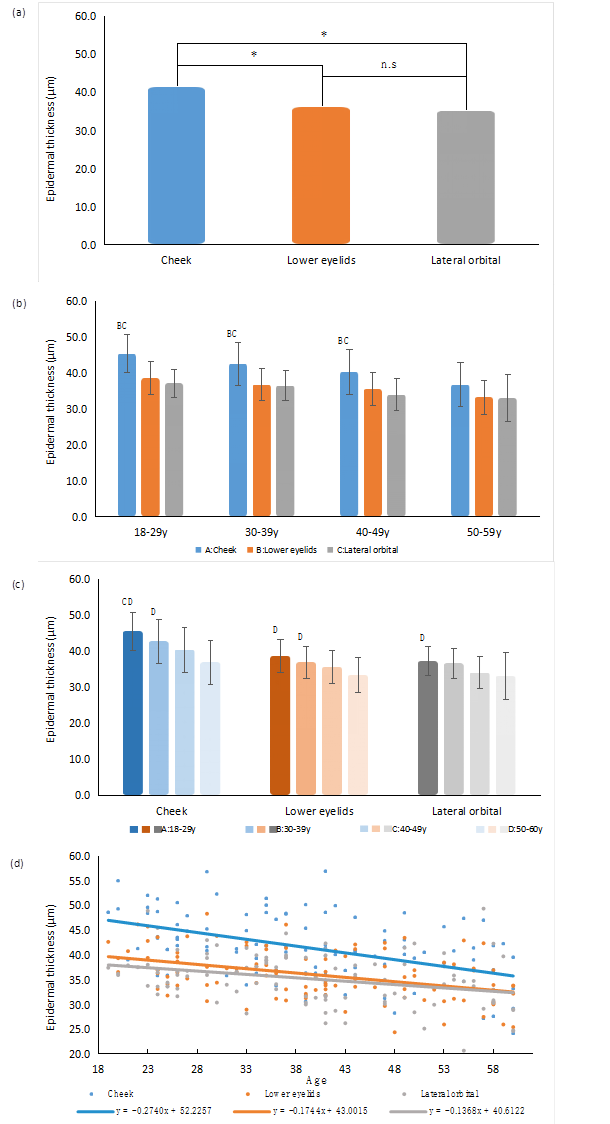
Figure 1: Distribution of ET depending on the age of the subject. Changes of ET in cheek, lateral orbital, and lower eyelids with ages. Significant values are determined as *p lessthan 0.05, **p lessthan 0.01 (Tukey’s multiple comparisons paired test). The regression line demonstrates the ET as a function of age.
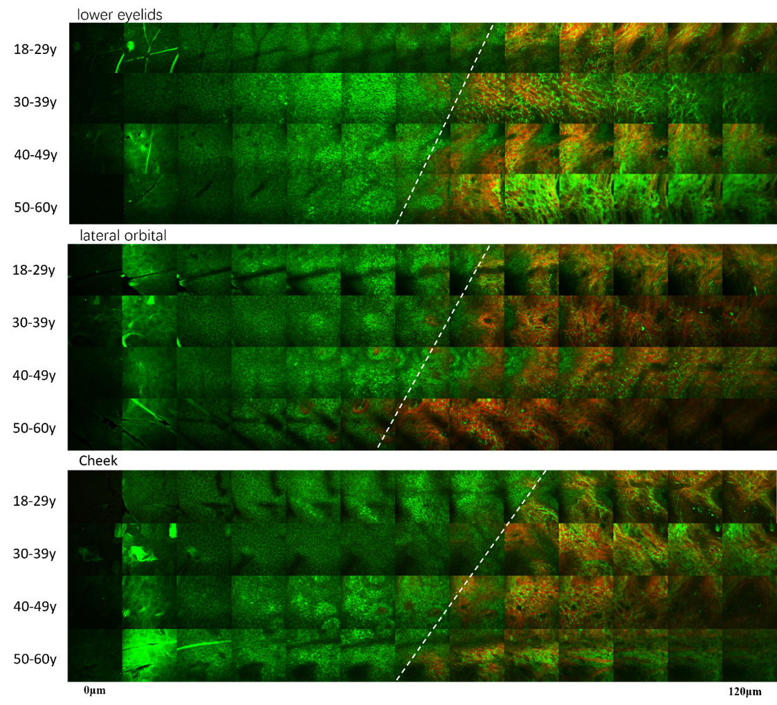
Figure 2: In vivo TPM images of normal human skin. In the illustration, the white dashed line delineates the boundary of the authentic epidermal junction. Typical epidermal thickness images were captured at the cheek, lateral orbital, and lower eyelids across the age groups, ranging from young to old. The epidermis gets thinner with age.
When disregarding age as a variable, no significant differences in the DEJ were observed among the three facial regions (cheeks, lower eyelids, and lateral orbital) (Figure 3a). However, upon incorporating age factors into the analysis, a distinct pattern became evident: DEJ values in the lower eyelids and lateral orbital exhibited a significant decline in individuals over 50 years of age compared to those in their 20s (p lessthan 0.05). More precisely, a marked decrease in DEJ was noted with advancing age, both in the lower eyelids (from 2.50 in the 18-29 age group to 2.12 in the 50-60 age group) and in the lateral orbital area (from 2.64 in the 18-29 age group to 2.05 in the 50-60 age group). Interestingly, DEJ values in the cheeks did not exhibit any significant change across age groups (Figure 3b). Furthermore, linear regression analyses of the three regions indicated a decrease in the normalized area of DEJ with age, reflecting alterations in DEJ morphology. Specifically, the undulations of DEJ gradually diminish with age (Figure 3c). Figure 4 illustrates the age-related variations in DEJ across the three facial regions, underscoring a general trend of DEJ ridge flattening in the aging process.

Figure 3: Distribution of DEJ depending on the age of the subject. Changes of DEJ in cheek, lateral orbital, and lower eyelids with ages. Significant values are determined as *p lessthan 0.05, **p lessthan 0.01 (Tukey’s multiple comparisons paired test). The regression line demonstrates the DEJ as a function of age.
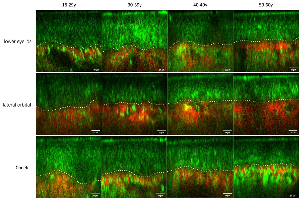
Figure 4: In vivo TPM images of normal human skin. Typical dermal-epidermal junction images were captured at the cheek, lateral orbital, and lower eyelids across the age groups, ranging from young to old. The flattening of DEJ with age is clearly visible.
TPM is capable of distinguishing between collagen and elastic fibers within the dermal matrix. This study introduced the SAAID as a metric to evaluate the skin aging process and to investigate the factors influencing SAAID values. Figure 5a presents the mean SAAID values along with their standard deviations, correlating these measurements to the ages of the subjects. A comparative analysis between the oldest participants, aged 50, and the youngest, in their 20s (each group comprising a quarter of the sample), revealed significant variations in SAAID across the three examined facial regions (p lessthan 0.05). Additionally, the study observed that the correlation between SAAID and age was moderated by specific facial sites, with a general decline in SAAID noted in the cheeks, lower eyelids, and lateral orbital areas with increasing age. Consequently, linear regression models were employed for each facial site (cheeks, lower eyelids, and lateral orbital) to quantitatively describe these relationships (Figure 5b), revealing that the predictive variable at the cheek accounted for 33.4 percent of the variance in SAAID (R²=0.334), while the lower eyelid and lateral orbital variables explained 27.1 percent (R²=0.251) and 25.1percent (R²=0.251) of the variance, respectively. The analysis indicated a more marked decline in SAAID with age in the cheek area. Ignoring age-related factors and using the cheek as a baseline, significant differences were noted in the periocular region (lower eyelid and lateral orbital) (Figure 5c).
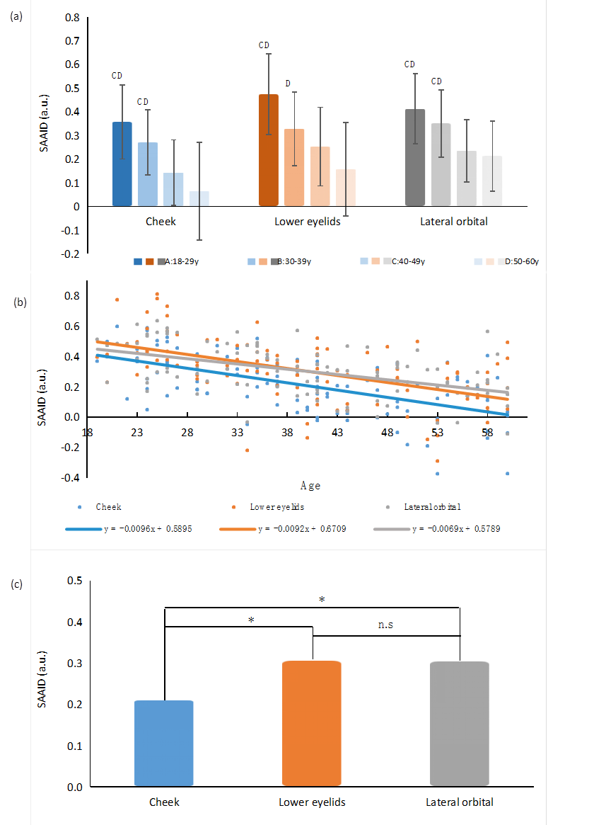
Figure 5: Distribution of the SAAID depending on the age of the subject. Changes of SAAID in cheek, lateral orbital, and lower eyelids with ages. Significant values are determined as *p lessthan 0.05, **p lessthan 0.01 (Tukey’s multiple comparisons paired test). The regression line demonstrates the SAAID as a function of age.
Figure 6 demonstrates that the distribution and clarity of elastic and collagen fibers, which are distinct in youthful skin, tend to become obscured as skin ages. This is further evidenced by the increase in autofluorescence values associated with elastic fiber density in aged skin, consistent with histological observations in the same cohort. The occurrence of localized high autofluorescence phenomena, resulting from the breaking and curling of fibers, contributes to elevated AF values. In contrast, second harmonic generation (SHG) values, indicative of collagen fiber density, did not significantly differ between older and younger participants. The study also documents a significant reduction in SAAID with advancing age, underscoring its potential as a reliable indicator of dermal aging.
Figure 6 demonstrates that the distribution and clarity of elastic and collagen fibers, which are distinct in youthful skin, tend to become obscured as skin ages. This is further evidenced by the increase in autofluorescence values associated with elastic fiber density in aged skin, consistent with histological observations in the same cohort. The occurrence of localized high autofluorescence phenomena, resulting from the breaking and curling of fibers, contributes to elevated AF values. In contrast, second harmonic generation (SHG) values, indicative of collagen fiber density, did not significantly differ between older and younger participants. The study also documents a significant reduction in SAAID with advancing age, underscoring its potential as a reliable indicator of dermal aging.

Figure 6: In vivo TPM images of normal human skin. Typical (a) Autofluorescence and (b) Second Harmonic Generation (SHG) images were captured at the cheek, lateral orbital, and lower eyelids across the age groups, ranging from young to old.
During the eight-week trial period of eye cream application, the product demonstrated its gentle and safe characteristics, as evidenced by the absence of localized erythema, swelling, edema, or systemic adverse skin reactions among the participants. Figure 7 provides a detailed summary of the clinical outcomes, highlighting significant improvements in periocular skin aging parameters. Notably, skin hydration levels exhibited a marked increase of 21.53 percent after 14 days, 32.17 percent after 28 days, and 38.07 percent by Day 56, reflecting a progressive enhancement in skin moisturization. Additionally, both skin elasticity and TEWL showed substantial improvement within the first 14 days of the eye cream application. TEWL decreased by 17.25 percent at Day 14, 23.25 percent at Day 28, and further to 25.38% by Day 56, underscoring the effectiveness of the eye cream in fortifying the skin barrier. The elasticity parameter, R2, registered a 15.07 percent increase at Day 14, 16.03 percent at Day 28, and 20.13 percent by Day 56, indicating a notable enhancement in skin elasticity.
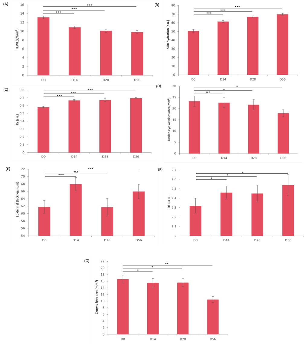
Figure 7: The example of in vivo anti-aging efficacy of the eye cream. (A: Trans epidermal water loss improvement, B: Skin hydration improvement, C: Skin elasticity improvement, D: Under-eye wrinkle reduction, E: Epidermal thickness improvement, F: Increase in DEJ undulation, G: Crow’s feet reduction). (*p≤0.05 vs. base line, **p≤0.01 vs. baseline, ***p≤0.001 vs. baseline).
The study also documented a continuous reduction in periocular wrinkles throughout the duration of the study. According to Primos-Lite assessment, under-eye wrinkles reduced by 6.70% at Day 28 and 22.82% at Day 56, while crow’s feet wrinkles diminished by 6.55% at Day 14, 6.25% at Day 28, and significantly by 36.92% at Day 56. Correspondingly, the DEJ index, a marker of skin aging, exhibited significant improvements of 6.03% at Day 14, 5.60% at Day 28, and 9.48% by Day 56. The ET index showed increases of 9.90% at Day 14 and 6.70% by Day 56, indicative of amelioration in skin aging parameters. Although the SAAID did not display significant enhancement, a positive trend was nonetheless observed. In summary, this clinical evaluation substantiates the eye cream's efficacy in anti-wrinkle action, skin firming, and improvement of epidermal thickness, thereby affirming its beneficial effects on periocular skin aging. Figure 8 presents representative examples of the eye cream's in vivo anti-aging efficacy.

Figure 8: The example of in vivo anti-aging efficacy of the eye cream. (A: Crow's feet reduction, B: Under-eye wrinkle reduction, C: Epidermal thickness improvement).
TPM stands out for its superior depth penetration and minimal tissue damage, thanks to its use of longer-wavelength photons. This technique excels in imaging specific structures like collagen fibers through SHG, providing valuable insights into complex biological samples [13]. While optical coherence tomography offers a broader field of view, its lower contrast makes delineating the DEJ challenging. In contrast, TPM's precise SHG signals enable accurate DEJ localization and detailed characterization of the dermal fiber network, aiding in the assessment of skin aging with parameters such as the SAAID [14, 15]. The label-free imaging capability of TPM eliminates the need for conventional histological processing, streamlining research workflows [16]. However, high-frequency ultrasound, when compared, tends to overestimate epidermal thickness, likely including dermal papillae in its measurements, and is less reliable for accurate thickness assessment.
This study observed a decrease in epidermal thickness with age, especially in the cheeks, lower eyelids, and lateral orbital areas, with the most significant reduction in the cheeks. Notably, individuals under 50 showed thicker epidermis in the cheeks compared to the periocular region, suggesting younger skin's greater resilience to mechanical stress. However, this difference narrows after age 50, reflecting a uniform thinning of skin across these areas with aging. These observations align with previous findings by Whitton and Everall et al. [17], indicating decreased variability in skin thickness across body parts in older age, implying a reduced skin protective capacity against environmental and mechanical stress due to decreased elasticity and the consequent impact on thickness measurement accuracy.
Significant declines in DEJ values in individuals over 50 were noted in the lower eyelids and lateral orbital areas, contrasting with those in the cheeks remain unchanged with age. Aging is generally associated with the flattening of DEJ undulations and a decrease in the normalized area of DEJ, reflecting shape alterations [18, 19]. However, no significant differences in DEJ values across the three facial areas were found in an all-age study, suggesting a consistent DEJ undulation pattern across the face despite age. In addition, cheek DEJ values remained stable across ages, indicating variations in aging mechanisms or resistance. These findings underline the complex nature of skin aging, particularly the stability of DEJ in the cheeks and the overall morphological changes, highlighting areas for future study.
The decrease in the SAAID with age is more pronounced in the cheek region, resulting in a progression from an organized to a blurred fiber arrangement in aged skin. AF values rise with age, reflecting higher elastic fiber density and corresponding to histological findings of broken, curled fibers showing localized high AF. In contrast, SHG values, indicating collagen fiber density, remain unchanged between older and younger individuals, suggesting elastin fiber density increases with age presumably due to elastosis [20]. Elastosis, characterized by the accumulation of abnormal elastic fibers in the dermis, arises from the degradation and disorganization of the skin's elastic fibers [21, 22]. This condition is primarily attributed to photoaging from UV radiation, which is more pronounced in the cheek area, likely due to greater sun exposure compared to the periocular region [23]. This contributes to signs of aging, such as wrinkles and a leathery texture. The decline in SAAID, alongside stable SHG but increased AF values, aligns with reduced epidermal thickness, illustrating the complex impact of aging on skin.
The anti-aging efficacy of the eye cream, infused with plant extracts, peptides, and antioxidants, was clinically tested. Within two weeks of use, significant improvements in periocular epidermal thickness were noted, largely due to increased stratum corneum hydration and reduced TEWL. Such improvements underscore the vital role of a robust skin barrier in facilitating keratinocyte proliferation and differentiation [24, 25]. Additionally, the DEJ index showed significant improvement following the eight-week application period. This, combined with increased skin elasticity and firmness, as well as a reduction in the area and length of wrinkles in the lower eyelids and lateral orbital regions, suggests that the anti-aging active ingredients within the eye cream, such as Tabebuia Impetiginous bark extract [26], acetyl hexapeptide-8 [27], dipeptide diaminobutyroyl benzylamide diacetate [28], arginine/lysine polypeptide [29], niacinamide [30], ergothioneine [31], and ectoine [32], may mitigate the undulation of the DEJ. However, no significant improvement was observed in the SAAID, for which two primary reasons are postulated. Firstly, elastin, which has a very low turnover rate and sees a decline in production post-maturity, is vulnerable to damage from various factors [33]. The active ingredients are ineffective at eliminating elastin fragments. Secondly, the eye cream formulation did not incorporate skin penetration enhancement techniques, likely inhibiting the ability of hydrophilic actives to sufficiently penetrate and stimulate elastin synthesis [34]. Future studies will explore formulations utilizing supramolecular skin penetration enhancement technologies to address these limitations. Furthermore, this investigation centered on a Chinese female cohort, focusing specifically on the periocular and cheek regions. To cultivate a comprehensive understanding of facial aging, future research should embrace a broader demographic and include regions prone to aging like the forehead and nasolabial folds. This broader scope would enhance our holistic grasp of facial aging across diverse skin types. Moreover, delving into the molecular underpinnings of changes in epidermal thickness, DEJ undulation, and skin autofluorescence, with a particular emphasis on the role of Sirtuin 1 [34-36], could unveil novel targets for anti-aging interventions, offering promising avenues for the development of more effective anti-aging strategies.
This study showcases the innovative use of a portable two-photon microscope for 3D skin analysis, revealing age-related reductions in epidermal thickness, most notably in the cheek region. Significant aging effects were observed in the dermal-epidermal junction of participants over 50, especially in the periocular areas, while cheek DEJ values remained consistent across ages. The cheek area also displayed a significant decline in the skin aging autofluorescence index, indicating a higher vulnerability to photoaging. Furthermore, our results confirm the effectiveness of an anti-aging eye cream in addressing epidermal thinning and DEJ alterations, highlighting its potential in anti-aging skincare.
Conflicts of Interest
The authors declare that they have no conflict of interest.
Dear Editorial Team, Clinical Medical Reviews and Reports. My experience with the journal was highly positive. The peer-review process was rigorous, constructive, and completed in a timely manner. The reviewers provided valuable comments that helped improve the quality and clarity of our manuscript. The editorial office was professional, responsive, and supportive throughout all stages of the publication process. Communication was clear and efficient, and any questions were addressed promptly. Overall, I found the journal to maintain high scientific standards and an excellent publication workflow. I would be pleased to consider submitting future work to this journal. Best wishes from, Elena Popa.

It was my pleasure to submit my testimonial concerning the Reviewer Board of our Scientific Journal “Brain and Neurological Disorders”. The Reviewers focused on some modifications and their contribution was helpful. The ladies of our Editorial Office were also supported my efforts. It was my honor to have such a co-operation and I am looking forward for more collaboration.

Dear Grace Pierce, Editorial Coordinator of Journal of Clinical Research and Reports, Thank you for the speedy and efficient peer review process. I appreciate the fact that your peer reviewers do not take months to respond like with some other journals. I would also like to thank the editorial office for responding quickly to my questions. It is an excellent journal. I plan to submit more manuscripts in the future. Best wishes from, Robert W. McGee

Dear Grace Pierce, Editorial Coordinator of Journal of Clinical Research and Reports, Working with you and your team on our recent publication in JCRR has been a truly wonderful and enjoyable experience. The responses were prompt, and the reviewers were patient, constructive, and highly professional. One reviewer in particular gave me the feeling that a professor was carefully reading and commenting on my coursework, which was deeply touching. The entire process was straightforward and hassle‑free, with no tedious online forms to complete. I highly recommend this journal. Best wishes from, DR Aibing Rao, Head of R&D

I Appreciate the Opportunity to Share my Experience with the Journal of Clinical Research and Reports. The peer review process was timely and constructive, and the feedback provided helped improve the quality of our manuscript. The editorial office was professional, responsive, and supportive throughout the process, ensuring smooth communication and efficient handling of the submission. Overall, it was a positive experience collaborating with your team.

Dear Mercy Grace, Editorial Coordinator of Obstetrics Gynecology and Reproductive Sciences, We would like to express our gratitude for your help at all stages of publishing and editing the article. The editors of the magazine answer all the necessary questions and help at every stage. We will definitely continue to cooperate and publish other works in the Obstetrics Gynecology and Reproductive Sciences! Best wishes from, Alla Konstantinovna Politova,
