
Case Report | DOI: https://doi.org/10.31579/2690-1919/258
Tribochemistry Consulting, Salt Lake City, UT 84117, USA and University of Economy, Biotribology Lab. Garbary 2, 85-229 Bydgoszcz, Poland.
*Corresponding Author: Zenon Pawlak. Tribochemistry Consulting, Salt Lake City, UT 84117, USA and University of Economy, Biotribology Lab. Garbary 2, 85-229 Bydgoszcz, Poland.
Citation: Zenon Pawlak. (2022). Deactivated Phospholipids in an Osteoarthritic Joints: Lost Surface Lubricity, J. Clinical Research and Reports, 11 (5) DOI: 10.31579/2690-1919/258.
Copyright: © 2022 Zenon Pawlak. This is an open-access article distributed under the terms of The Creative Commons Attribution License, which permits unrestricted use, distribution, and reproduction in any medium, provided the original author and source are credited.
Received: 10 July 2022 | Accepted: 15 August 2022 | Published: 10 October 2022
Keywords: cartilage; osteoarthritis (oa); β2-glycoprotein i (β2-gpi); active and deactivated phospholipid
This study is designed to determine phospholipids (PLs) dysfunction on cartilage surface and in synovial fluid (SF) in an osteoarthritic (OA) joints. The (PLs) in (SF) were measured for normal samples, with early eOA and late lOA stages of (OA) and rheumatoid arthritis (RA). During cartilage inflammation enzymatically activated β2-Glycoprotein I was transformed into antibody conformation (see Fig. 3). Deactivated PLs molecule lost ability to form bilayers and liposomes content in synovial fluid (SF) in osteoporotic joints is significantly higher (2-3 times) above the normal concentration of PLs. Additionally, the osteoporotic cartilage surface showed surface lubricity (f) 0.015 about three times higher, compared to the normal cartilage.
Phospholipids, PLs, molecules with the capacity to adsorb to solid surfaces and/or fluid interface a property that allows them to act as multifunctional chemical compound. The concept that surface-active phospholipids (SAPL) provide effortless sliding of many tissues, including joints, pleura, pericardium and peritoneum was pioneered by Hills research [1, 2]. Phospholipids are highly self-organized biomolecules in aqueous media, and their structure allows them to form spontaneously vesicles, lamellar phases, and surface membranes function as efficient lubricant, Fig. 1. The analyses of the composition of the SAPLs in the human knee joints reveal that they mostly contain unsaturated phospholipids;30% palmitoyllinoleoylphosphatidylcholine (PLPC), 23% dilinoleoyl-phosphatidylcholine (DLPC), 17.5% palmitoyl-oleoyl-phosphatidylcholine (POPC) and 16% stearoyl-linoleoylphosphatidylcholine (SLPC), 8% saturated dipalmitoyl-phosphatidylcholine (DPPC) [3].
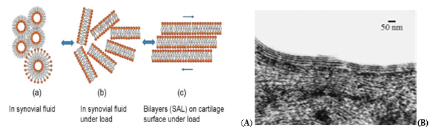
Figure 1: Formation of phospholipid phases in synovial fluid and bilayers on cartilage surface under load at a pH ~ 7.4. A (a) The lipid spheres and vesicles in synovial fluid, A(b) formations of lamellar phases separate opposing cartilage surfaces and A(c) cartilage upper surface bilayers facilitating lamellar-repulsive lubrication with lamellar slippage of the bilayers. (B) Morphology of the articular surface of human knee showing adsorbed phospholipid bilayers [2].
Strongly adsorb and cohesive SAPL linings to act as lubricant, barrier against abrasion, corrosion and possibly against invasion by microorganisms. As the 'sealant', it could be the true barrier rather than the cells providing its mechanical support [1, 2]. The multilamellar structure of phospholipids, namely the surface amorphous layer (SAL), covers the natural surface of articular cartilage found in diarthrodial joints [4, 5]. A very high porosity (75 to 80 %) to be a critical factor in providing excellent hydration, more specifically hydration lubrication properties displayed by articular cartilage [2, 3]. Morphology of the articular surface of human knee showing adsorbed phospholipid bilayers, Fig.1 (B) [2].
The surface-active phospholipids (SAPL) coating on the articular surface possesses highly desirable lubricating properties for efficient joint function. The inflammation and breakdown of cartilage cause bony and surface deterioration and was name osteoarthritis disease. The uppermost lipid bilayer (i.e., surface amorphous layer (SAL)) of articular cartilage surface is degraded during cartilage inflammation osteoarthritic disease [4, 5, 6]
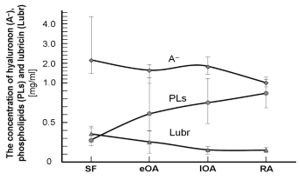
Figure 2: The levels of hyaluronan (A-), phospholipids (PLs) and lubricin (Lubr) in human synovial fluid (SF) in patients with healthy joints control (SF) and joint diseases with early osteoarthritis (eOA), late osteoarthritis (lOA) and rheumatoid arthritis (RA) were 2 to 3 times higher above the normal range.
Compared to patients with healthy joints control (SF), patients with early (eOA) and those with late (lOA) had higher levels of most PLs species (2 to 3 times) above the normal range (see Fig. 2). The concentration of components of synovial fluid (SF), such as hyaluronan (A-), lubricin, and surface-active phospholipids, from unaffected controls (or normal), (eOA), (lOA), and RA in the human synovial fluid are shown in Fig. 2. During osteoarthritis, OA, and rheumatoid arthritis, RA, contain less hyaluronan (A-) and lubricin and are contain more with phospholipids. Also, the MW distribution of (A-) shifted toward the lower range in OA and RA SF. These results indicate that activities in OA and RA SF are enhanced, leading to decreased levels of lubricin and hyaluronan (A-) [4 -7].
The phospholipid species were quantified by mass spectrometry (ESI-MS/MS) [4]. The research was carried out using synovial fluid derived from undamaged controls and patients with early and late osteoarthritis and rheumatoid arthritis. After comparing control synovial fluids, SF of patient with early and late OA had higher levels of most PLs species. Most of the PLs data for this paper was taken from Kosinska et al [4, 5].
Molecules of β2-Glycoprotein I, (β2-GPI) (MW of 50 kDa) circulate in the body in the range 50-500 μg/ml and autoimmune virus transforms β2-GPI in an antibody, Figure 3. The (β2-GPI) participates in antiphospholipid antibody syndrome (APS) through binding of (β2-GPI) to the anionic charged phospholipid (-PO4-) group. At a pH around 7, (β2-GPI) amino acids (arginine, lysine and tryptophan) are positively charged (-NH3+): an acid-base interaction occurs between the protonated amino acid functional group (-NH3+) and the phosphate (–PO4-) membrane group: (β2-GPI-NH3+) + (PLs–PO4-) → (-NH3+ PO4-) interaction and electrostatic attractions is strong enough to destroy thePLs bilayer on the articular surface and deactivate all phospholipids in the synovial fluid (SF). β2-GPI is a highly adhesive protein that binds to various cell receptors, β2-GPI binds with negatively charged phosphate groups (-PO4-) at pH ~ 7.4. During cartilage inflammation enzymatically activated β2-Glycoprotein I was transformed into antibody an open hockey-stick-like conformation (see Figure 3) [8, 9].
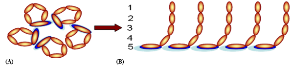
Figure 3; (A) Conversion of β2-Glycoprotein I (β2-GPI) of the circular conformation (closed molecules) into (B) an open hockey-stick-like conformation, each molecule has five domains (1-5).
In Figure 3 (A) the closed circular conformation of plasma (β2-GPI) as it circulates in plasma in a healthy joint, and (B) the open hockey stick-like
conformation in APS syndrome and (β2-GP I) is binding to negatively charged (-PO4-) phospholipids. The autoantibodies will bind and stabilize (β2-GPI) in its hockey-stick-like conformation.
Phospholipid binding takes place in the domain (5) of β2-GPI, Figure 3 (A), which contains positively charged groups (-NH3+) at pH ~ 7. In the blood plasma, the β2-GPI molecule is in a closed form in which the domain (1) interacts with the domain (5). For a pH of about 7, amino acids in the domain (5) (arginine, lysine, and tryptophan) with a functional group (-NH2) are positively charged (-NH3+), hence the acid-base interaction between the group of protonated amino acids (-NH3+), and the phosphate group (-PO4-):
(β2-GPI(-NH3+) + PL(-PO4-) → β2-GPI (-NH3+) (-PO4-) PL (1)
The electrostatic interactions are strong enough to destroy the PL bilayer on the cartilage surface and deactivate all phospholipid molecules in the synovial fluid (Figure 4), whose association constant, Kass = 105 [7, 10].
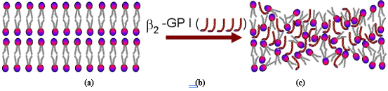
Figure 4 : (a) PL bilayers on the cartilage surface, (b) open stick-hockey conformation of β2-glycoprotein I, (c) phospholipid molecules deactivated by stick-hockey conformation of β2-glycoprotein I
Wettability measurement of the contact angle (theta) clearly demonstrated a highly significant decrease in hydrophobicity, falling from 103 degrees for 13 bovine controls (78 degrees for five human controls) to 56 degrees for arthritic hips. These changes were reflected in the quantities of SAPL (and proteolipid) recovered from the same articular surfaces by solvent rinsing, yields of SAPL being 60% lower from osteoarthritic hips [1, 11]. These results indicate that the outermost lubricating layer of SAL deposited onto lubricating surfaces is deficient in OA.
PLs are the foundation in joints boundary lubrication (Fig. 1). In conclusion, we have provided mechanism deactivation functional group (-PO4-) of PLs by β2-GPI is believed to totally destroy the cartilage surface amorphous layer (SAL). Deactivated PLs molecules form a new insoluble compound β2-GPI (-NH3+) (-PO4-) PL) with no ability to lubricate.
PL (-PO4-) → β2-GPI (-NH3+) (-PO4-) PL
(Active PL at pH 7.4 with (-PO4-)functional group) (Deactivated PL at pH 7.4)
Additionally, hyaluronan and lubricin are unable to take supportive function of lubricant without active phospholipids presence. This fact is in support Hills hypothesis that hyaluronan and lubricin are molecular carriers for insoluble SAPL [1]. Cartilage degradation of membrane anionic phospholipids by antibodies (β2- Glycoprotein I) belong to Anti Phospholipid Syndrome (APS) disorder. The work showed surface active phospholipid adsorbed to the articular surface contributes to joint lubrication, but deactivation functional group (-PO4-) of PLs ruins their smartness.
Dear Editorial Team, Clinical Medical Reviews and Reports. My experience with the journal was highly positive. The peer-review process was rigorous, constructive, and completed in a timely manner. The reviewers provided valuable comments that helped improve the quality and clarity of our manuscript. The editorial office was professional, responsive, and supportive throughout all stages of the publication process. Communication was clear and efficient, and any questions were addressed promptly. Overall, I found the journal to maintain high scientific standards and an excellent publication workflow. I would be pleased to consider submitting future work to this journal. Best wishes from, Elena Popa.

It was my pleasure to submit my testimonial concerning the Reviewer Board of our Scientific Journal “Brain and Neurological Disorders”. The Reviewers focused on some modifications and their contribution was helpful. The ladies of our Editorial Office were also supported my efforts. It was my honor to have such a co-operation and I am looking forward for more collaboration.

Dear Grace Pierce, Editorial Coordinator of Journal of Clinical Research and Reports, Thank you for the speedy and efficient peer review process. I appreciate the fact that your peer reviewers do not take months to respond like with some other journals. I would also like to thank the editorial office for responding quickly to my questions. It is an excellent journal. I plan to submit more manuscripts in the future. Best wishes from, Robert W. McGee

Dear Grace Pierce, Editorial Coordinator of Journal of Clinical Research and Reports, Working with you and your team on our recent publication in JCRR has been a truly wonderful and enjoyable experience. The responses were prompt, and the reviewers were patient, constructive, and highly professional. One reviewer in particular gave me the feeling that a professor was carefully reading and commenting on my coursework, which was deeply touching. The entire process was straightforward and hassle‑free, with no tedious online forms to complete. I highly recommend this journal. Best wishes from, DR Aibing Rao, Head of R&D

I Appreciate the Opportunity to Share my Experience with the Journal of Clinical Research and Reports. The peer review process was timely and constructive, and the feedback provided helped improve the quality of our manuscript. The editorial office was professional, responsive, and supportive throughout the process, ensuring smooth communication and efficient handling of the submission. Overall, it was a positive experience collaborating with your team.

Dear Mercy Grace, Editorial Coordinator of Obstetrics Gynecology and Reproductive Sciences, We would like to express our gratitude for your help at all stages of publishing and editing the article. The editors of the magazine answer all the necessary questions and help at every stage. We will definitely continue to cooperate and publish other works in the Obstetrics Gynecology and Reproductive Sciences! Best wishes from, Alla Konstantinovna Politova,
