
Clinical Report | DOI: https://doi.org/10.31579/2643-6612/032
* Faculty of Medicine, Department of Dentistry, Monastir University, Tunisia.
*Corresponding Author: Nissaf Daoauhi, Faculty of Medicine, Department of Dentistry, Monastir University, Tunisia.
Citation: Farah Chawali, Asma Nakhli, Nissaf Daoauhi, Zohra Nouira, Belhassen Harzallah, et al. (2022). Creating a New Smile using Ceramic Restoration associated with Laser Depigmentation: Clinical Report. J Dentistry and Oral Maxillofacial Surgery, 5(2); DOI:10.31579/2643-6612/032
Copyright: © 2022, Nissaf Daoauhi. This is an open access article distributed under the Creative Commons Attribution License, which permits unrestricted use, distribution, and reproduction in any medium, provided the original work is properly cited.
Received: 05 March 2022 | Accepted: 28 March 2022 | Published: 11 April 2022
Keywords: smile; central incisor; diode laser; gingival hyperpigmentation; ceramics; CAD
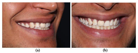
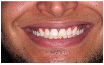
The article describes a clinical situation of managing a non harmonious smile caused by old metal ceramic crown on central incisor associated with gingiva hyperpigmentation. It concerns a 23-year-old nonsmoking patient of brown race with a chief complaint of poor aesthetics. He was bothered about the dark-color of upper gum and the discrepancy between the maxillary central incisors. The treatment procedure started by gingival depigmentation using laser diode, followed then by Lithium Disilicate Ceramic crown.
Thanks to a well-planned multi-disciplinary approach, the result was esthetically acceptable and the patient was satisfied.
Smile esthetics is determined by the color shade, shape, and position of the teeth as well as the gingiva which is considered as the most pigmented tissue in the oral cavity. According to literature, several factors such as thickness of the epithelium, the keratinization quality of the gingiva, the volume of pigments in the tissue can affect gingival color [1]. Creating a new smile in patients with pigmented gingiva is considered as a challenge especially when it is associated with other defects concerning the harmony of teeth caused by old restorations in the aesthetic zone [2].
Gingival depigmentation can be performed by different methods. The selected method depends on the patient’s preference as well as the expertise and experience of the clinician. Recent studies demonstrated that the use of diode laser is a safe and effective treatment modality that provides optimal aesthetics with minimal discomfort in patients with gingival hyperpigmentation [7,8].
During the smile, the central incisor should be the most dominant displayed tooth. Starting points for aesthetic management of maxillary central incisor are shape, size, shade, incisal edge position and proportions [a,b]. According to authors, the width/Length ratio is expected to be between 75% and 78% [3-6].
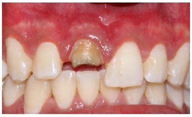
This clinical presentation is dealing with aesthetic rehabilitation of the smile associating laser diode depigmentation of gingiva and all ceramic crown in central incisor . It concerns a 23-year-old nonsmoking patient of brown race with a chief complaint of poor aesthetics. He was bothered about the dark-color of upper gum and the discrepancy between the maxillary central incisors which was caused by old metal ceramic crown restoring the right one. He was complaining about the form, the shade and the greyish transparency of the crown metal margin through the marginal gingiva. Aesthetic analysis revealed a convex profile with parallel commissural and mid-pupillary lines, squared teeth form, No harmonious smile lines. size discrepancies between central incisors were noticed. Photographs were taken using Canon 7OOD Camera.
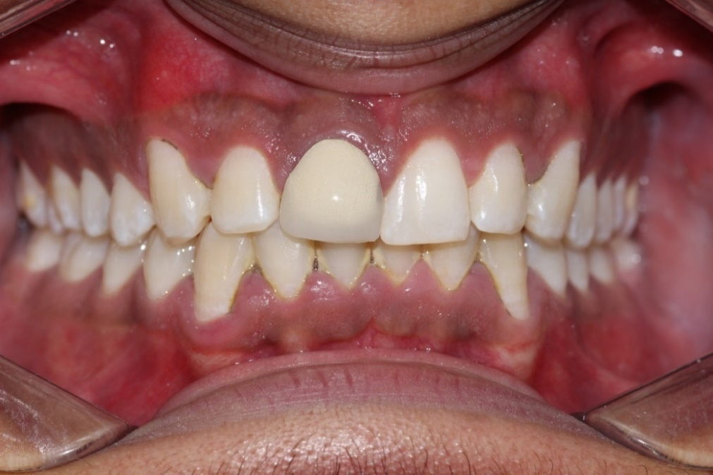
Periodontal probing revealed a thick gingival biotype. bilateral melanin pigmentation was noticed. According to the pigmentation index of Kumar et al. the score was diagnosed as “3” (diffuse brown to black pigmentation, marginal, and attached) [7].
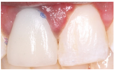
The treatment procedure started by gingival depigmentation, followed then by aesthetic build up of the core and Lithium Disilicate Ceramic crown.
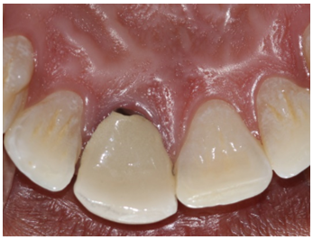
Firstly, the old crown was removed. a handmade provisional restoration, with precisely fitting and highly polished margins was performed using resin material Texton (SS White, New Jersey, USA). It established correct form and proportions of the central incisor.
Secondly, a semiconductor diode surgical laser unit (Elexxion pico 808 nm diode laser, Elexxion AG, Singen, Germany) was used for depigmentation. it consists on digitally pulsed diode laser with a frequency of 20,000 Hz, a peak power of 5 W, and a pulse width of 26 microseconds.
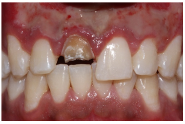
Finally, the core was built up and the crown on central incisor was, performed using Lithium Disilicate Ceramic material IPS e.max CAD (Ivoclar Vivadent, Schaan, Liechtenstein); moderate translucency block was used. it was managed according aesthetics guidelines and references.
An aesthetic try in of the crown before glazing was necessary. It allows the assessment of the color, the form and proportions. The incisal edge position, the midline, the axial inclination, at this stage, should be checked.
The Rehabilitation of the smile remains a challenge especially when it is associated with gingival problems such as gingival hyperpigmentation.
The morphological features of incisors and facial proportions are closely correlated. According to Williams et al. the shape of central incisor is determined by facial form. This teeth should be restored according to the concept of dominance. Some studies are speaking about more attractive tapered incisors. [1,13,14]. The incisal edge should be parallel to mid-pupillary line. The Characterization of central incisor surface texture is as important as the shade, the form and dimensions .
Various methods have been used for Gingival depigmentation; they include gingivectomy [8], gingivectomy with free gingival autografting [9], electrosurgery [10], Cryosurgery [11], chemotherapy [12]. But, some of these techniques are prone to complications [13].
Recently lasers have been used to gingival depigmentation [8]. Semi-conductor diode, Er: YAG Nd: YAG laser, and CO2 laser are commonly used for de-epithelization. Compared to the erbium laser, melanin shows a strong absorption of the diode wavelengths. According to recent studies [10], it guarantees a shorter treatment procedure with the diode [12]. Figures (1-6)
The Rehabilitation of the smile remains a challenge for prosthodontists, especially when it is associated with gingival problems such as gingival hyperpigmentation. A diode laser today seems to be an effective and safe technique for melanin depigmentation.
Dear Editorial Team, Clinical Medical Reviews and Reports. My experience with the journal was highly positive. The peer-review process was rigorous, constructive, and completed in a timely manner. The reviewers provided valuable comments that helped improve the quality and clarity of our manuscript. The editorial office was professional, responsive, and supportive throughout all stages of the publication process. Communication was clear and efficient, and any questions were addressed promptly. Overall, I found the journal to maintain high scientific standards and an excellent publication workflow. I would be pleased to consider submitting future work to this journal. Best wishes from, Elena Popa.

It was my pleasure to submit my testimonial concerning the Reviewer Board of our Scientific Journal “Brain and Neurological Disorders”. The Reviewers focused on some modifications and their contribution was helpful. The ladies of our Editorial Office were also supported my efforts. It was my honor to have such a co-operation and I am looking forward for more collaboration.

Dear Grace Pierce, Editorial Coordinator of Journal of Clinical Research and Reports, Thank you for the speedy and efficient peer review process. I appreciate the fact that your peer reviewers do not take months to respond like with some other journals. I would also like to thank the editorial office for responding quickly to my questions. It is an excellent journal. I plan to submit more manuscripts in the future. Best wishes from, Robert W. McGee

Dear Grace Pierce, Editorial Coordinator of Journal of Clinical Research and Reports, Working with you and your team on our recent publication in JCRR has been a truly wonderful and enjoyable experience. The responses were prompt, and the reviewers were patient, constructive, and highly professional. One reviewer in particular gave me the feeling that a professor was carefully reading and commenting on my coursework, which was deeply touching. The entire process was straightforward and hassle‑free, with no tedious online forms to complete. I highly recommend this journal. Best wishes from, DR Aibing Rao, Head of R&D

I Appreciate the Opportunity to Share my Experience with the Journal of Clinical Research and Reports. The peer review process was timely and constructive, and the feedback provided helped improve the quality of our manuscript. The editorial office was professional, responsive, and supportive throughout the process, ensuring smooth communication and efficient handling of the submission. Overall, it was a positive experience collaborating with your team.

Dear Mercy Grace, Editorial Coordinator of Obstetrics Gynecology and Reproductive Sciences, We would like to express our gratitude for your help at all stages of publishing and editing the article. The editors of the magazine answer all the necessary questions and help at every stage. We will definitely continue to cooperate and publish other works in the Obstetrics Gynecology and Reproductive Sciences! Best wishes from, Alla Konstantinovna Politova,
