
Research Article | DOI: https://doi.org/10.31579/2690-4861/037
cardiovascular thoracic surgery, Apollo hospitals Visakhapatnam, India
*Corresponding Author: Sujit Kumar Mohanty, cardiovascular thoracic surgery, Apollo hospitals Visakhapatnam, India.
Citation: Sujit Kumar Mohanty. (2020) Coronary Sinus Atrial Septal Defect without Persistent Left Superior Vena Cava: Single Vessel Coronary Artery Disease, Surgically Managed – A Rarest Case Report. International Journal of Clinical Case Reports and Reviews. 3(3); DOI: 10.31579/2690-4861/037
Copyright: © 2020 Sujit Kumar Mohanty, This is an open-access article distributed under the terms of the Creative Commons Attribution License, which permits unrestricted use, distribution, and reproduction in any medium, provided the original author and source are credited.
Received: 30 July 2020 | Accepted: 29 August 2020 | Published: 05 September 2020
Keywords: 60 years old female; hypotension; sinus atrial septal; single vessel coronary artery
Coronary sinus defect is a spectrum of cardiac anomalies in which part or all of the common wall between the coronary sinus and left atrium is absent.By morphology, it may present completely unroofed coronary sinus or completely unroofed coronary sinus without persistent left superior vena cava also it may present like partially unroofed midportion or terminal portion of coronary sinus. Depending upon the morphology cyanosis is mild to severe.
We are reporting a case of partial coronary sinus defect in an adult female patient with coronary artery disease.
True understanding of the morphology of the syndrome awarded the classic paper by raghib, Edwards and colleagues in 1965. The descriptive phrase unrefined coronary sinus was first used by Helseth and Peter in 1974. Unroofed coronary sinus has it meet common major association cardiac anomaly in the atrioventricular septal defect.
The partially unroofed midportion of coronary sinus also called the biatrial opening of the coronary sinus or coronary sinus-to-left atrial window or fenestration. Diagnosis of the unroofed coronary sinus in made by echocardiography and confirmed at operation.
The degree of cyanosis or desaturation depends on the presence of persistent left superior vena cava and extend of unroofing of the coronary sinus.
A 60 years old female presented with moderate effort intolerance, pulse oximeter saturation of 96% moderate cardiomegaly, a wide, fixed split second heart sound and systolic outflow murmur. a chest radiography showed Cardio megaly, dilated pulmonary arteries and plethora . Electrocardiogram showed sinus rhythm with normal cardiac axis and RSR pattern in V1.
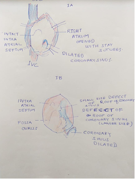
Echocardiography revealed an enlarged ostium of the coronary sinus, which was shunting left to right there was an unroofing of the terminal portion of the coronary sinus, where the left atrial blood entered the dilated coronary sinus. The right atrium and the right ventricle were dilated without evidence of pulmonary arterial hypotension.
We did also coronary angiography which was showing left anterior descending coronary artery normal. Left circumflex normal but rt. Coronary artery has 90% lesion.

So after taking to operation theatre and drapping over, midsternotomy done.pericardiotomy and stay suture taken pericardium also harvested simultaneously saphenous vein harvested from leg then after heparinization. Aortic, SVC, IVC cannulation done, pt was put on cardiopulmonary bypass. We grafted rt posterior descending artery with reversed saphenous vein graft followed by proximal to aorta on pump beating heart we put antegrade cardioplegic canula, temperature drift to 32 degrees centigrade.
SVC, IVC snugged with umblical tape. Then cross clamp on cardioplegic given heart arrested RA open, a dilated coronary sinus seen. then we open interatrial septum ,there is two separate opening in the mid terminal part of coronary sinus , no left svc found we also confirm with by given cardioplegia through antigrade , blood coming from these two openings. Both the opening we closed with separate e pericardial patch (as shown in fig 2 & fig 3) after that it is reconfirmed.

There is no leakage to left atrium after giving cardioplegia, interatrial septum closed directly then. Right atrium closed in two layers. rewarming done umblical tape desnugged after deairing cross clamp taken out heart beating started blood pressure and heart rate came to normal with minimal inotrope supports gradual weaning from cardio pulmonary bypass ,we also did transesophgeal echocardiography (TEE) showing no left to right shunt through coronary sinus..
Defects located posteroinferior to the oval fossa are extremely rare interatrial communications and are often associated with PLSVC. This is caused by a defect in the embryonic left -atrio venous fold which forms a common wall between coronary sinus and the left atrium. they may be unrecognizable, when they occur in any form of atrioventricular septal defect .complete unroofing of coronary sinus is described as type 1 or 2 in the presence or absence of PLSVC, respectively .partial unroofing is described as type 3 or 4 when the mid or terminal portion of the coronary sinus is involved .our patients is, an example type 3, 4 defect .then as outline decannulation done. And after Protamin given and hemostasis maintained. Sternum closed with steel wire, wound closed in layer, patient shifted to ICU in stable condition, patient did well and discharge from hospital on 11th post-operative day.
DECELERATION OF PATIENT CONSENT - the authors certify that they have obtained all appropriate patient consent forms .in the form the patients has/ have given his /her / their consent for his /her /their images and other clinical information to be reported in the journal. The patient understand that their names and initials will not be published and due efforts will be made to conceal their identity, but anonymity cannot be guaranteed.
FINANCIAL SUPPORT AND SPONSORSHIP – nil
CONFLICTS OF INTERST - There are no conflicts of interest.
Dear Editorial Team, Clinical Medical Reviews and Reports. My experience with the journal was highly positive. The peer-review process was rigorous, constructive, and completed in a timely manner. The reviewers provided valuable comments that helped improve the quality and clarity of our manuscript. The editorial office was professional, responsive, and supportive throughout all stages of the publication process. Communication was clear and efficient, and any questions were addressed promptly. Overall, I found the journal to maintain high scientific standards and an excellent publication workflow. I would be pleased to consider submitting future work to this journal. Best wishes from, Elena Popa.

It was my pleasure to submit my testimonial concerning the Reviewer Board of our Scientific Journal “Brain and Neurological Disorders”. The Reviewers focused on some modifications and their contribution was helpful. The ladies of our Editorial Office were also supported my efforts. It was my honor to have such a co-operation and I am looking forward for more collaboration.

Dear Grace Pierce, Editorial Coordinator of Journal of Clinical Research and Reports, Thank you for the speedy and efficient peer review process. I appreciate the fact that your peer reviewers do not take months to respond like with some other journals. I would also like to thank the editorial office for responding quickly to my questions. It is an excellent journal. I plan to submit more manuscripts in the future. Best wishes from, Robert W. McGee

Dear Grace Pierce, Editorial Coordinator of Journal of Clinical Research and Reports, Working with you and your team on our recent publication in JCRR has been a truly wonderful and enjoyable experience. The responses were prompt, and the reviewers were patient, constructive, and highly professional. One reviewer in particular gave me the feeling that a professor was carefully reading and commenting on my coursework, which was deeply touching. The entire process was straightforward and hassle‑free, with no tedious online forms to complete. I highly recommend this journal. Best wishes from, DR Aibing Rao, Head of R&D

I Appreciate the Opportunity to Share my Experience with the Journal of Clinical Research and Reports. The peer review process was timely and constructive, and the feedback provided helped improve the quality of our manuscript. The editorial office was professional, responsive, and supportive throughout the process, ensuring smooth communication and efficient handling of the submission. Overall, it was a positive experience collaborating with your team.

Dear Mercy Grace, Editorial Coordinator of Obstetrics Gynecology and Reproductive Sciences, We would like to express our gratitude for your help at all stages of publishing and editing the article. The editors of the magazine answer all the necessary questions and help at every stage. We will definitely continue to cooperate and publish other works in the Obstetrics Gynecology and Reproductive Sciences! Best wishes from, Alla Konstantinovna Politova,
