
Review Article | DOI: https://doi.org/10.31579/2690-8808/309
Department of Marine Engineering, Chabahar Maritime University, Chabahar, Iran.
*Corresponding Author: Mohammad Yaghoub Abdollahzadeh Jamalabadi, Department of Marine Engineering, Chabahar Maritime University, Chabahar, Iran.
Citation: Abdollahzadeh Jamalabadi MY, (2026), Computational Porous Media Techniques for Biomechanical Simulation: Advances in Tissue Engineering, Joint Mechanics, and Personalized Medicine, J, Clinical Case Reports and Studies, 7(3); DOI:10.31579/2690-8808/309
Copyright: ©, 2026, Mohammad Yaghoub Abdollahzadeh Jamalabadi. This is an open access article distributed under the Creative Commons Attribution License, which permits unrestricted use, distribution, and reproduction in any medium, provided the original work is properly cited.
Received: 17 February 2026 | Accepted: 03 March 2026 | Published: 10 March 2026
Keywords: porous media; biomechanical simulation; poroelasticity; tissue mechanics; fluid-structure interaction; computational modeling; cartilage; bone; machine learning
Background: Biological tissues exhibit complex multiphase mechanical behavior arising from their hierarchical porous microstructure, where solid matrix and interstitial fluid interact across multiple spatial and temporal scales. Understanding these interactions is critical for advancing orthopedic treatments, tissue engineering strategies, and clinical diagnostics.
Objective: This review synthesizes recent advancements (2000-2025) in porous media modeling techniques applied to biomechanical systems, with emphasis on theoretical foundations, computational implementations, experimental validation approaches, and clinical translation pathways.
Methods: We critically examine computational approaches including poroelastic theory, poroviscoelastic formulations, multiple-network models, and multiphase flow simulations. The review encompasses applications in bone mechanics, cartilage biomechanics, tissue engineering scaffolds, and pathological conditions, with particular focus on osteoarthritis and implant design. Integration of experimental validation techniques with advanced imaging modalities (micro-CT, MRI, multiphoton microscopy) and emerging machine learning frameworks is systematically evaluated.
Key Findings: Porous media frameworks successfully predict tissue behavior across scales from cellular (~10 μm) to organ levels (~10 cm), capturing time-dependent phenomena including fluid pressurization, consolidation, and stress relaxation. Recent advances in physics-informed neural networks enable 10-100× computational acceleration while maintaining physical consistency. Patient-specific models derived from clinical imaging data demonstrate clinical applicability for surgical planning and implant optimization. Multiphysics coupling incorporating electrokinetic effects, osmotic pressure, and biochemical transport significantly enhances physiological relevance.
Conclusions: Porous media techniques have matured from theoretical constructs to clinically relevant computational tools. Integration of machine learning with physics-based models, coupled with high-resolution experimental validation, positions these approaches as essential infrastructure for precision medicine and regenerative medicine applications. Critical challenges remain in parameter identification from limited clinical data, computational efficiency for real-time applications, and regulatory pathway establishment for clinical decision-support systems.
FEM – Finite Element Method
FSI – Fluid-Structure Interaction
ECM – Extracellular Matrix
GAG – Glycosaminoglycan
BMD – Bone Mineral Density
CFD – Computational Fluid Dynamics
3D – Three-Dimensional
TPMS – Triply Periodic Minimal Surface
MPET – Multiple-Network Poroelastic Theory
Biological tissues exhibit complex mechanical behaviors arising from their hierarchical porous structures, where solid and fluid phases interact in sophisticated ways. The fundamental understanding of these interactions is critical for advancing orthopedic treatments, tissue engineering strategies, and clinical diagnostics. Traditional biomechanical models often oversimplify tissue behavior by treating them as single-phase materials, which limits their physiological relevance and predictive accuracy.
Porous media theory provides a robust mathematical framework for capturing the multiphase nature of biological tissues. This approach acknowledges that tissues contain interconnected pores saturated with interstitial fluid, whose behavior significantly influences mechanical properties and mass transport phenomena. Recent technological advances in imaging, computational power, and experimental techniques have enabled detailed investigations of porous media phenomena at unprecedented resolution and complexity.
The application of porous media techniques to biomechanical systems has grown exponentially over the past two decades. Researchers have developed sophisticated computational models incorporating poroelasticity, poroviscoelasticity, and multiphase flow mechanics to simulate phenomena ranging from cartilage deformation under loading to fluid transport in bone. These advances have direct implications for understanding degenerative diseases such as osteoarthritis and for designing more effective tissue engineering scaffolds.
This review synthesizes current knowledge on porous media techniques in biomechanical simulation, with emphasis on theoretical foundations, computational implementations, and clinical applications. We discuss recent innovations in model development, validation strategies, and integration with experimental data. Understanding these advances is essential for researchers and clinicians aiming to harness porous media approaches for improved diagnosis, treatment, and regeneration of biological tissues.
The application of porous media theory to biomechanical simulation has revolutionized our understanding of biological tissues and their mechanical behavior. This review examines recent contributions that have shaped the field, from foundational biphasic theory to contemporary applications in clinical diagnostics, tissue engineering, and machine learning integration. The theoretical foundation for porous media approaches in biomechanics was established by Mow et al. [1], who introduced the biphasic theory for articular cartilage. Their seminal work demonstrated that cartilage could be modeled as a mixture of solid matrix and interstitial fluid, successfully predicting creep and stress relaxation behavior under compression. This biphasic formulation provided the first rigorous framework for understanding how fluid pressurization contributes to load support in hydrated soft tissues. Building upon this foundation, Armstrong et al. [2] extended the analysis to unconfined compression configurations, deriving analytical solutions that enabled experimental validation of biphasic theory. Their work elucidated the role of exudation and inflow in determining the time-dependent response of cartilage, establishing critical benchmarks for subsequent numerical implementations. Simon [3] provided a comprehensive review of multiphase poroelastic finite element models for soft tissue structures, synthesizing developments in the field and establishing mathematical frameworks that would guide computational implementations for decades. This work was instrumental in transitioning poroelastic theory from analytical solutions to numerical simulations capable of handling complex geometries and boundary conditions. Accurate determination of material properties is essential for predictive biomechanical simulations. Athanasiou et al. [4] conducted comparative studies of human acetabular and femoral head cartilage, revealing site-specific variations in mechanical properties that have important implications for joint mechanics and tissue engineering strategies. Chung and Mansour [5] introduced regression models for determining poroelastic properties, offering efficient alternatives to computationally expensive inverse analysis. Their approach demonstrated that statistical relationships between experimental measurements and material parameters could accelerate property characterization while maintaining accuracy. Subsequently, Chung and Mansour [6] advanced the field by combining constrained optimization with finite element analysis, enabling robust determination of poroelastic properties from experimental data. This methodology addressed the inherent non-uniqueness in parameter estimation, providing more reliable material characterization for computational models. Kotelsky et al. [10] introduced an innovative approach linking poroelastic material properties to cellular responses. By quantifying loading-induced cell death, they established direct relationships between mechanical behavior and biological outcomes, opening new avenues for understanding mechanotransduction in health and disease.
The clinical relevance of poroelastic modeling is perhaps most evident in osteoarthritis research. Elahi et al. [7] employed in silico approaches to investigate the differential contributions of collagen degradation and proteoglycan depletion to cartilage degeneration. Their computational study revealed distinct mechanisms underlying primary and secondary osteoarthritis, providing insights that could guide targeted therapeutic interventions. Uzuner S et al. [11] conducted numerical analyses of poroelastic cartilage models specifically focused on osteoarthritis progression. By systematically varying material properties to simulate degenerative changes, they demonstrated how alterations in permeability and stiffness affect load distribution and fluid pressurization, contributing to our understanding of disease mechanisms. The complexity of biological tissues necessitates multi-physics frameworks that account for coupled phenomena. Gu et al. [8] extended biphasic theory to include ionic effects, developing triphasic models that captured the charged nature of hydrated tissues. Their work on streaming potential data provided experimental validation for electrokinetic phenomena in cartilage, essential for understanding mechanoelectrochemical transduction. Hui et al. [9] adopted systems biology perspectives to analyze synovial joint lubrication across scales. Their integrative framework connected molecular mechanisms to tissue-level function, demonstrating how poroelastic and boundary lubrication mechanisms interact to maintain joint health and how their disruption leads to pathology.
The principles of poroelasticity have been extensively applied to scaffold design for regenerative medicine. Paul et al. [12] reviewed design strategies for biomimetic porous scaffolds in bone tissue engineering, emphasizing the importance of matching mechanical properties and mass transport characteristics to native tissue requirements. Su et al. [13] demonstrated advanced manufacturing approaches by fabricating 3D-printed porous Ti6Al4V scaffolds with bioactive coatings. Their work highlights how additive manufacturing enables precise control over pore architecture while surface modifications enhance biological performance. Mamuti et al. [14] systematically analyzed mechanical characteristics and permeability of triply periodic minimal surface (TPMS) and Voronoi porous structures. Their computational study provided quantitative relationships between architectural parameters and functional performance, guiding rational scaffold design. Loha et al. [15] integrated mechanoregulatory algorithms with finite element analysis to predict bone ingrowth into porous hip stems. Their predictive framework enables optimization of implant designs to promote osseointegration while maintaining mechanical stability. Ntousi et al. [20] provided a comprehensive narrative review of computational modeling approaches for bone tissue engineering scaffolds, synthesizing current methodologies and identifying challenges in translating computational predictions to clinical applications.
Recent advances have witnessed convergence between poroelastic modeling and machine learning. Donmazov et al. [16] reviewed machine learning applications in soft tissue biomechanics and biomaterials, highlighting how data-driven approaches can accelerate simulations, enhance parameter estimation, and discover hidden patterns in biomechanical data.
Haider et al. [17] developed integrated approaches combining poroelastic models with machine learning for stiffness estimation and classification of biological cells using constriction microchannels. Their hybrid methodology demonstrates the power of combining physics-based and data-driven models for cellular mechanophenotyping. Stetter and Stein [18] explored machine learning applications in human movement analysis, illustrating how data-driven approaches complement physics-based musculoskeletal models for clinical gait analysis and sports performance optimization. Dindorf C et al. [19] addressed the challenge of limited training data in biomechanical machine learning by generating realistic synthetic posture data using generative artificial intelligence. This approach has significant implications for developing robust predictive models when experimental data is scarce.
Jamalabadi [21-25] investigated porosity effects on surface temperature for porous blocks under coupled thermal-fluid conditions, demonstrating the importance of porous media theory for understanding heat transfer in biological and bioinspired systems. Jamalabadi [22] explored nanoscale effects in porous media through studies of graphene layer configurations, and [23] developed numerical frameworks for nonlinear ultrasound propagation in tissue phantoms, extending porous media concepts to wave propagation problems. Finally Jamalabadi [24] provided a comprehensive review of computational modeling in tumor and brain disorders, with particular focus on ablation therapies. This work highlights the expanding application of porous media concepts to pathological tissues and therapeutic interventions. Table 1 shows the overview of Porous Media Models in Biomechanics
| Model Type | Key Features | Governing Equations | Primary Applications | Limitations | Representative Studies |
|---|---|---|---|---|---|
| Biphasic | Solid + fluid phases; linear elasticity | Equilibrium + Darcy flow | Cartilage compression, stress relaxation | No viscosity; small strain | Mow et al. 1980 [1] |
| Triphasic | Solid + fluid + ions; electrokinetic effects | Biphasic + Nernst-Planck | Charged tissues (cartilage); streaming potential | Increased complexity | Gu et al. 1993 [8] |
| Poroviscoelastic | Solid + fluid; time-dependent solid properties | Viscoelasticity + Darcy | Dynamic loading; frequency-dependent behavior | Parameter identification challenges | Multiple studies |
| Multiple-network (MPET) | Multiple fluid compartments; distinct permeabilities | Multiple continuity equations | Brain tissue; compartmentalized systems | High computational cost | Recent developments |
| Finite deformation | Large strain capability; nonlinear | Nonlinear kinematics + flow | Joint mechanics; soft tissue trauma | Complex implementation | Modern FEM studies |
Table 1: Overview of Porous Media Models in Biomechanics
This review paper aims to synthesize and critically evaluate the recent advancements in computational porous media techniques for biomechanical simulation, spanning theoretical developments, numerical implementations, and clinical applications from 2000 to 2025. The paper is structured to first establish the theoretical foundations of poroelasticity, poroviscoelasticity, and multiphase models, followed by an examination of computational methods and their implementation. It then systematically explores key clinical and research applications in cartilage mechanics, osteoarthritis, bone scaffolds, and tissue engineering, before culminating in an analysis of emerging technologies such as multiscale multiphysics coupling, machine learning integration (including physics-informed neural networks), and advanced imaging validation techniques. The review concludes by identifying current challenges and future directions, demonstrating how these computational frameworks have evolved from theoretical constructs into clinically relevant tools poised to advance precision medicine and regenerative therapies.
2.1 Poroelastic Theory
Poroelasticity, originally developed by Biot in the 1940s, provides the mathematical foundation for understanding coupled solid-fluid deformation. In biological applications, this theory models tissues as a solid matrix saturated with fluid. The fundamental equations describe stress-strain relationships in the solid phase while accounting for fluid pressure and flow through the porous network. In Table 2 there are some material properties of biological tissues.
| Tissue | Porosity (%) | Permeability (10⁻¹⁵ m²) | Aggregate Modulus (MPa) | Poisson’s Ratio (Drained) | Poisson’s Ratio (Undrained) | Key References |
|---|---|---|---|---|---|---|
| Articular Cartilage | 70-85 | 0.5-5.0 | 0.5-2.0 | 0.10-0.20 | ~0.50 | [1,2,4] |
| Meniscus | 65-75 | 1.0-10.0 | 0.3-0.8 | 0.15-0.25 | ~0.50 | Multiple |
| Trabecular Bone | 50-90 | 10-1000 | 50-500 (anisotropic) | 0.20-0.30 | ~0.45 | Multiple |
| Cortical Bone | 5-10 | 0.01-0.1 | 10,000-20,000 | 0.30-0.35 | ~0.45 | Multiple |
| Tendon | 50-70 | 0.1-1.0 | 50-200 (direction-dependent) | 0.40 (transverse) | ~0.50 | Multiple |
| Intervertebral Disc | 70-85 | 0.5-5.0 (nucleus), 0.01-0.1 (annulus) | 0.1-1.5 | 0.10-0.30 | ~0.50 | Multiple |
Table 2: Representative Material Properties of Biological Tissues
The classical poroelastic model yields several key predictions relevant to biomechanics: undrained stiffness exceeds drained stiffness, stress relaxation occurs due to fluid redistribution, and consolidation processes govern time-dependent behavior. These phenomena are particularly important in articular cartilage, where fluid flow dynamics significantly influence mechanical properties during loading. Recent extensions of Biot's theory have incorporated nonlinear effects, large deformations, and chemical interactions between solid and fluid phases. Advanced formulations such as multiple-network poroelastic theory have been developed to better represent complex biological tissues with distinct fluid compartments and transport mechanisms.
The fundamental governing equations for poroelastic media in biomechanical applications consist of coupled equilibrium and continuity equations:
Equilibrium equation (solid phase):
∇·σ + ρb = 0
where σ is the total stress tensor, ρ is the bulk density, and b represents body forces.
Constitutive relationship:
σ = σ_eff - αpI
where σ_eff is the effective stress in the solid skeleton, α is the Biot coefficient (typically ~1 for biological tissues), p is the pore fluid pressure, and I is the identity tensor.
Effective stress (linear poroelasticity):
σ_eff = λ(∇·u)I + 2μ∇ˢu
where λ and μ are Lamé parameters, u is the displacement field, and
∇ˢu = (∇u + ∇uᵀ)/2
is the symmetric gradient operator.
Continuity equation (fluid phase):
∂ε_v/∂t + ∇·q = Q_s
where ε_v = ∇·u is the volumetric strain, q is the fluid flux, and Q_s represents source/sink terms.
Darcy’s law:
q = -(k/μ_f)∇p
where k is the permeability tensor (units: m²), μ_f is the fluid dynamic viscosity (units: Pa·s).
Consolidation equation (combining above):
∇²p = (λ + 2μ)/(αk/μ_f) · ∂ε_v/∂t = c·∂ε_v/∂t
where c is the consolidation coefficient (units: m²/s).
Key dimensionless parameters:
1. Poisson’s ratio (drained vs. undrained):
– ν_drained = λ/(2(λ + μ))
– ν_undrained ≈ 0.5 (nearly incompressible)
2. Normalized moduli:
– Undrained modulus: E_u = E(1 - ν)/(1 + ν)(1 - 2ν)
– Drained modulus: E_d = E
3. Characteristic time scales:
– Diffusion time: t_diff = L²/c (where L is characteristic length)
– For articular cartilage (L ~ 2mm, c ~ 10⁻⁶ m²/s): t_diff ~ 1000 s
Biological tissue considerations:
• Large deformation effects: For strains > 10%, finite deformation poroelasticity must be employed
• Anisotropy: Fibrous tissues (cartilage, tendon) require transversely isotropic or orthotropic formulations
• Inhomogeneity: Spatial variation in material properties (e.g., depth-dependent in cartilage)
• Nonlinearity: Strain-dependent permeability: k(ε) = k₀ exp(M·ε) where M ~ 10-40 for cartilage
2.2 Poroviscoelastic Models
Biological tissues exhibit both poroelastic and viscoelastic behaviors. Poroviscoelastic models combine fluid-phase effects with time-dependent mechanical properties of the solid matrix. These models prove essential for tissues like cartilage and tendon, which display frequency-dependent mechanical responses. The solid matrix's viscoelasticity arises from internal friction and rearrangement of molecular structures, particularly involving collagen fibers and proteoglycans.
Solid matrix viscoelasticity (generalized Maxwell model):
σ_eff(t) = ∫₀ᵗ G(t-τ) · ∂ε(τ)/∂τ dτ
where G(t) is the relaxation modulus:
G(t) = G_∞ + Σᵢ Gᵢ exp(-t/τᵢ)
• G_∞: long-term (equilibrium) modulus
• Gᵢ: modulus contributions from different relaxation mechanisms
• τᵢ: relaxation times (typically ranging from 0.1-1000 s for biological tissues)
Coupling with fluid flow, The total stress in poroviscoelastic media:
σ_total = σ_viscoelastic - αp I
Frequency-dependent behavior:
Storage modulus:
G’(ω) = G_∞ + Σᵢ Gᵢω²τᵢ²/(1 + ω²τᵢ²) Loss modulus: G’’(ω) = Σᵢ Gᵢωτᵢ/(1 + ω²τᵢ²)
where ω is the angular frequency.
Poroviscoelastic formulations typically employ constitutive relationships incorporating strain-rate dependency and stress relaxation. Advanced models utilize multiple relaxation times to capture the full spectrum of tissue behavior across physiological loading frequencies. Experimental validation through dynamic mechanical analysis has confirmed the superior predictive accuracy of poroviscoelastic models compared to purely poroelastic or viscoelastic approaches. At the nanoscale, poroelasticity has been identified as the dominant mechanism underlying frequency-dependent mechanical behavior, even at deformation amplitudes of nanometers. Figure 1 provides a comprehensive rheological characterization of a material that exhibits both poroelastic and viscoelastic behavior. Through a series of graphs, it illustrates the key time-dependent responses: stress relaxation showing a biphasic decay, creep compliance with distinct instantaneous and delayed components, and the frequency-dependent storage and loss moduli. The final graph of the loss angle provides a measure of the material's damping or energy dissipation, highlighting the combined effects of fluid flow and solid matrix viscosity.
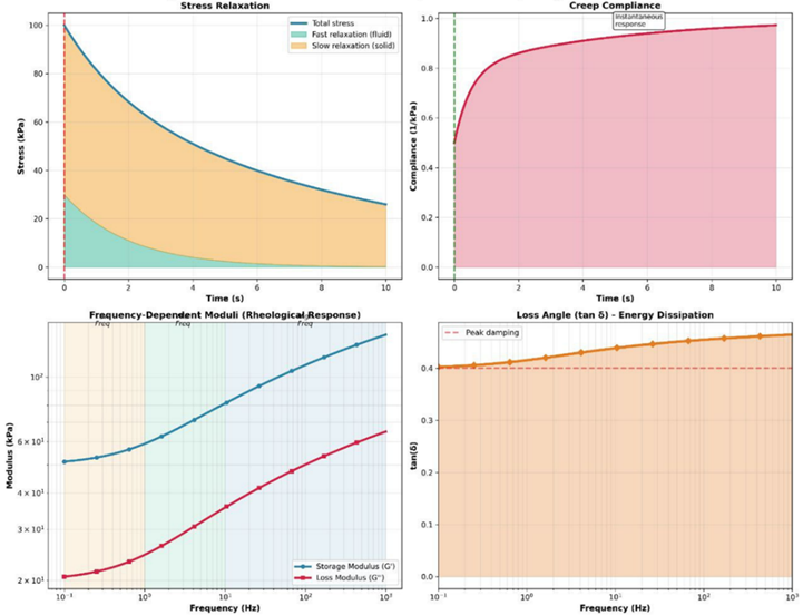
Figure 1: Poroviscoelastic Material Response. Rheological characterization showing stress relaxation with biphasic response, creep compliance with instantaneous and delayed components, storage/loss moduli versus frequency, and loss angle showing energy dissipation.
This comprehensive rheological portrait in Figure 1, is essential for validating computational models, as it provides the key viscoelastic and poro-elastic benchmarks—such as the distinct relaxation phases and frequency-dependent damping—that any accurate simulation of soft tissue must replicate.
2.3 Computational Implementation and Numerical Methods
Finite element method (FEM) implementations of porous media theory have become standard in biomechanical research. These approaches discretize the tissue domain and solve coupled differential equations governing solid deformation and fluid pressure simultaneously. U-P formulations, where displacement and pressure are primary variables, represent the most widely adopted approach in biomechanics. TABLE 3 shows the computational implementation approaches.
| Method | Formulation | Advantages | Disadvantages | Typical Applications | Software Examples |
|---|---|---|---|---|---|
| u-p FEM | Displacement-pressure | Most widely used; well-established | Pressure oscillations in nearly incompressible materials | General poroelastic problems | ABAQUS, COMSOL, FEBio |
| u-p-U FEM | Displacement-pressure-fluid velocity | Improved stability | Higher DOF; increased cost | Complex flow problems | Custom implementations |
| Stabilized FEM | Pressure stabilization terms | Eliminates oscillations | More complex implementation | Nearly incompressible tissues | Research codes |
| Mixed formulations | Enhanced variable spaces | Superior accuracy at interfaces | Implementation complexity | Multi-material problems | Advanced FEM codes |
| Meshless methods | Kernel approximations | No mesh; large deformations | Computational cost; boundary conditions | Fracture, extreme deformation | SPH, RKPM implementations |
| Multiscale FEM | Coupled micro-macro | Links scales explicitly | Very high computational cost | Scaffold design; bone mechanics | Research platforms |
Table 3: Computational Implementation Approaches
Recent advances include stabilized finite elements addressing pressure oscillations in nearly incompressible materials, assumed strain methods improving computational efficiency, and mixed formulations enhancing accuracy near material discontinuities. Coupling with multiphase flow algorithms enables simulation of nutrient transport, waste removal, and ion diffusion through tissue pores. Higher-order elements and adaptive mesh refinement strategies have significantly improved solution accuracy while managing computational costs.
Integration of imaging data with computational models has advanced considerably. Segmentation algorithms extract tissue geometry from CT and MRI scans, while image-based meshing generates patient-specific models.
Machine learning approaches increasingly assist in parameter identification and model calibration, reducing the need for extensive experimental testing. Physics-informed neural networks represent a promising frontier, constraining deep learning with governing equations to improve accuracy with limited training data. Figure 2 shows the pore pressure and Darcy velocity (arrows) at two different times: at t = 15 s, when the sample is compressed (left), and at t = 45 s, when the sample is stretched (right). The pore pressure increases under compression and results in fluid outflow (red arrows) and volume decrease (left). Conversely, the pore pressure is negative under tension, indicating fluid inflow (blue arrows) and volume increase (right).
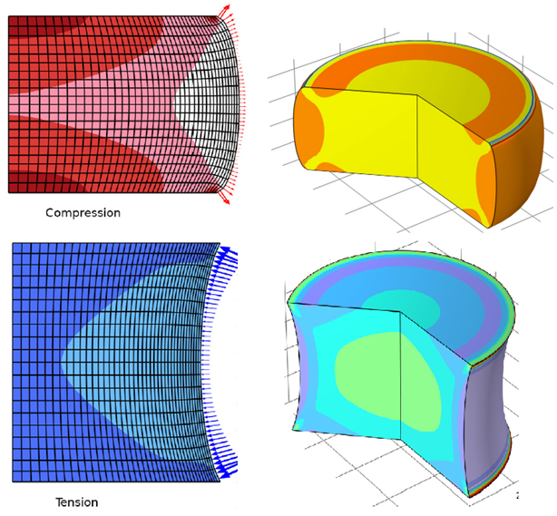
Figure 2: Poroviscoelastic pressure Response and Darcy velocity under compression and tension.
This visualization of pressure fields and fluid velocity under opposing loading conditions directly illustrates the fundamental "pump-like" mechanism of load support and fluid redistribution in hydrated tissues, a process critical for understanding both healthy joint lubrication and the failure modes in degenerative disease.
3.1 Cartilage Mechanics and Osteoarthritis
Articular cartilage provides an ideal application domain for porous media techniques due to its complex multiphase structure. Cartilage consists of a collagen-proteoglycan matrix saturated with fluid, making it fundamentally porous. Poroelastic and poroviscoelastic models successfully predict cartilage behavior under compression, tension, and combined loading at both macroscopic and nanoscale levels. TABLE 4 shows some clinical applications and validation studies.
Figure 3 provides a visual and quantitative analysis of the degenerative changes in cartilage associated with osteoarthritis (OA). It shows a
progression from healthy tissue, through early-stage OA degradation, to advanced OA, with accompanying charts that quantify the changes in key material properties. As the disease progresses, the chart illustrates a decrease in tissue stiffness, an increase in permeability (allowing fluid to flow out more easily), and a significant loss of proteoglycans from the matrix, all of which contribute to the tissue's mechanical dysfunction.
Recent research has utilized porous media simulations to investigate mechanisms of cartilage degeneration in osteoarthritis. Models incorporating changes in matrix composition, altered permeability, and modified mechanical properties reproduce observed disease progression. These predictive models inform development of disease-modifying therapies and surgical intervention strategies. Integration with biochemical models tracking collagen and proteoglycan turnover provides comprehensive understanding of degenerative processes. Advanced techniques enable determination of biphasic material properties from experimental creep and stress-relaxation data, facilitating patient-specific model development.
| Application Domain | Clinical Problem | Modeling Approach | Key Outcomes/Metrics | Validation Method | Clinical Impact | Representative Studies |
|---|---|---|---|---|---|---|
| Osteoarthritis | Cartilage degeneration | Poroelastic FEM with degradation | Stress distribution, contact pressure | Experimental creep/relaxation; MRI T1ρ mapping | Disease mechanism insight; therapeutic targets | Elahi et al. 2023 [7]; Uzuner et al. 2024 [11] |
| Joint Replacement | Implant design, wear | Poroelastic + contact mechanics | Contact area, fluid film thickness | Experimental tribology; retrieval analysis | Implant optimization | Multiple studies |
| Bone Scaffold | Osseointegration, stability | Poroelastic + mechanoregulation | Tissue differentiation, bone ingrowth | Animal studies; μCT imaging | Scaffold design optimization | Loha et al. 2024 [15]; Paul et al. 2024 [12] |
| Tissue Engineering | Cell viability, construct maturation | Coupled poroelastic-biochemical | O₂ concentration, cell density | Bioreactor experiments; histology | Bioreactor protocol optimization | Multiple studies |
| Diagnostic Imaging | Tissue property mapping | Inverse poroelastic analysis | Stiffness, permeability maps | MRI elastography; ultrasound | Patient-specific assessment | Emerging applications |
| Surgical Planning | Outcome prediction | Patient-specific poroelastic | Post-operative stress, alignment | Prospective clinical trials | Personalized treatment | Emerging clinical trials |
Table 4: Clinical Applications and Validation Studies.
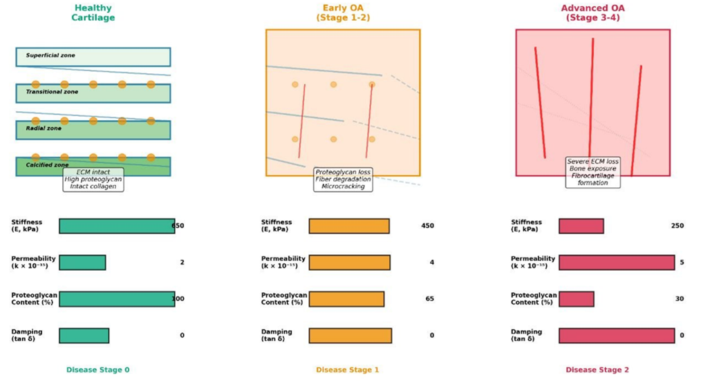
Figure 3: Osteoarthritis Progression. Disease progression analysis showing structural changes from healthy cartilage through early OA degradation to advanced OA, with quantified property changes including stiffness decrease, permeability increase, and proteoglycan loss. The quantified decline in stiffness coupled with a rise in permeability explains the clinical observation of faster, more painful joint deformation under load, as the tissue loses its ability to maintain the high fluid pressure necessary for effective load support (see Figure 3).
The figures presented illustrate the application of porous media theory to biomechanical systems. Figure 4 demonstrates the fundamental biphasic nature of articular cartilage by showing the solid matrix constituent and interstitial fluid phase, with accompanying stress-strain curves that illustrate the critical distinction between undrained and drained mechanical responses—a hallmark prediction of poroelastic theory. As well, Figure 4 illustrates the fundamental biphasic nature of articular cartilage, depicting its composition as a porous solid matrix of collagen and proteoglycan saturated with an interstitial fluid phase. The accompanying stress-strain curves visualize a key prediction of poroelastic theory: the material's stiffness is significantly higher under rapid, undrained loading conditions where fluid has no time to escape, compared to slow, drained loading where fluid flow can occur. This foundational concept explains the time-dependent mechanical behavior critical to cartilage function.
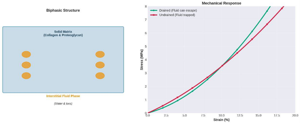
Figure 4: Poroelastic Model of Articular Cartilage – Schematic illustration of cartilage as a biphasic material showing the solid matrix (collagen and proteoglycan) and fluid phase, with stress-strain response curves demonstrating undrained vs. drained stiffness behavior.
This stark contrast between the undrained and drained responses underpins the time-dependent nature of cartilage function, explaining, for instance, why a quick impact can cause different damage patterns compared to a sustained, static load.
3.2 Bone and Scaffold Design
Trabecular bone exhibits marked porosity with fluid-saturated lacunar-canalicular network. Porous media models successfully predict bone's anisotropic mechanical behavior and fluid transport essential for osteocyte mechanotransduction. These simulations reveal how porosity, mineralization, and microstructural organization collectively determine macroscopic properties.
Porous media theory has revolutionized understanding of implant osteointegration and bone scaffold design. Computational models simulate tissue growth around implants, fluid shear stress effects on bone cells, and long-term stability predictions. Triply periodic minimal surface (TPMS) scaffolds, designed using porous media principles, demonstrate superior
mechanical properties and biological performance. Recent advances employ finite element analysis coupled with computational fluid dynamics to optimize scaffold porosity, permeability, and mechanical properties for enhanced bone regeneration. These computational approaches reduce reliance on costly animal studies while providing mechanistic insights into critical design parameters. Figure 5 presents contemporary scaffold design optimization using triply periodic minimal surfaces (TPMS), showcasing both the geometric architecture and corresponding computational finite element mesh, alongside simulated fluid velocity streamlines that visualize nutrient transport pathways critical for bone regeneration. Figure 5 showcases the application of porous media principles in tissue engineering through the design of a bone scaffold. It presents the intricate geometry of a triply periodic minimal surface (TPMS) scaffold, designed to mimic the porous architecture of natural bone, alongside its corresponding finite element mesh used for computational analysis. The simulated fluid velocity streamlines visualize the nutrient transport pathways through the scaffold's interconnected pores, demonstrating how computational fluid dynamics is used to assess and optimize the scaffold's permeability for cell survival and growth.
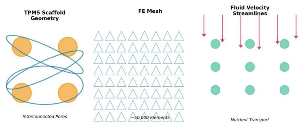
Figure 5: Bone Scaffold Architecture and Fluid Flow – Triply periodic minimal surface (TPMS) scaffold design showing pore geometry, with corresponding finite element mesh and simulated fluid velocity streamlines through interconnected pores demonstrating nutrient transport pathways
By quantifying the flow velocity streamlines, such simulations allow researchers to identify regions of the scaffold that may suffer from poor nutrient delivery, guiding iterative design improvements to ensure uniform cell growth throughout the entire implant.
3.3 Tissue Engineering and Bioreactors
Scaffold design for tissue engineering benefits significantly from porous media simulations. Predicting nutrient diffusion, oxygen transport, and metabolic waste removal through scaffolds remains critical for engineering functional tissues. Porous media models incorporating reactive transport equations guide optimization of pore size, porosity, and material properties for enhanced cell viability and proliferation.
Computational models enable in silico evaluation of scaffold designs before experimental fabrication, dramatically accelerating development cycles. Coupled biomechanical-biochemical simulations predict how mechanical loading influences cell fate and tissue formation within scaffolds. Bioreactor simulations using porous media frameworks determine optimal flow conditions and mechanical stimuli to promote desired tissue differentiation. These advances have substantially improved regenerative medicine outcomes across multiple tissue types, demonstrating clinical translation potential. Figure 6 illustrates the coupled biomechanical-biochemical complexity of tissue engineering bioreactors, depicting oxygen concentration distributions and perfusion flow fields within a scaffold, demonstrating how computational models predict cell viability zones and guide design optimization. Furthermore, this image depicts a computational model of a porous scaffold within a perfusion bioreactor, a system designed to culture cells under controlled fluid flow. The simulation shows the scaffold seeded with cells, while contour plots overlay critical biochemical and physical information: oxygen concentration gradients (top) reveal how nutrient distribution can vary, and velocity fields (bottom) map the perfusion flow that delivers nutrients and removes waste. This integrated analysis is essential for predicting cell viability zones and optimizing flow conditions to ensure uniform cell proliferation throughout the scaffold.
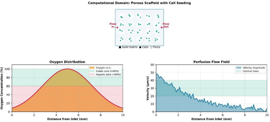
Figure 6: Bioreactor Scaffold Simulation – Computational domain showing a porous scaffold with cell seeding, coupled with contour plots of oxygen concentration (top) and velocity fields from perfusion bioreactor flow demonstrating nutrient transport efficiency for cell proliferation
The ability to predict zones of hypoxia (low oxygen) and stagnant flow within the scaffold is critical for pre-emptively addressing a major bottleneck in tissue engineering—ensuring cell survival deep within a construct before it can develop its own vascular network.
3.4 Recent Advancements and Emerging Technologies
3.4.1 Multiscale and Multiphysics Coupling
Contemporary porous media approaches increasingly couple mechanical, fluid, chemical, and electrical phenomena across scales. Multiscale models bridge cellular to tissue-level processes, linking molecular signaling with macroscopic tissue behavior. These integrative frameworks prove particularly valuable for understanding mechanotransduction where cellular responses to loading ultimately alter tissue properties.
Advanced coupling strategies employ homogenization techniques to transfer information between scales efficiently. Machine learning accelerates these
upscaling procedures through surrogate model development. Multiple-network poroelastic theory provides sophisticated frameworks for modeling distinct fluid compartments with different transport characteristics, particularly valuable for brain tissue mechanics and cerebrospinal fluid dynamics. Emerging multiphysics frameworks incorporating electrokinetic effects, osmotic pressure, and surface tension interactions continue expanding predictive capabilities.
Figure 7 outlines a hierarchical approach to modeling biological tissues, bridging phenomena across multiple length scales. It starts at the nanoscale with molecular bonding interactions, moves up to the microscale where individual fiber mechanics are considered, continues to the mesoscale to model tissue-level fluid-solid coupling, and finally reaches the macroscale for simulating the response of a whole organ. Homogenization techniques are conceptually shown as the method for transferring information between these scales, enabling predictions at the organ level based on fundamental material properties.
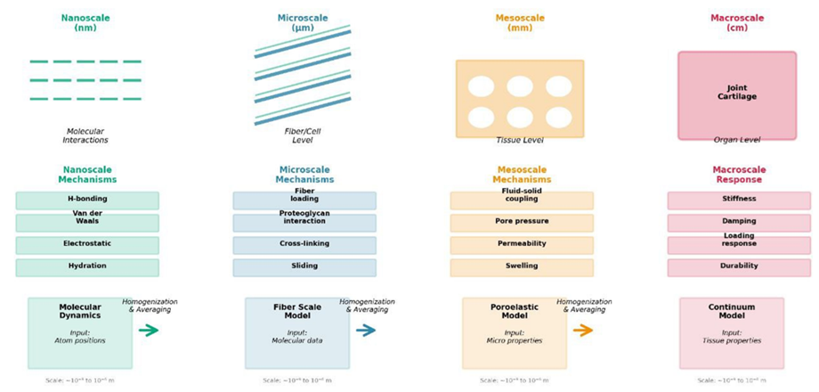
Figure 7: Multiscale Modeling Framework. Hierarchical representation bridging nanoscale (molecular bonding) through microscale (fiber mechanics) and mesoscale (tissue-level fluid-solid coupling) to macroscale (organ-level response) via homogenization.
By quantifying how nanoscale interactions (like molecular bonding) and microscale architecture (fiber alignment) collectively determine the macroscopic failure risk of an organ, this hierarchical framework provides a rational basis for designing biomaterials that function seamlessly from the cellular level up.
3.4.2 Machine Learning Integration
Machine learning has recently transformed porous media biomechanics through enhanced parameter identification, model calibration, and inverse
problem solving. Neural networks trained on experimental or computational data enable rapid material property prediction from imaging data. Physics-informed neural networks enforce porous media governing equations while learning from sparse data, dramatically reducing computational requirements and improving accuracy even with limited training samples. For the use of machine learning has a good knowledge of problem physics help more accurately simulation. Figure8 shows a fluid flow through porous media
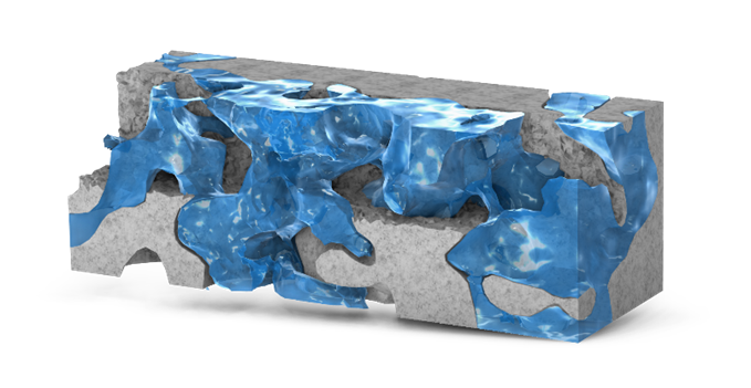
Figure 8: fluid flow through porous media
The complex flow patterns visualized here are governed not only by the pore geometry but also by the fluid's interaction with the solid matrix, and machine learning models are now being trained on such data to rapidly predict permeability from simple microstructural images. Generative models including variational autoencoders create synthetic tissue microstructure datasets for model training and uncertainty quantification. Convolutional neural networks facilitate rapid image segmentation for finite element mesh generation. Reinforcement learning optimizes scaffold designs and surgical planning strategies. These data-driven approaches complement traditional mechanics-based models, leveraging their complementary strengths. Deep learning techniques have demonstrated ability to reduce computation time for biomechanical models while improving accuracy of image segmentation and mesh generation. Figure 9 integrates machine learning advances by
presenting a physics-informed neural network (PINN) workflow that combines experimental imaging data, finite element models, and deep learning architectures for accelerated parameter identification, with quantitative comparisons demonstrating improved prediction accuracy over traditional inverse analysis methods. Figure 9 diagram presents a modern machine learning approach to biomechanical modeling, illustrating a physics-informed neural network (PINN) workflow. It shows how imaging data is integrated with a finite element model to inform a neural network architecture. The network is trained to solve inverse problems, such as identifying material properties, by being constrained by the physical laws (governing equations) of poroelasticity. The comparison graph of predictions vs. experimental data demonstrates the high accuracy achievable with this hybrid approach, which is faster than traditional iterative methods.
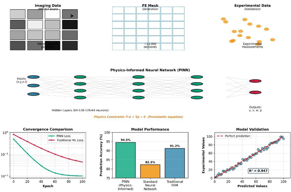
Figure 9: Physics-Informed Neural Network Workflow – Schematic showing integration of imaging data, finite element model, and neural network architecture for parameter identification, with comparison of predictions vs. experimental data demonstrating accuracy improvements
This hybrid approach overcomes the traditional limitations of purely data-driven models by embedding physical laws directly into the learning process, ensuring that the AI's predictions remain physically plausible even when extrapolating beyond the specific conditions of the training data.
3.4.3 Advanced Imaging and Validation
High-resolution imaging modalities including synchrotron X-ray microcomputed tomography and multiphoton microscopy now provide unprecedented detail of tissue porous architecture. These imaging data directly inform computational model geometry and material property distributions. Confocal microscopy tracks fluid transport and cell behavior within tissue models, enabling quantitative validation of poroelastic predictions.
Digital volume correlation and other image-based kinematics techniques measure deformation fields during mechanical testing, providing comprehensive validation datasets for computational models. Integration of imaging with mechanical testing creates powerful experimental validation frameworks, substantially increasing confidence in model predictions and expanding their clinical applicability. Advanced imaging techniques enable creation of image-based poroelastic models reflecting patient-specific tissue microstructure, advancing personalized medicine approaches. Figure 10 presents a complete 10-step workflow for creating patient-specific computational models from medical images. The process begins with clinical imaging (MRI/CT) and proceeds through segmentation of the target tissue, surface reconstruction, CAD modeling, mesh generation, and assignment of material properties and boundary conditions. The final steps show the finite element solver computing the results, which are visualized as stress and pressure distributions, demonstrating a direct pathway from diagnostic imaging to personalized biomechanical analysis.
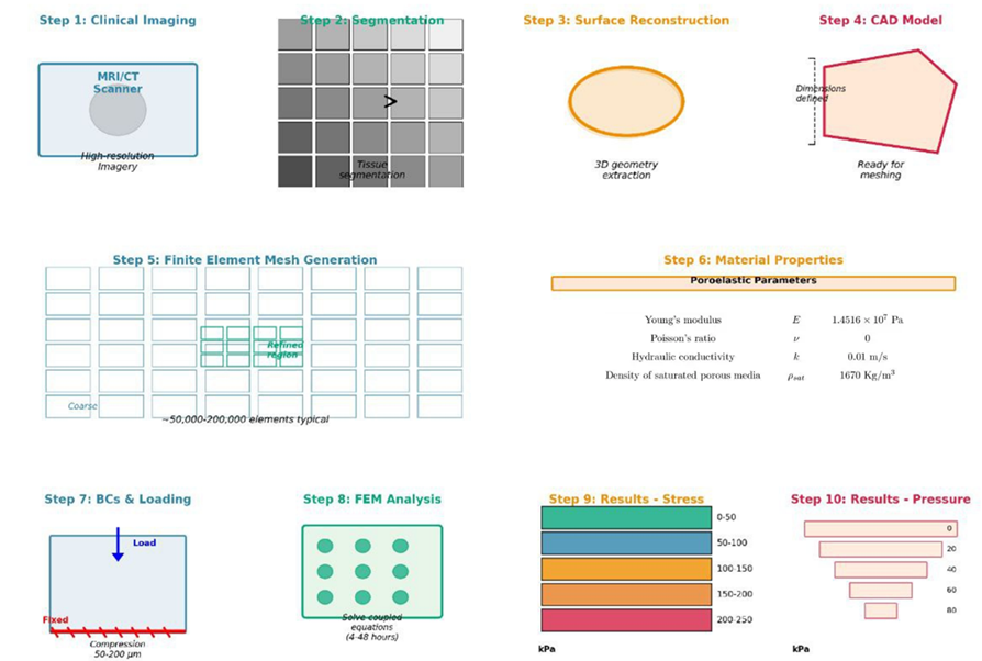
Figure 10: Image-Based Computational Pipeline. 10-step complete workflow from MRI/CT imaging through segmentation, surface reconstruction, CAD modeling, mesh generation, material property assignment, boundary conditions, FEM solver, to final stress and pressure results.
This fully integrated pipeline represents a significant step toward clinical translation, as it creates a direct, automatable pathway from a patient's standard-of-care medical images to a personalized computational model that could be used to virtually test different surgical or therapeutic strategies.
3.5 Challenges and Future Research Directions
Despite substantial progress, several challenges remain in applying porous media techniques to clinical biomechanics. Accurate characterization of tissue heterogeneity and anisotropy across scales continues limiting model accuracy. Identifying material parameters from limited experimental data remains problematic, necessitating robust inverse problem methodologies. The integration of machine learning with physics-based models shows promise for addressing these challenges, yet requires careful validation to ensure biological relevance.
Computational efficiency remains a constraint for clinical applications requiring real-time predictions or extensive parameter studies. Novel discretization approaches, accelerated algorithms, and machine learning surrogates address these limitations. Integration of patient-specific data from clinical imaging into personalized biomechanical models represents a critical frontier, with applications in surgical planning, implant design optimization, and disease prognosis.
Future research should emphasize closed-loop approaches where computational predictions guide experiments, and experimental observations refine models iteratively. Standardization of validation protocols would facilitate comparison across studies and increase clinical acceptance. Investment in open-source computational frameworks and benchmark datasets will democratize access to porous media simulation tools. These advances promise to transform biomechanical simulation from research tools into clinical decision-support systems.
The reviewed literature demonstrates the remarkable evolution of porous media techniques in biomechanical simulation, from foundational biphasic theory to contemporary multi-scale, multi-physics, and data-integrated approaches. Early work by Mow and colleagues established the theoretical framework [1-3], while subsequent studies refined material characterization methods [4-6,10] and extended applications to clinical problems [7,11].
The translation of poroelastic principles to tissue engineering [12-15,20] represents a significant achievement, enabling rational design of scaffolds that mimic native tissue mechanical and transport properties. Concurrently, the integration of machine learning [16-19] promises to accelerate simulations, improve parameter estimation, and enable personalized medicine applications.
Emerging trends include multi-physics formulations incorporating electrokinetic [8], biochemical [7,9], and thermal [21-23] phenomena. The convergence of advanced manufacturing, computational modeling, and artificial intelligence positions porous media techniques at the forefront of biomechanical innovation.
Porous media techniques have fundamentally advanced biomechanical simulation, enabling accurate prediction of complex tissue behaviors and physiological processes. From foundational poroelastic theory through contemporary multiphysics approaches, these frameworks increasingly capture the intricate coupling between solid and fluid phases central to biological function. Applications spanning cartilage, bone, and tissue engineering demonstrate substantial clinical relevance.
Recent technological convergence—advanced imaging, machine learning, and computational power—has dramatically accelerated progress. Integration of patient-specific data with personalized computational models promises transformation of clinical practice. Continued investment in methodological development, experimental validation, and clinical translation will position porous media biomechanics as essential infrastructure for precision medicine and regenerative medicine advancement.
The authors declare no conflicts of interest.
Dear Editorial Team, Clinical Medical Reviews and Reports. My experience with the journal was highly positive. The peer-review process was rigorous, constructive, and completed in a timely manner. The reviewers provided valuable comments that helped improve the quality and clarity of our manuscript. The editorial office was professional, responsive, and supportive throughout all stages of the publication process. Communication was clear and efficient, and any questions were addressed promptly. Overall, I found the journal to maintain high scientific standards and an excellent publication workflow. I would be pleased to consider submitting future work to this journal. Best wishes from, Elena Popa.

It was my pleasure to submit my testimonial concerning the Reviewer Board of our Scientific Journal “Brain and Neurological Disorders”. The Reviewers focused on some modifications and their contribution was helpful. The ladies of our Editorial Office were also supported my efforts. It was my honor to have such a co-operation and I am looking forward for more collaboration.

Dear Grace Pierce, Editorial Coordinator of Journal of Clinical Research and Reports, Thank you for the speedy and efficient peer review process. I appreciate the fact that your peer reviewers do not take months to respond like with some other journals. I would also like to thank the editorial office for responding quickly to my questions. It is an excellent journal. I plan to submit more manuscripts in the future. Best wishes from, Robert W. McGee

Dear Grace Pierce, Editorial Coordinator of Journal of Clinical Research and Reports, Working with you and your team on our recent publication in JCRR has been a truly wonderful and enjoyable experience. The responses were prompt, and the reviewers were patient, constructive, and highly professional. One reviewer in particular gave me the feeling that a professor was carefully reading and commenting on my coursework, which was deeply touching. The entire process was straightforward and hassle‑free, with no tedious online forms to complete. I highly recommend this journal. Best wishes from, DR Aibing Rao, Head of R&D

I Appreciate the Opportunity to Share my Experience with the Journal of Clinical Research and Reports. The peer review process was timely and constructive, and the feedback provided helped improve the quality of our manuscript. The editorial office was professional, responsive, and supportive throughout the process, ensuring smooth communication and efficient handling of the submission. Overall, it was a positive experience collaborating with your team.

Dear Mercy Grace, Editorial Coordinator of Obstetrics Gynecology and Reproductive Sciences, We would like to express our gratitude for your help at all stages of publishing and editing the article. The editors of the magazine answer all the necessary questions and help at every stage. We will definitely continue to cooperate and publish other works in the Obstetrics Gynecology and Reproductive Sciences! Best wishes from, Alla Konstantinovna Politova,
