
Research Article | DOI: https://doi.org/10.31579/2640-1045/052
1 Department of Endodontics, Vassouras University, Vassouras, Rio de Janeiro, Brazil
2 Department of Endodontics, Vassouras University, Vassouras, Rio de Janeiro, Brazil
3 Department of Endodontics, Vassouras University, Vassouras, Rio de Janeiro, Brazil
4 School of Dentistry, Iguaçu University, Nova Iguaçu, Rio de Janeiro, Brazil.
5 Faculty of Dentistry, São José University, Rio de Janeiro, RJ
6 Department of Endodontics Iguaçu University, Nova Iguaçu, Brazil
7 Department of Endodontics, Vassouras University, Vassouras, Rio de Janeiro, Brazil
8 Veterinarian Faculty, Rural Federal Rio de Janeiro University, Seropédica, RJ, Brazil.
9 Medical school, Iguaçu University, Nova Iguaçu, Brazil.
10 Medical school Iguaçu University, Nova Iguaçu, Brazil.
11 Medical school graduate, Iguaçu University, Nova Iguaçu, Brazil.
12 Department of Restorative Dentistry, Londrina State University, Londrina, Brasil
13 Department of Endodontics, Iguaçu University, Nova Iguaçu, Brazil.
*Corresponding Author: Marília FaguryMarceliano-Alves, Department of Endodontics, Iguaçu University, Nova Iguaçu, Brazil.
Citation: L F C Toledo, L F G R Oliveira, J C F Silveira, E O P B França, M P W Galhardi, V Ronquete, Alexandre S Cunha, Lidiane C Soares, M Orsini, Jacqueline F Nascimento, Pablo A Silva and M F V M Alves. (2020). Comparative analysis of the antimicrobial potential of different intra-canal medication to microorganisms present in the failure of the endodontic therapy: In vitro study. Endocrinology and Disorders. 4(1); DOI:10.31579/2640-1045/052
Copyright: © 2020, Marília FaguryMarceliano-Alves: This is an open access article distributed under the Creative Commons Attribution License, which permits unrestricted use, distribution, and reproduction in any medium, provided the original work is properly cited.
Received: 04 August 2020 | Accepted: 24 August 2020 | Published: 01 September 2020
Keywords: endodontic, bacteria, persistent infection, intra-canal medication
Microorganisms that infect the root canals system are the main etiologic factor of the periapical pathologies. Some microorganisms are resistant to the antimicrobial treatment and may survive in the root canal after the chemical mechanical preparation and intra-canal medication, characterizing a persistent infection. In cases of failure of the endodontic treatment, a new approach may be done using additional measures that involve this infectious process control through the elimination or maximum reduction of microorganisms. Therefore, this article aims to evaluate the antimicrobial potential of different formulations of intra-canal medication compared to strains of Enterococcus faecalis, Pseudomonas aeruginosa and Staphylococcus aureus in Petri plates. It was used diffusion test in agar where each Petri plate with the inoculated bacteria presented 5 wells that were filled with each medication. The diameters of the bacterial inhibition zones were measured and registered to each tested medication at the period of 24 hours, 48 hours, 1 week and 2 weeks. All the medications promoted inhibition halos; however, a higher elimination of micro-organisms can be significantly achieved through the association of different substances in the formulation of an intra-canal medication, with emphasis to HPG and Ca(OH)2 + CHX.
The endodontic therapy presents predictable results when is done according to the recommended precepts through attendance protocols based in scientific evidences that guarantee a high rate of success[1]. Therefore, even some cases of well filled canals can shelter an infection that usually is the cause of the maintenance of a periapical lesion that leads to the need to a re-intervention by the professional [2].
Microorganisms that infect the root canals system are the main etiologic factor of the periapical pathologies [3]. For this reason, infectious diseases should be treated seeking to the maximum elimination of the microorganisms. That is, it should eliminate the endodontic infection or reduce it significantly to the treatment or re-treatment succeed [2].
The microbiota associated to the endodontic infections is heterogeneous, presenting different compositions to each type of infection and patients. The determination of the microbial population of the endodontic infections is very important to define the best strategy to the microbial control in the endodontic treatment [4].
The primary infection has polymicrobial nature, with strict anaerobic bacteria clearly dominating this condition [5]. Some microorganisms are resistant to the antimicrobial treatment and can survive in the root canal after the chemical mechanical prepare and intra-canal medication, characterizing a persistent infection [4, 6].
The composition of microorganisms present in the persistent infection is characterized as a microorganism’s association found in the primary and secondary infection [7].
The persistent infection is considered as the main cause of the majority of the endodontic problems, as persistent exudation and symptomatology, flare-up and failure of the endodontic treatment [2]. Studies have been demonstrated that the success index of tooth with filled canals in presence of detectable levels of cultivable bacteria is significantly shorter than in cases of negative culture [3, 8]. The results confirm that when bacteria stay viable into the root canals even when a fill is suitable, they continue to obtain nutrient and survive in an enough number to perpetuate a periapical lesion [6].
The reduction of microorganisms into the root canal system promoted by the chemical mechanical prepare is, most times, enough to allow that the mechanisms of periapical tissues occur. However, in cases where the perpetuation of a periapical lesion caused by the persistence of an endodontic infection demonstrates that the immediate instituted therapy there was not enough to the success of the treatment.
In the failure cases of the endodontic treatment, a new approach may be done using additional measures that involve the control of this infectious process through the elimination or maximum reduction of microorganisms, using in the endodontic treatment an intra-canal medication with determined chemical substances that may be efficient, as it was demonstrated in different studies [3, 9, 10, 11, 12].
Therefore, this article aims to evaluate the antimicrobial potential of different formulations of intra-canal medication compared to strains of Enterococcus faecalis, Pseudomonas aeruginosa and Staphylococcus aureus in Petri plates.
Materials and methods
Medicaments
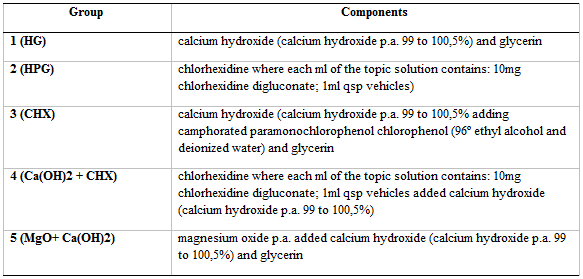
The medicaments include a) calcium hydroxide powder paste mixed with glycerin in the 2:1 proportion (vol:vol); b) calcium hydroxide powder paste mixed with camphorated paramonochlorophenol and glycerin in 3:1:1 proportion; c) chlorhexidine digluconate (2% gel; Floral Manipulation Pharmacy, Vassouras, Rio de Janeiro, Brazil); d) chlorhexidine digluconate paste (2% gel; Floral Manipulation Pharmacy, Vassouras, Rio de Janeiro, Brazil) mixed with calcium hydroxide powder in 2:1 proportion and e) magnesium oxide powder paste mixed with calcium hydroxide powder mixed with glycerin in 1:1:1 proportion. All the pastes were prepared aiming to a similar consistence to toothpastes (Chart 1).
Bacterial sample
The antibacterial activities of the medicaments were evaluated against facultative anaerobic bacteria, Staphylococcus aureus (ATCC 9144), Enterococcus faecalis (ATCC 29212) and aerobic, Pseudomonas aeruginosa (ATCC 27583).
Experimental procedure
Preparation of bacterial strains
The bacterial strains previously frozen in brain and heart infusion broth (BHI) with 20% glycerol were thawed and streaked on Müeller Hinton agar (MH) containing 5% defibrinated lamb blood (SPlabor, Presidente Prudente, São Paulo, Brazil) . The incubation took place at 37 ° C for 24 h to assess its viability and morphotintorial characteristics.
Inoculum preparation
The phenotypic detection of drug resistance was performed according to the recommendations of the Clinical and Laboratory Standards Institute (CLSI). After 18 to 24 hours of incubation at 35°C on non-selective nutrient agar, colonies were suspended in brain and heart infusion broth (BHI) until a turbidity equivalent to the McFarland 0.5 scale was obtained, which corresponds to a concentration of approximately 1.5 x 10⁸ microorganisms / ml.
The facultative anaerobic microorganisms were maintained in brain and heart infusion (BHI) broth (SPlabor, Presidente Prudente, São Paulo, Brazil) and the aerobic bacterium was maintained in BHI broth with heminine (5mg/L) and menadione (0,5mg/L) (SPlabor, Presidente Prudente, São Paulo, Brazil). Afterward, 1 ml of culture of the bacteria to be tested was placed into the agar plates.
Antimicrobial susceptibility test
It was used agar diffusion test. Petri plates containing BHI agar supplemented with heminine and menadione were inoculated with the tested bacteria using sterile appliers with cotton tip that were rubbed from the centers to the edges. Growth of the bacteria was observed at the end of the period of incubation by checking the morphology of colonies grown onto agar plates. In each Petri plate with the inoculated bacteria it was perforated 5 wells with 5mm of profundity and 6mm of diameter and it were filled with the tested medicaments. Positive control plates were streaked with bacteria, but no medicament was used. These wells were filled with each medicine through 100µl pipettes, and each drop contained 25µl. The plates were incubated in 37°C for 14 days. After, the diameters of the bacterial inhibition zones were measured and registered to each tested material, with the 6mm diameter as cut-off value in the 24 hours, 48 hours, 1-week e 2-weeks count. To fill each well of each Petri plate, 4 25µl drops of each medication were deposited. Three agar plates were used for each bacterial strain tested.
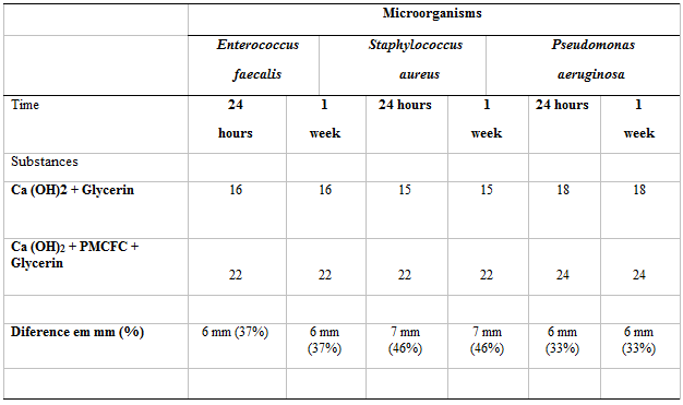
In the Table 1, there are the averages of the inhibition halos (in mm) propitiated by the materials in function of the microorganism in the aerobic condition measured in 24, 48 hours, 1 week and 2 weeks (Figures 1 to 3).


The groups 2 and 4 presented big zones of inhibition against the tested bacteria. The groups 1 and 3 also presented inhibitor halos against all the strains; but, generally, it was not more efficient than the groups 2 and 4. The group 5, that presents a new formulation, was efficient against all the tested microorganisms. It was more efficient that group 1 in all the samples and more efficient that the group 3, except against Pseudomonas aeruginosa where they presented equivalent results. The group 1 presented the worst results against all the bacteria strains used in this experiment. Bacteria survived in saline (control group) until the end of the experiment (two weeks).

Microorganisms are the main etiologic agents of the endodontic infections6. The reduction or elimination of the bacteria seems a logic objective to the success of the endodontic treatment and indeed, the literature supports this concept. The microorganisms used in this work are commonly found in cases of persistent infection [13, 14].
After the chemical mechanical prepare done diligently, some canals can shelter detectable levels of cultivable bacteria. In Shuping et al [10] study the authors relate that in the instrumentation of canals irrigated with sodium hypochlorite (NaOCl), 61.9% of the canals were free of bacteria.
When a medication based in calcium hydroxide was used for minimally 1 week, 92.5% of the canals do not present bacteria. This reduction was considered significantly when compared with the instrumentation and irrigation with NaOCl, in isolation. The authors concluded that even the chemical mechanical prepare is an important step in the reduction of the bacteria during the endodontic treatment, the addition of a medication containing calcium hydroxide may be used to reach a more predictable result.
In 2007, Siqueira et al [14] concluded that the instrumentation of the canals using 2.5% sodium hypochlorite as irrigator reduced significantly the number of bacteria in the canal. However, bacteria stay cultivable in more than half the cases (54.5%). The using of calcium hydroxide intra-canal medication associated to camphorated paramonochlorophenol in inert vehicle for 7 days increased significantly the number of cases with negative cultures (90.9%). According to the authors of this study, even calcium hydroxide combined with an inert vehicle can eliminate the majority of the endodontic bacteria when in direct contact, these effects cannot be as pronounced in the environment of the root canal, in what that contact cannot be always reached or, given the low solubility of the calcium hydroxide, the concentration of hydroxyl ions may not reach enough levels to a broad elimination of bacteria in biofilms at the untouched canal walls, dentin tubules, irregularities and other anatomic variations.
The physical-chemical limitations of the calcium hydroxide and the resistance reported by some microbial species motivated the association of Ca(OH)2 with other antimicrobial, as the camphorated paramonochlorophenol(PMCFC). PMCFC has strong antibacterial activity, with the action in distance in reason of the volatility of the paramonochlorophenol and satisfactory biocompatibility due to its lent release when associated to the Ca(OH)2 and glycerin [15, 16].
In 1996, Siqueira, Uzeda [15] accomplished a study aiming to compare the antibacterial effect of calcium hydroxide in an inert vehicle and in a biologically active vehicle (PMCFC) against facultative anaerobic bacteria. The infected specimens were exposed to the medications of calcium hydroxide mixed with saline solution (inert vehicle) or camphorated paramonochlorophenol for 1 hour, 1 day and 1-week periods. The viability of the bacteria after these exposition times was evaluated by incubation of samples in culture medium to compare the efficiency. The Ca(OH)2 + PMCFC association was efficient to eliminate bacteria after 1 hour of exposition, except by Enterococcus faecalis that was eliminated only after one day of exposition. In contrast, Ca(OH)2 in inert vehicle was ineffective against all the microorganisms tested even after 1 week of exposition. The results showed that PMCFC increased the antibacterial effects of the calcium hydroxide. These data are confirmed in this study because the inhibitor halos formed by HPG are higher than HG against all the tested microorganisms (Table 2). Other studies obtained equivalent results, where HPG presented higher potential to eliminate different species of microorganisms when compared to Ca(OH)2 in inert vehicles, as glycerin or saline [2, 16].
The combination of 2% Chlorhexidine in gel associated with calcium hydroxide (Ca(OH)2) obtained similar results to HPG group. However, the inhibitor halos of HPG were almost 3mm higher than Staphylococcus aureus in the period of 24 hours and 2mm after 2 weeks. In the plates that contain Pseudomonas aeruginosa strains, this difference was shorter, almost 2mm and 1mm after 24 hours and 1-week periods, respectively. In 2003, Siqueira et al. [2] also reported equivalent results when HPG and Ca(OH)2 + CHX were compared. In this study, a discreet difference in the antibacterial action of HPG was perceived when compared to Ca(OH)2 + CHX. But both presented the highest inhibition halos.
When compared with all the other groups, the group with the Ca(OH)2 + CHX combination presented higher inhibition halos, except when achieved the same inhibition halo than the group that contains MgO+ Ca(OH)2 in the period of 2 weeks. The superior antibacterial action of Ca(OH)2 + CHX when compared with the groups 1 and 3 also was reported in the study of Manzur et. al. [17]. The results highlighted by Manzur et al. [17]. Revealed presence of microorganisms in 18%, 45% and 1% of the samples when Ca(OH)2 in an inert vehicle, 2% CHX and the combination of 2% Ca(OH)2 + CHX were used, respectively. In this study, the combination of calcium hydroxide associated to magnesium oxide (MgO+ Ca(OH)2) in inert vehicle (glycerin) presented higher inhibition halo than 2% chlorhexidine in gel in the period of 24 hours and 1 week in the Petri plates containing Enterococcus faecalis and Staphylococos aureus. In the plates with the Pseudomonas aeruginosa bacterium, the difference of 1mm in the inhibition halo favoring MgO+ Ca(OH)2 was perceived in the period of 1 week. There was not found in the literature, MgO+ Ca(OH)2 comparative analysis in relation to other medications when used culture methods or through collected samples into the root canals system. When compared to Ca(OH)2 in inert vehicle, MgO+ Ca(OH)2 presented better results in all the samples.
Among the metallic oxides, the magnesium oxide (MgO) is presented as stable in adverse conditions and can be considered safe to human beings [18, 19]. In medicine, MgO is used to combat heartburn due to this high pH (almost 12) and in the bony regeneration [20, 21]. Recently, MgO particles presented highlighted potential as antimicrobial agent. Makhlufet al. [22] demonstrated that small particles of MgO presented satisfactory antibacterial activity against Escherichia coli and Staphylococcus aureus. Though the antibacterial mechanism of MgO is not defined, three antibacterial mechanisms have been reported. Reactive oxygen formation that leads to damages to the bacterial AND, antioxidant action and damages to the cell metabolism: damages to the cell wall and the alkaline effect [23, 24]. These authors highlight also the promising antibacterial potential of MgO, but it is necessary more studies about the MgO particles activity in relation to other microorganism’s species.
According to the information exposed above, the chemical mechanical prepare reduces significantly the bacterial concentration into the root canal. However, the application of intra-canal medication for 1 week seems to be recommended to increase the bacterial elimination. For a more predictable result in the endodontic therapy in cases of infected canals, the results of this study corroborate many previous studies [11,15,16,17,18] suggesting that a higher elimination of microorganisms can be significantly achieved through the association of different substances in the formulation of an intra-canal medication, with focus in HPG e Ca(OH)2 + CHX. There are the inhibition halos average (in mm) propitiated by the materials in function of the microorganism in the aerobic condition measured in 24 hour, 48 hours, 1 week and 2 weeks.
Dear Editorial Team, Clinical Medical Reviews and Reports. My experience with the journal was highly positive. The peer-review process was rigorous, constructive, and completed in a timely manner. The reviewers provided valuable comments that helped improve the quality and clarity of our manuscript. The editorial office was professional, responsive, and supportive throughout all stages of the publication process. Communication was clear and efficient, and any questions were addressed promptly. Overall, I found the journal to maintain high scientific standards and an excellent publication workflow. I would be pleased to consider submitting future work to this journal. Best wishes from, Elena Popa.

It was my pleasure to submit my testimonial concerning the Reviewer Board of our Scientific Journal “Brain and Neurological Disorders”. The Reviewers focused on some modifications and their contribution was helpful. The ladies of our Editorial Office were also supported my efforts. It was my honor to have such a co-operation and I am looking forward for more collaboration.

Dear Grace Pierce, Editorial Coordinator of Journal of Clinical Research and Reports, Thank you for the speedy and efficient peer review process. I appreciate the fact that your peer reviewers do not take months to respond like with some other journals. I would also like to thank the editorial office for responding quickly to my questions. It is an excellent journal. I plan to submit more manuscripts in the future. Best wishes from, Robert W. McGee

Dear Grace Pierce, Editorial Coordinator of Journal of Clinical Research and Reports, Working with you and your team on our recent publication in JCRR has been a truly wonderful and enjoyable experience. The responses were prompt, and the reviewers were patient, constructive, and highly professional. One reviewer in particular gave me the feeling that a professor was carefully reading and commenting on my coursework, which was deeply touching. The entire process was straightforward and hassle‑free, with no tedious online forms to complete. I highly recommend this journal. Best wishes from, DR Aibing Rao, Head of R&D

I Appreciate the Opportunity to Share my Experience with the Journal of Clinical Research and Reports. The peer review process was timely and constructive, and the feedback provided helped improve the quality of our manuscript. The editorial office was professional, responsive, and supportive throughout the process, ensuring smooth communication and efficient handling of the submission. Overall, it was a positive experience collaborating with your team.

Dear Mercy Grace, Editorial Coordinator of Obstetrics Gynecology and Reproductive Sciences, We would like to express our gratitude for your help at all stages of publishing and editing the article. The editors of the magazine answer all the necessary questions and help at every stage. We will definitely continue to cooperate and publish other works in the Obstetrics Gynecology and Reproductive Sciences! Best wishes from, Alla Konstantinovna Politova,
