
Commentary Article | DOI: https://doi.org/10.31579/2692-9562/069
Professor & Chair: Div. Oral & Maxillofacial Radiology, University of British Columbia
*Corresponding Author: David MacDonald, Professor & Chair: Div. Oral & Maxillofacial Radiology, University of British Columbia.
Citation: David MacDonald. (2023). Commentary on “a Retrospective Study of Incidental Findings Occurring in a Consecutive Case Series of Lateral Cephalograms of 12 to 20-Year-Old Patients Referred for Routine Orthodontic Treatment” [1]. Journal of Clinical Otorhinolaryngology, 5(2); DOI: 10.31579/2692-9562/069
Copyright: © 2023 David MacDonald. This is an open access article distributed under the Creative Commons Attribution License, which permits unrestricted use, distribution, and reproduction in any medium, provided the original work is properly cited
Received: 03 November 2022 | Accepted: 14 January 2023 | Published: 28 January 2023
Keywords: hypopharyngeal; heterogeneous diseases
As the lateral cephalogram (Figure 1) covers the nasal cavity, cervical vertebra and the entire pharynx (from the base of the skull right down to the larynx), findings in these areas are relevant to the oto-rhino-laryngologist. Although nasal endoscopy is the best choice for evaluation of nasopharyngeal tonsil (adenoid) size.
As the lateral cephalogram (Figure 1) covers the nasal cavity, cervical vertebra and the entire pharynx (from the base of the skull right down to the larynx), findings in these areas are relevant to the oto-rhino-laryngologist. Although nasal endoscopy is the best choice for evaluation of nasopharyngeal tonsil (adenoid) size [2], the lateral cephalogram exhibits very good diagnostic accuracy for the diagnosis for hypertrophic nasopharyngeal tonsils and for posterior upper airway obstruction [3]. In addition to sleep apnea hyperplasia of the tonsils may be associated with mouth-breathing [4].
The lateral cephalogram (also called lateral cephalometric radiograph), has been used for nearly a century, routinely prescribed by orthodontists for cephalometric measurement as an essential part of orthodontic treatment planning. The lateral cephalogram permits the evaluation of dimensions and position of craniofacial structures and adjacent soft-tissues. and the identification of skeletal anomalies and/or pathology [5]. The lateral cephalogram in an orthodontic context is almost always accompanied by a dental panoramic radiograph (DPR) to display better panoramically the whole dentition. Although the DPR does experience secondary artifacts that are superimposed on the primary image, these are well understood by the dentist. The study, the subject of this commentary, focused solely on incidental findings of those areas of the lateral cephalograms which are outside the jaws and not displayed on the DPR, namely the cranium and vertebra as these are outside the normal anatomic sphere of dental practice, excepting that of the orthodontist. The overlap of bilateral structures is infrequently a problem for the orthodontist as most structures of interest are in the midline, such as the sella (S), nasion (N) and A and B points (Figure 1). Cone-beam computed tomography (CBCT) imparts a significantly higher radiation dose to the more radiation-sensitive child patient and is unlikely to replace the lateral cephalogram and DPR in routine orthodontics [6]. Indeed, many graduate programs in orthodontics restrict prescription of large field-of-view CBCT to patients with craniofacial anomalies (including those with cleft lip and palates) where cross-sectional imaging is required prior to surgery. As many of these cases require multiple surgeries, multiple CBCTs are preferred to multiple multidetector computed tomography (MDCT) as the former imparts a much-reduced radiation dose burden to the child than the latter.
This retrospective study was performed on the lateral cephalograms of 1765 consecutive 12-to-20-year-old patients (800 males and 965 females) presenting for treatment to a North American graduate orthodontic program. All these patients were considered normal, because no abnormalities were found neither in their medical history nor on their clinical examination. Therefore, none had cleft-lip and palates, craniofacial anomalies nor any serious or potentially serious medical condition.
Overall prevalence of incidental findings was 18.8% of which 10.3% were ponticulus posticus and 4.2%, bridging of sella turcica. The importance of these is that ponticulus posticus by its envelopment of the vertebra artery may case dizziness, head and neck pain whereas the latter though generally associated with syndromes is now reported in the normal general population. Further reflection upon this finding may or may not reveal hitherto undetected disease; time with tell. A similar experience was realized with taurodontism, a dental anomaly which subsequently found in normal populations, may also be an indicator of as of yet undetected cases of Klinefelter’s syndrome [7]. Although the occipital spurs and for ponticulus posticus were more prevalent in males, the size of the sella turcicas did not differ between sexes. Of the 1156 patients completing treatment about 2 years later, only one lateral cephalogram displayed progression of its ponticulus posticus in that time.
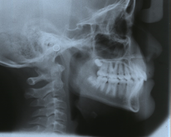
Figure 1: Displays a lateral cephalogram that excluded most of the cranium and is typical in most countries outside North America.
Even there, there has a gradual movement away from making lateral cephalograms that include the whole cranium, in the interests of reducing the area irradiated. This trend may mean that, going forward, enlarged parietal foramina, rare as they are, may become less noticeable on lateral cephalograms because of this exclusion of the top of the skull. But as this report revealed all of the incidental findings outside the jaws, namely the aforementioned 18.8% of all lateral cephalograms including in the study, would not have been detected simply because the orthodontists did not notice them. Although the vast majority of children presenting for orthodontic treatment are overtly normal, some dental anomalies may suggest an underlying medical condition, such as a genetic or an endocrine disorder [8]. All those lesions appearing in the jaws were identified on the accompanying DPR for each case.
While this study performed on overtly normal juveniles did not display nasopharyngeal masses simply because of their age [9], too old for hyperplastic nasopharyngeal tonsils and not old enough for nasopharyngeal carcinomas (NPC), the increasing extension of orthodontic treatment or retreatment to adult populations increases the likelihood of the latter being present, particularly in those communities of Southeast Asian extraction, namely those originating from the southern Chinese provinces of Quangdong, Guangxi, Hunan, Fujian, Jiangxi and Hongkong and Macao [10]. Quangdong and Quangxi have the highest mortalities from NPC[11]. Although NPC is rare with a global incidence of about 1:100,000 people, it is as high as 30:100,000 in South China & Southeast Asia [12,13]. Seventy-seven percent of those diagnosed with NPC in British Columbia were of Asian ethnicity; the majority had non-keratinizing NPC (81%) [14].
Almost of fifth of lateral cephalogram of normal orthodontic 12-to-20-year-old patients displayed incidental findings; of these are the ponticulus posticus, vertebral fusion, and enlarged parietal foramina were clinically significant. Hyperplastic nasopharyngeal tonsils were not observed in this age group, but would be an important observation when adults are being considered for orthodontic treatment or retreatment.
Lateral cephalogram displaying the most important and frequently used cephalometric ‘points’ on the base of the skull, nose and dentoalveolar apparatus. The lateral head position is held perfectly perpendicular to the central ray by the cephalostat (made of plastic). Its ‘ear plugs’ are placed within the external acoustic meatuses. The metal rings which denote the external acoustic meatus are precisely superimposed upon each other. The age of the patient from the degree of dental development is 12 to 14 years of age, the usual age for orthodontic treatment. The size of soft-tissue outline of the nasopharynx, silhouetted against the air-filled nasopharynx is normal in this otherwise normal patient seeking orthodontic treatment.
‘S’ point denotes the centre of the sella turcica; ‘N’ point, the nasion; ‘A’ point, the maximum concavity of the maxilla and ‘B’ point, the maximum concavity of the mandible. Due to the long film/receptor-focal distance used the superimposed right and left sides appear almost perfectly superimposed, except for the lower borders of the mandible and floors of the orbits.
Dear Editorial Team, Clinical Medical Reviews and Reports. My experience with the journal was highly positive. The peer-review process was rigorous, constructive, and completed in a timely manner. The reviewers provided valuable comments that helped improve the quality and clarity of our manuscript. The editorial office was professional, responsive, and supportive throughout all stages of the publication process. Communication was clear and efficient, and any questions were addressed promptly. Overall, I found the journal to maintain high scientific standards and an excellent publication workflow. I would be pleased to consider submitting future work to this journal. Best wishes from, Elena Popa.

It was my pleasure to submit my testimonial concerning the Reviewer Board of our Scientific Journal “Brain and Neurological Disorders”. The Reviewers focused on some modifications and their contribution was helpful. The ladies of our Editorial Office were also supported my efforts. It was my honor to have such a co-operation and I am looking forward for more collaboration.

Dear Grace Pierce, Editorial Coordinator of Journal of Clinical Research and Reports, Thank you for the speedy and efficient peer review process. I appreciate the fact that your peer reviewers do not take months to respond like with some other journals. I would also like to thank the editorial office for responding quickly to my questions. It is an excellent journal. I plan to submit more manuscripts in the future. Best wishes from, Robert W. McGee

Dear Grace Pierce, Editorial Coordinator of Journal of Clinical Research and Reports, Working with you and your team on our recent publication in JCRR has been a truly wonderful and enjoyable experience. The responses were prompt, and the reviewers were patient, constructive, and highly professional. One reviewer in particular gave me the feeling that a professor was carefully reading and commenting on my coursework, which was deeply touching. The entire process was straightforward and hassle‑free, with no tedious online forms to complete. I highly recommend this journal. Best wishes from, DR Aibing Rao, Head of R&D

I Appreciate the Opportunity to Share my Experience with the Journal of Clinical Research and Reports. The peer review process was timely and constructive, and the feedback provided helped improve the quality of our manuscript. The editorial office was professional, responsive, and supportive throughout the process, ensuring smooth communication and efficient handling of the submission. Overall, it was a positive experience collaborating with your team.

Dear Mercy Grace, Editorial Coordinator of Obstetrics Gynecology and Reproductive Sciences, We would like to express our gratitude for your help at all stages of publishing and editing the article. The editors of the magazine answer all the necessary questions and help at every stage. We will definitely continue to cooperate and publish other works in the Obstetrics Gynecology and Reproductive Sciences! Best wishes from, Alla Konstantinovna Politova,
