
Research Article | DOI: https://doi.org/10.31579/2694-0248/051
1 Chelsea and Westminster Hospital NHS Foundation Trust, 369 Fulham Road, London.
2 Continental Hospitals Nanakramguda, Gachibowli, India.
3 University Hospitals of Leicester Leicester, United Kingdom.
*Corresponding Author: Aditi Pandey, Chelsea and Westminster Hospital NHS Foundation Trust, 369 Fulham Road, London.
Citation: Aditi Pandey, Deepthi Nandan Adla, Radhakant Pandey, (2023), Clinical Tests show High Diagnostic Accuracy in the Assessment of Degenerative Full-Thickness Rotator Cuff Tears, J. Clinical Orthopedics and Trauma Care, 5(1); DOI:10.31579/2694-0248/051
Copyright: © 2023, Aditi Pandey. This is an open access article distributed under the Creative Commons Attribution License, which permits unrestricted use, distribution, and reproduction in any medium, provided the original work is properly cited.
Received: 08 November 2022 | Accepted: 02 January 2023 | Published: 13 January 2023
Keywords: rotator cuff injuries; predictive value of tests; arthroscopy; shoulder
Introduction
Currently, the diagnosis of full-thickness rotator cuff tears (FTCT) relies heavily on imaging. We suggest that clinical examination can reliably be used as a substitute for diagnostic imaging, particularly in relatively older patients who are undergoing conservative management. Our study evaluates the diagnostic value of 5 clinical tests in the assessment of FTCT in secondary care.
Methods
115 patients were examined by a consultant shoulder surgeon for suspected FTCT and underwent diagnostic imaging. Clinical examination included the Empty Can test, Resisted External Rotation test, External Rotation Lag test, Belly-press test and Lift-off test. 52 of these patients were referred for shoulder arthroscopy, while the rest were managed conservatively. The sensitivity, specificity, positive predictive value, negative predictive value and diagnostic accuracy of these tests were calculated, comparing clinical results with arthroscopic findings.
Results
We show that in combination these 5 special tests have high diagnostic value for FTCT, with an overall accuracy of 90%. Both the Empty Can test and the Resisted External Rotation test had a sensitivity of 97%. While the External Rotation Lag test had poor accuracy, it was 100% successful at ruling-in tears. The Belly-press and Lift-off tests were 100% sensitive and specific for full-thickness tears of the subscapularis.
Discussion and Conclusion
Clinical tests for the diagnosis of full-thickness rotator cuff tears have high diagnostic value, comparable to imaging modalities explored in the literature. Overreliance on MRI and ultrasonography may not be justified, particularly in a relatively older population when a rotator cuff tear repair is not scheduled.
Rotator cuff disease is one of the four most common causes of shoulder pain in the community [1]. Correct diagnosis of FTCT would ensure prompt management of this condition, improving outcomes and reducing disability [2]. A comprehensive physical exam is essential as rotator cuff tears are difficult to diagnose based on history-taking alone [3].
Special clinical tests have been developed to specifically examine each component of the rotator cuff 3. Hermans et al. describe over 25 physical examination manoeuvres which have all been endorsed for this task. However, there is a sparsity of high-quality primary studies evaluating the diagnostic accuracy of these specific clinical examinations [4] . Systematic reviews comment on the heterogeneity of these primary studies, leading to difficulty in conducting meta-analysis of the data [5]. Currently BMJ: best practice suggests that a combination of 4 physical tests should be used to assess the rotator cuff. These include: the Empty Can test, the External Rotation test, the Lift-off test and the Belly-press test [6].
In light of this controversy, current management approaches rely heavily on imaging, with MRI and ultrasonography becoming almost mandatory in the diagnosis of rotator cuff tears. The use of diagnostic imaging may also be increasing because of the practice of defensive medicine and other medico-legal reasons [7]. Imaging requirements now form part of the rotator cuff repair criteria in medical coverage firms in North America [8] . Subsequently, reliance on clinical examination is decreasing. This is despite resources being wasted organising imaging to assess rotator cuff tears even when no surgical management is planned.
We hypothesise that a thorough clinical examination is an accurate diagnostic tool for FTCT when it is carried out by an experienced orthopaedic surgeon. Thus, heavy dependence on MRI and ultrasonography may not be justified, particularly when surgical repair is not scheduled. Primarily, we aim to evaluate the diagnostic value of clinical examination for FTCT in comparison with arthroscopy. We will also analyse the data of patients who had FTCT but did not have arthroscopy.
This study was a retrospective review of prospectively collected data from 2010 to 2013. It was conducted in accordance with the Declaration of Helsinki and Good Clinical Practice.
Patient Selection
141 patients were referred by their General Practitioners to a tertiary shoulder unit with suspected rotator cuff pathology. Patients with a history of trauma to the concerned shoulder were excluded, including acute rotator cuff tears. Other exclusion criteria included: previous surgery to the same shoulder, glenohumeral arthritis, shoulder instability, frozen shoulder or a previous cuff tear diagnosed by ultrasonography or MRI. After these criteria were applied, 115 patients were included in our analysis.
Clinical Examination
All patients were examined by a single fellowship-trained consultant shoulder specialist. These patients were clinically examined for FTCT using five tests: three tests for supraspinatus/ infraspinatus tears and 2 tests for subscapularis tears. Each test was recorded as positive or negative for FTCT. The tests were regarded as positive if there was weakness or weakness associated with pain in comparison to the other shoulder. In accordance with the literature, pain alone was not taken as a positive test 9. The following clinical tests were performed.
Empty Can test
The shoulder was elevated to 900 in plane of the scapula with the elbow fully extended and the shoulder internally rotated with thumb pointing downwards. The patient resisted a downward force on the distal forearm by the examiner.
Resisted External Rotation test
The patient’s arm was positioned by their side with the elbow flexed to 900. The patient’s attempt at external rotation was resisted by the examiner.
External Rotation Lag test
The patient’s arm was positioned by their side with the elbow flexed to 900. The examiner moved the patient’s shoulder to the location of maximal external rotation, passively. The patient was then asked to hold that position.
Belly-press test
The patient was told to press the palmar surface of their hands on their abdomen and instructed to bring their elbows in front of their abdomen, keeping the arm in maximal internal rotation. The examiner applied gentle pressure on the elbows from the front.
Lift-off test
The patient was instructed to put the dorsal surface of the hand on the ipsilateral buttock and then lift the hand off the buttock by a few inches. The examiner applied resistance to the hand from behind.
Following clinical examination, all 115 patients had a radiograph, followed by an MRI (75 patients) or ultrasound scan (40 patients). After undergoing clinical examination and investigations, 52 patients required arthroscopic surgery to their shoulder. These patients had the 5 clinical tests repeated on the morning of their surgery. The rotator cuff findings on clinical examination were then compared with arthroscopic findings. The 63 patients who did not undergo arthroscopy formed the non-arthroscopy cohort. The rotator cuff findings on clinical examination were then compared with the results of diagnostic imaging.
Shoulder arthroscopy
All shoulder arthroscopies were performed under general anaesthesia and an interscalene block in a lateral position with traction by the consultant surgeon or under his supervision. The rotator cuff was thoroughly inspected and findings were recorded. A FTCT was diagnosed when a hole/ defect in part of a tendon insertion communicated through to the sub-acromial space. The sub-acromial space was then assessed for confirmation of the FTCT.
The data analysis included the calculation of sensitivity, specificity, positive predictive value, negative predictive value and overall accuracy. These values were determined as follows, using a 2x2 table [10,11] . In our investigation, only FTCT were considered positive. Since both input and output variables were categorical variables, we chose the Fisher’s exact test to complete statistical analysis. [12 13 14]. Our findings were regarded as significant for P-values less than 0.05.
Sensitivity
Sensitivity was analysed by dividing true positive (TP) tests by the total false negative (FN) tests and TP tests. The formula is TP/ (TP + FN).
Specificity
Specificity was calculated by dividing true negative (TN) tests by the total false positive (FP) tests and TN tests. The formula is TN/ (TN + FP).
Positive Predictive Value (PPV)
The PPV was analysed by dividing TP tests by the total TP and FP tests. The formula is TP/ (TP + FP).
Negative Predictive Value (NPV)
The NPV was determined by dividing TN tests by the total TN and FN tests. The formula is TN/ (TN + FN).
Accuracy
Accuracy was determined by dividing the sum of TP and TN tests by the sum of all tests. The formula is (TP + TN)/ (TP + TN + FP + FN).
The selection procedure for our patient sample is displayed in Figure 1.
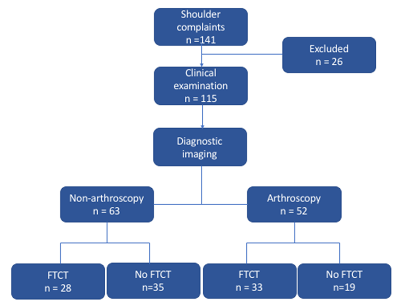
Figure 1: Diagram demonstrating the selection procedure of our study. n = the number of patients assessed at each stage.
141 patients were referred by their GPs to a tertiary shoulder unit with suspected rotator cuff pathology. 115 patients entered the study after exclusion criteria were applied. Of the 52 patients who required arthroscopic surgery, 33 patients had full-thickness tears while 19 patients did not. However, on clinical examination 36 patients were thought to have FTCT, indicating that there were 3 falsely positive clinical examinations in this cohort. Patient demographics are displayed in Table 1. The non-arthroscopy cohort had an older age range, reflecting clinical practice to refer younger patients for shoulder surgery while managing older patients conservatively.
| Arthroscopy cohort | Non-arthroscopy cohort | |
| Number of patients | 52 | 63 |
| Age range (years) | 40 - 68 (Mean = 51) | 62 - 79 (Mean = 68) |
| Gender | 33 males and 19 females | 39 males and 24 females |
| Duration of symptoms (months) | 6 - 18 (Mean = 9) | 6 - 24 (Mean = 10) |
Table 1: Patient demographic information
The diagnostic value of overall clinical examination for FTCT is displayed in Table 2. The results show that as a whole, clinical examination has statistically significant diagnostic value in the diagnosis of FTCT. With a very high sensitivity and NPV, clinical examination was particularly successful at ruling-out FTCT in our sample population. Overall, physical examination has a diagnostic accuracy of 90%.
| Test | Sensitivity | Specificity | Positive Predictive Value | Negative Predictive Value
| Accuracy
| P-value |
| Overall clinical examination | 0.97 | 0.79 | 0.89 | 0.94 | 0.90 | <0> |
Table 2: The diagnostic values of overall clinical examination for FTCT.
The reference standard was shoulder arthroscopy. The P-value was calculated using Fisher’s exact test comparing clinical test results to arthroscopic findings
The diagnostic value of the 5 different clinical tests for FTCT are displayed in Table 3. The Empty Can test, Resisted External Rotation test and External Rotation Lag test assess the integrity of the supraspinatus, infraspinatus and teres minor. The Belly-press test and Lift-off test assess the subscapularis.Both the Resisted External Rotation test and Empty Can test were very accurate when ruling-out FTCT: they both have a sensitivity of 97%. However, the Resisted External Rotation test had a higher specificity and PPV than the Empty Can test, indicating it was more capable of ruling-in tears. The External Rotation Lag test had the highest specificity of the 3 tests (100%). However, it had a sensitivity of 17% and poor diagnostic accuracy. A combination of tests had higher specificity (P-value <0> Positive Predictive Value Negative Predictive Value Accuracy Test Sensitivity Specificity P-value Empty Can test 0.97 0.83 0.86 0.95 0.90 <0> Resisted External Rotation test 0.97 0.91 0.93 0.95 0.94 <0> External Rotation Lag test 0.17 1.00 1.00 0.49 0.54 0.0586 Lift-off test 1.00 1.00 1.00 1.00 1.00 <0> Belly-press test 1.00 1.00 1.00 1.00 1.00 <0>
Table 3: The diagnostic values of individual clinical examinations for FTCT.
The reference standard was shoulder arthroscopy. The P-value was calculated using Fisher’s exact test comparing clinical test results to arthroscopic findings.
Our results show that full-thickness tears of the subscapularis are not common; only 4 out of the 33 FTCT were tears of the subscapularis. Therefore, the Belly-press test and Lift-off test may have limited utility as a universal screening tool for FTCT due to the low incidence of positive test results. However, both tests had a sensitivity and specificity of 100%: all 4 patients that had full-thickness subscapularis tears clinically were confirmed to have full-thickness subscapularis tear arthroscopically. In this respect, both the Belly-press test and the Lift-off test are highly accurate at screening for full-thickness subscapularis tears.
Additionally, we analysed the patients in the non-arthroscopy group who were clinically positive for FTCT. Of the 63 patients who did not have arthroscopy, 25 had FTCT diagnosed clinically while 28 had FTCT diagnosed by imaging modalities, indicating that there were 3 falsely negative clinical examinations in this cohort. The diagnostic values of clinical examination for FTCT in this cohort are displayed in Table 4. Even when diagnostic imaging was used as the reference standard, clinical examination had high diagnostic value for the assessment of FTCT.
| Test | Sensitivity | Specificity | Positive Predictive Value
| Negative Predictive Value
| Accuracy
| P-value |
| Overall clinical examination | 0.89 | 1.00 | 1.00 | 0.92 | 0.95 | <0> |
Table 4: The diagnostic values of overall clinical examination for FTCT.
The reference standard was diagnostic imaging. The P-value was calculated using Fisher’s exact test comparing clinical examination findings to the results of diagnostic imaging.
Our results show that clinical examination (using the combination of special tests described in our methods) is an accurate diagnostic tool for the diagnosis of FTCT when compared to arthroscopy. This remained true in the non-arthroscopy cohort when our reference standard was diagnostic imaging.
One test to rule them all?
As the current literature stands, there is little evidence recommending one special test over another in the assessment of rotator cuff disease. Key systematic reviews have worked to identify the most accurate individual clinical test but variation in their methodology alongside poor quality primary studies has prevented consensus [4, 5, 15-18].
In agreement with our findings, the Empty Can test, External Rotation Lag test and Belly-press test have been shown to have diagnostic utility by these systematic reviews. Individually, our results show that the Resisted External Rotation test was the best test to use as a screening tool for FTCT. Alternatively, if any one of the External Rotation Lag test, Belly-press test or Lift-off test was positive we were quite likely to find a tear on arthroscopy. Although highly specific, the External Rotation Lag test showed poor sensitivity and NPV in our study. Notably, the 5 patients in the arthroscopic cohort with a positive External Rotation Lag test all had large cuff tears. Similarly, the 11 patients with a positive External Rotation Lag test in the non-arthroscopic cohort all had large cuff tears on MRI. Therefore, it is possible that this test is particularly sensitive at detecting large to massive rotator cuff tears.
Do multiple tests increase diagnostic accuracy?
While individual tests may fall short, our results show that a thorough clinical examination by an experienced orthopaedic surgeon still provides a highly accurate diagnosis. Moreover, an experienced clinician is unlikely to rely on merely one manoeuvre to assess the shoulder: the diagnostic process involves the amalgamation of several clinical tests to arrive at a conclusion.
Sgroi et al. demonstrated that diagnostic capability increased when 3 or more clinical tests were used to identify tears in the supraspinatus muscle. The authors went on to establish the same when 2 or more clinical tests were used to identify tears in the infraspinatus muscle [10,19] . In agreement with our findings, the literature demonstrates that combining multiple clinical tests improves their diagnostic value in the assessment of rotator cuff disease [20,21].
Murrell et al. found that a combination of 3 clinical features: weakness in abduction, weakness in external rotation and impingement, had a PPV of 98% when predicting rotator cuff tears [22]. Notably, patients had the same PPV if they had 2 out of 3 of these clinical features and were also over the age of 60 years. There is limited exploration of the diagnostic value of patient information for rotator cuff tears in the literature. However, increasing age and night pain are examples of patient characteristics & history which have significant diagnostic value in this condition [23,11]. In future research, it would be interesting to evaluate the diagnostic accuracy of history-taking and physical examination in combination.
Diagnostic imaging in rotator cuff disease
Clinicians rely heavily on MRI/ultrasonography for the diagnosis of rotator cuff disease. However, there are disadvantages to this diagnostic approach. Due to the high prevalence of asymptomatic rotator cuff tears in the population, interpreting diagnostic imaging can be complex. This is particularly true in older patients; over 50% of asymptomatic individuals have rotator cuff tears on diagnostic imaging when they are over 65 years of age [24] . Furthermore, MRI is expensive and it cannot be used in obese, claustrophobic patients or patients with devices such as pacemakers. In comparison with MRI, ultrasonography is an inexpensive and convenient method of evaluating the shoulder. However, the type of ultrasound equipment as well as operator experience affect its accuracy in identifying tears [25].
In light of these disadvantages, we suggest that elderly patients who are being scheduled for surgical rotator cuff repair should only have diagnostic imaging after failing conservative management. Patients with tears in the non-arthroscopic cohort were treated non-operatively as they were relatively older than patients scheduled for arthroscopy (Table 1). Nevertheless, clinical examination had a high overall diagnostic accuracy in both cohorts, regardless of the age difference. Our results show that an accurate diagnosis for FTCT can be made clinically by a skilled orthopaedic surgeon in secondary care. There were 3 falsely negative patients who displayed FTCT on imaging but not examination. However, these were small full-thickness tears in relatively older patients and were treated non-operatively leading to asymptomatic patients.
Both MRI and ultrasonography have been shown to have similar levels of efficacy in identifying rotator cuff tears. A Cochrane review commented that MRI had an estimated sensitivity of 98% and specificity of 79% 26. This is nearly identical to our results for the sensitivity and specificity of overall clinical examination, which were 97% and 79% respectively. However, ultrasonography can have a sensitivity and specificity as low as 66% and 54% respectively [27].
Similar to clinical examination, both imaging modalities are poorer at identifying partial-thickness tears than full-thickness tears [25 28]. However, the majority of the time partial- thickness tears do not need surgical management. For these reasons, we aimed to diagnose only full-thickness tears on clinical examination.
Clinical relevance and future implications
Hanchard et al. expand on the merits of having sensitive and specific physical manoeuvres for the diagnosis of musculoskeletal conditions [5]. They do not require additional time and resources to organise and they can be completed during routine secondary care consultation with an orthopaedic consultant, yielding immediate results. Since they rely on reproducing symptoms, they will not superfluously detect asymptomatic tears.
Although clinical examinations can be conducted in primary or secondary care, we suggest that they may only be diagnostically accurate when performed by specialists; in this way they can reliably be used as a substitute for diagnostic imaging. As one author eloquently puts it: “Respect must be shown for the physical exam [8].
Like primary studies which have come before us, there are some key limitations to our methodology. We have used the QUADAS tool to aid our retrospection [29]. Firstly, we acknowledge that the diagnostic value of a test transforms with changes in disease prevalence. Since our sample size was highly selective, it is likely the prevalence of FTCT was higher than in the general population [30]. Secondly, our shoulder surgeon was not blinded to the results of the clinical tests, which may have led to bias when the arthroscopy was performed. This may have caused him to overestimate the diagnostic value of clinical examination.
Clinical tests for the diagnosis of FTCT have high sensitivity and specificity, comparable to imaging modalities explored in the literature. When used in combination, these tests are valuable and reliable diagnostic tools. In conclusion, over-reliance on MRI and ultrasonography may not be justified, particularly in a relatively older population when a rotator cuff tear repair is not scheduled.
This study was conducted in accordance with the Declaration of Helsinki. AP and RP did the literature search, analysed and interpreted the data and wrote the manuscript. NA and RP did the study design and collected and analysed the data. All authors contributed to final approval of draft of the manuscript. All listed authors meet authorship criteria. This research did not receive any specific grant from funding agencies in the public, commercial, or not-for-profit sectors. No author has any competing interests to declare. Data privacy laws have been followed. Patients or the public were not involved in the design, or conduct, or reporting, or dissemination plans of our research. The manuscript is an accurate, and transparent account of the study.
Dear Editorial Team, Clinical Medical Reviews and Reports. My experience with the journal was highly positive. The peer-review process was rigorous, constructive, and completed in a timely manner. The reviewers provided valuable comments that helped improve the quality and clarity of our manuscript. The editorial office was professional, responsive, and supportive throughout all stages of the publication process. Communication was clear and efficient, and any questions were addressed promptly. Overall, I found the journal to maintain high scientific standards and an excellent publication workflow. I would be pleased to consider submitting future work to this journal. Best wishes from, Elena Popa.

It was my pleasure to submit my testimonial concerning the Reviewer Board of our Scientific Journal “Brain and Neurological Disorders”. The Reviewers focused on some modifications and their contribution was helpful. The ladies of our Editorial Office were also supported my efforts. It was my honor to have such a co-operation and I am looking forward for more collaboration.

Dear Grace Pierce, Editorial Coordinator of Journal of Clinical Research and Reports, Thank you for the speedy and efficient peer review process. I appreciate the fact that your peer reviewers do not take months to respond like with some other journals. I would also like to thank the editorial office for responding quickly to my questions. It is an excellent journal. I plan to submit more manuscripts in the future. Best wishes from, Robert W. McGee

Dear Grace Pierce, Editorial Coordinator of Journal of Clinical Research and Reports, Working with you and your team on our recent publication in JCRR has been a truly wonderful and enjoyable experience. The responses were prompt, and the reviewers were patient, constructive, and highly professional. One reviewer in particular gave me the feeling that a professor was carefully reading and commenting on my coursework, which was deeply touching. The entire process was straightforward and hassle‑free, with no tedious online forms to complete. I highly recommend this journal. Best wishes from, DR Aibing Rao, Head of R&D

I Appreciate the Opportunity to Share my Experience with the Journal of Clinical Research and Reports. The peer review process was timely and constructive, and the feedback provided helped improve the quality of our manuscript. The editorial office was professional, responsive, and supportive throughout the process, ensuring smooth communication and efficient handling of the submission. Overall, it was a positive experience collaborating with your team.

Dear Mercy Grace, Editorial Coordinator of Obstetrics Gynecology and Reproductive Sciences, We would like to express our gratitude for your help at all stages of publishing and editing the article. The editors of the magazine answer all the necessary questions and help at every stage. We will definitely continue to cooperate and publish other works in the Obstetrics Gynecology and Reproductive Sciences! Best wishes from, Alla Konstantinovna Politova,
