
Case Report | DOI: https://doi.org/10.31579/2690-1897/114
Maxillofacial surgery department, hospital of specialities, Rabat-Morocco
*Corresponding Author: Othmane bouanani, Maxillofacial surgery department, hospital of specialities, Rabat-Morocco.
Citation: Othmane bouanani*, Rajaa Elazzouzi and Malik Boulaadas. (2022). Clear cell variant of squamous cell carcinoma of skin: A case report and review of the literature Journal of Surgical Case Reports and Images 5(4); DOI: 10.31579/2690-1897/114
Copyright: © 2021, Othmane bouanani, This is an open access article distributed under the Creative Commons Attribution License, which permits unrestricted use, distribution, and reproduction in any medium, provided the original work is properly cited.
Received: 18 May 2022 | Accepted: 15 June 2022 | Published: 20 August 2022
Keywords: carcinoma ; skin tumor ; clear cell carcinoma
Clear cell squamous cell carcinoma (SCC) is a rare variant of SCC of skin in which ultraviolet radiation has been suggested as possible etiology. We report a case of a 51-year-old male patient who presented with a 4 months history of a non-healing ulcer on the left side of his face. Histopathology and immunohistochemistry studies showed Clear cell variant of squamous cell carcinoma. This case showed an aggressive and bizarre clinical presentation but more report of cases are needed to have a better characterization of the clinical presentation and prognosis of this variant of this squamous cell carcinoma variant.
Squamous cell carcinoma (SCC) is the second most common type of skin cancer, with basal cell carcinoma being the most common [1]. SCC is predisposed for by excessive ultraviolet light exposure, hence, its association with advancing age and cumulative sun exposure, exposed anatomic sites and the highest incidence in sunny geographic localities [2]. Several histological subtypes of SCC have been described.
Clear cell SCC is a rare entity and only few cases were previously reported in the literature [3]. We present a case of clear cell SCC of skin with an exophytic mass on the face in order to add to the scarce literature of this rare variant of SCC.
A case of a 51year-old male patient, a farmer by profession, known as diabetic with poor medication adherence with no other known systemic disease or history of tobacco or alcohol use.
Who presented at the emergency room of oral and maxillo-facial surgery on account of a non-healing ulcer on the left side of his face of 4 months duration. The lesion was said to have started as a small firm painless swelling in the left infra-orbital region, which gradually increased in size until 2 months later when it became ulcerated with associated pain.
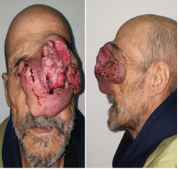
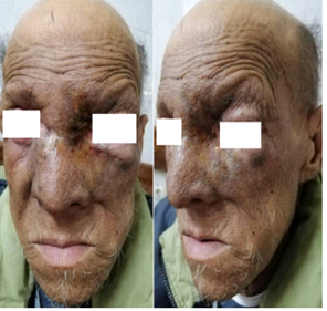
Physical examination revealed a cachectic and pale man with an obvious facial asymmetry due to a fleshy exophytic ulcerative budding mass on the left side of the face. The mass measured about 14 cm in its widest diameter and occupied the left orbito-naso jugale region such that the left eye globe could not be visualized involving also the entire nasal pyramid. With the presence of cervical lymphadenopathy Figure 1.
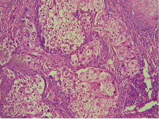
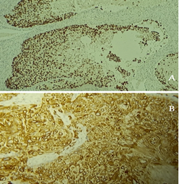
A biopsy was performed, the histopathologic evaluation supplemented by an immunohistochemical study revealed a Clear cell variant of squamous cell carcinoma. Microscopic examination of the biopsy specimen revealed a squamous nature of the tumor's profileration with hydropic appearance of tumor cells's cytoplasm. Keratinizing type. Immunohistochemistry then revealed positive staining results for Anti-Cytokeratin 7 antibody (Anti CK5/6), Anti-p63 Antibody (Anti P63). and negative results for Anti-Cytokeratin 7 antibody (Anti CK7). Figures 2-3
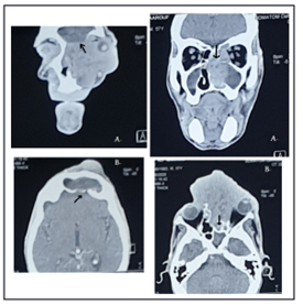
A cerebral and cervico-facial CT scan was performed showing an enormous aggressive facial tumor with sinonasal and intracranial extension. Figure 4
After a multidisciplinary consultation meeting, the decision was to set up a treatment with chemoradiotherapy. 6 months later, a subtotal clinical regression of the tumor was noted, yet the patient did not show up for the CT appointment and was lost to follow-up .Figure 5
Squamous cell carcinoma (SCC) is the second most frequent form of skin cancer superseded in occurrence only by basal cell carcinoma. However, some argue that an actinic keratosis should be considered as an SCC that is superficial, If so, then SCC could be considered the most common type of skin cancer [4].
The disease has been linked to immunosuppression, arsenic exposure, radiation, chronic ulceration, and human papillomavirus (HPV) infection [5].
There exists a wide histopathologic diversity of SCCs, many of which are associated with markedly different clinical behaviors. The histologic subtype has also been considered as a factor in determining the prognosis. Several histologic subtypes of SCC are described, That can range from indolent tumors with low metastatic potential, to remarkably aggressive tumors with high invasive potential [6-9].
We note, acantholytic, spindle cell, verrucous, clear cell, papillary, signet ring, pigmented, and desmoplastic SCC.We present in our review a case of clear cell SCC of skin with an exophytic mass on the face in order to add to the scarce literature of this rare variant of SCC.
Clear-cell SCC is an extremely rare variant of SCC. It is commonly referred to as hydropic SCC due to the extensive hydropic degeneration of neoplastic cells, and the accumulation of intracellular fluid.
It was first described by Kuo in 1980 [10] as a variant of SCC with extensive hydropic change. The hydropic degeneration of neoplastic cells and the accumulation of intracellular fluid, not the accumulation of glycogen, lipid, or mucin, results in its clear cell appearance.,
Kuo reported six cases occurring in the head and neck region of elderly Caucasian males with histories of excessive sun exposure. Clinically, the lesions appear as nodules or ulcerated masses, and can easily be confused with sebaceous neoplasms [1].
Other differential diagnosis include clear cell acanthoma, clear cell hidradenoma, clear cell hidradenocarcinoma, tricholemmoma, pilar tumor, balloon cell nevus, balloon cell melanoma, and metastatic renal cell carcinoma. The clear cells of clear cell acanthoma, clear cell hidradenoma, clear cell hidradenocarcinoma, tricholemmoma, pilar tumor all have a high content of cytoplasmic glycogen [10].
Kuo further classified the six cases of clear cell carcinoma into the major histologic types: Keratinizing (type I)
Nonkeratinizing (type II)
Pleomorphic (type III).
Type I is characterized by sheets or islands of tumor cells in a fibrotic stroma, with a sparse lymphocytic infiltrate. The tumor cells appear clear, with peripherally displaced nuclei, Some cells may also appear to have a “bubbled” cytoplasm. Distinguishing features include foci of keratinization and keratin pearls.
Type II lesions are predominantly dermal in origin, with no clear connection to the overlying epidermis, Unlike type I, this variant does not demonstrate evidence of keratinization. is characterized by parallel or anastomosing cords of tumor cells in a compressed fibrotic stroma with a dense, inflammatory infiltrate composed of plasma cells and lymphocytes. Central necrosis may be evident within tumor cords. The tumor cells appear to have central nuclei with finely reticulated clear cytoplasm.
Type III demonstrates marked pleomorphism with extensive vascular and perineural invasion. Foci of squamous differentiation and microcysts with acantholytic tumor cells may be seen.
In all three types, none has evidence of either glycogen or mucin in tumor cells [10].
we note a Keratinizing type in our case by the presence of dyskeratosis type of the squamous maturation. It is difficult to determine the prognosis of clear cell SCC based on the few cases that have been reported in the literature.
In our case, the patient received chemoradiotherapy, 6 months later, a subtotal clinical regression of the tumor was noted , yet the patient did not show up for the CT appointment and was lost to follow-up.
Clear cell squamous cell carcinoma (SCC) is an extremly rare and agressive variant of SCC of skin, with only few cases were previously reported in the literature which makes a treatment of choice and the prognosis hard to determine.
Dear Editorial Team, Clinical Medical Reviews and Reports. My experience with the journal was highly positive. The peer-review process was rigorous, constructive, and completed in a timely manner. The reviewers provided valuable comments that helped improve the quality and clarity of our manuscript. The editorial office was professional, responsive, and supportive throughout all stages of the publication process. Communication was clear and efficient, and any questions were addressed promptly. Overall, I found the journal to maintain high scientific standards and an excellent publication workflow. I would be pleased to consider submitting future work to this journal. Best wishes from, Elena Popa.

It was my pleasure to submit my testimonial concerning the Reviewer Board of our Scientific Journal “Brain and Neurological Disorders”. The Reviewers focused on some modifications and their contribution was helpful. The ladies of our Editorial Office were also supported my efforts. It was my honor to have such a co-operation and I am looking forward for more collaboration.

Dear Grace Pierce, Editorial Coordinator of Journal of Clinical Research and Reports, Thank you for the speedy and efficient peer review process. I appreciate the fact that your peer reviewers do not take months to respond like with some other journals. I would also like to thank the editorial office for responding quickly to my questions. It is an excellent journal. I plan to submit more manuscripts in the future. Best wishes from, Robert W. McGee

Dear Grace Pierce, Editorial Coordinator of Journal of Clinical Research and Reports, Working with you and your team on our recent publication in JCRR has been a truly wonderful and enjoyable experience. The responses were prompt, and the reviewers were patient, constructive, and highly professional. One reviewer in particular gave me the feeling that a professor was carefully reading and commenting on my coursework, which was deeply touching. The entire process was straightforward and hassle‑free, with no tedious online forms to complete. I highly recommend this journal. Best wishes from, DR Aibing Rao, Head of R&D

I Appreciate the Opportunity to Share my Experience with the Journal of Clinical Research and Reports. The peer review process was timely and constructive, and the feedback provided helped improve the quality of our manuscript. The editorial office was professional, responsive, and supportive throughout the process, ensuring smooth communication and efficient handling of the submission. Overall, it was a positive experience collaborating with your team.

Dear Mercy Grace, Editorial Coordinator of Obstetrics Gynecology and Reproductive Sciences, We would like to express our gratitude for your help at all stages of publishing and editing the article. The editors of the magazine answer all the necessary questions and help at every stage. We will definitely continue to cooperate and publish other works in the Obstetrics Gynecology and Reproductive Sciences! Best wishes from, Alla Konstantinovna Politova,
