

Research Article | DOI: https://doi.org/10.31579/2690-8794/331
1Department of Internal Medicine I, Cancer Research, Medical University of Vienna, Vienna, Austria.
2Department of Internal Medicine II, Division of Pulmonary Medicine, Medical University of Vienna, Medical School, Vienna, Austria.
3Pulmonary Cell Research, Department Biomedicine, University Hospital Basel, Switzerland.
4Ordensklinikum Linz Elisabethinen, Dept. Pneumology, Linz, Austria.
†In Memoriam
*Corresponding Author: Wilhelm Mosgoeller. MD, Medical University of Vienna, Borschkegasse 8a A-1090 Vienna, Austria.
Citation: Wilhelm Mosgoeller, Ventzislav Petkov, Bernhard Burian, Michael Roth, Christopher Lambers, et al, (2026), Chronic Obstructive Pulmonary Disease: Effective treatment with Vasoactive Intestinal Peptide (VIP), Clinical Medical Reviews and Reports, 8(8); DOI:10.31579/2690-8794/331
Copyright: © 2026, Wilhelm Mosgoeller. This is an open access article distributed under the Creative Commons Attribution License, which permits unrestricted use, distribution, and reproduction in any medium, provided the original work is properly cited.
Received: 29 June 2026 | Accepted: 20 July 2026 | Published: 18 August 2026
Keywords: COPD; chronic inflammation; lung biopsy; vasoactive intestinal peptide
Rationale: Chronic obstructive pulmonary disease (COPD) is characterized by progressive airflow limitation and chronic inflammation including the increased expression of Interleukin 8 (IL-8) and tumor necrosis factor (TNF, previously known as tumor necrosis factor alpha). We describe a novel physiological role of the Vasoactive Intestinal Peptide (VIP) and its therapeutical potential in COPD. Our rationale is based on a deficiency of VIP in the serum of COPD patients.
Objectives: The aim of this study was to evaluate a new therapy of COPD.
Methods: Molecular biological and immunochemical methods were used to characterize the action of VIP at the cellular level. A double-blind, placebo-controlled, randomized study was undertaken to evaluate the potential clinical role of VIP in COPD.
Measurements and Main Results: VIP treatment downregulates the expression of IL-8 and TNF that coincides with the inhibition of transcription factors AP-1, Stat3 and NF-κB. VIP treatment improves lung function and prognostic parameters of COPD. In a clinical study with 34 COPD patients, forced expiratory volume in 1s significantly improved with VIP by 0.107l and vital capacity by 0.155l compared with placebo (P<0.01).
According to the short 36-item questionnaire and to St. George’s respiratory questionnaire, the improvement in quality of life was greater with VIP (4.08; -5.25) than with placebo (0.20; 1.72). Similarly, exercise capacity increased by 24.1 m (P<0.05) in the VIP group as compared to placebo (-8.6m) using the six minutes walking test.
Conclusions: This study is the first to show the immunoregulatory effect of VIP in COPD patients, and supports the notion of inhaled VIP as an attractive future therapy to dampen exaggerated immune responses in chronic lung disorders. Our data provide “proof of concept” for further investigation of VIP and its potential role in COPD.
Chronic obstructive pulmonary disease (COPD) is the third leading cause of death worldwide, causing 3.4 million deaths in 2023, approximately 6% of all global deaths. COPD is the overall term for a group of chronic conditions that are associated with the obstruction of lungs' airways, usually referring to the following disorders: Chronic bronchitis, Bronchiectasis and Emphysema. The most common symptoms of COPD include shortness of breath, chronic coughing, chest tightness, greater effort to breathe, increased mucus production and frequent clearing of the throat.
The mainstay medication consists of anticholinergic and beta-adrenergic bronchodilators. Addition of corticosteroid therapy may enhance bronchodilator responses and reduce exacerbations. An important feature of COPD is the ongoing chronic inflammatory process in the airways as indicated by the current GOLD (Global Initiative for Chronic Obstructive Lung Disease) definition. Neutrophil granulocytes, macrophages, and T-cells are involved in the cellular mechanisms of inflammation in COPD and are associated with an increased release of pro- and anti-inflammatory mediators such as e.g. IL-8 and TNF, a protease-antiprotease imbalance, oxidative stress, viral infections, and a genetic predisposition (e.g. Alpha-1 Antitrypsin Deficiency) [8, 13, 14, 23, 26, 30, 34, 35, 36, 39, 40, 42, 43, 45, 46].
We were looking for a drug that provides the combined potential advantages of lowering inflammation of the lung tissues, lowering pulmonary arterial pressure, reducing bronchoconstriction, improving blood circulation to the heart and lung, enhancing healing of bronchial epithelial cells, and improving the cardiovascular deficiencies in COPD.
Vasoactive Intestinal Peptide (VIP) is an abundant biologically active peptide endogenous in humans as well as in other mammalian species. It is produced by neurons in the peripheral and central nervous system, by endocrine cells, T-lymphocytes, and B-lymphocytes. This natural peptide is one of the signal molecules of the neuroendocrine-immune network comprising vasodilating, anti-proliferative, anti-inflammatory, and immune-regulatory features. Its predominant biological activity is performed in the lungs, and a vast body of experimental, pharmacological as well as clinical evidence suggests VIP to be an attractive candidate for the treatment of COPD [3, 9, 11, 12, 16, 18, 53, 54].
VIP’s biological effects are mediated through the drug’s pharmacological activity on VPAC1, VPAC2, and PAC-1 receptors (7-membrane-spanning G-protein coupled receptors) at the transcriptional level via modulation of NF-κB, AP-1, Stat3, and cAMP responsive element (CRE)-binding, or ets-2 complexes in target cells [5, 12, 50]. The presence of VPAC receptors has been shown on airway epithelial cells, on macrophages surrounding capillaries and in the subintima of pulmonary arteries and veins [5, 50]. Stimulation of VPAC receptors activates pathways which have been shown to mediate the action of prostacyclins, nitric oxide, and phosphodiestrase inhibitors [19, 52].
We and others [21], noted that in patients with the lowest VIP serum levels, a more rapid formation of COPD was observed. The smallest values of VIP and the greatest changes in cardiopulmonary parameters were found in the cohort of COPD-patients with concomitant hypertension [21].
As a preferred route of administration, mouth inhalation was chosen. Previously we showed that after intravenous administration VIP is rapidly distributed to the lungs, where it gets completely metabolized into biologically inactive parts by neutral endopeptidase, mast cell tryptase and mast cell chymase [37]. In general, inhaled drugs intended for lungs act quickly, minimize undesired negative side effects, avoid the hepatic first-pass metabolism, and act locally. As the size variability among adult lungs is smaller than the overall body size variability, the dosing reliability is also improved when inhaling.
We and others have shown that inhaled VIP decreases pulmonary artery pressure and pulmonary vascular resistance in patients with pulmonary hypertension, and exerts immunoregulatory and anti-inflammatory effects in sarcoidosis [37, 59, 60]. However, the exact mode of action of VIP is not yet completely understood.
After producing cigarette smoke-induced COPD in a mouse model, and after treatment of those mice with VIP, the levels of antioxidant-related factors (malondialdehyde (MDA) and superoxide dismutase (SOD), fibrotic factors (hydroxyproline (HP) and TGF-β), pro-inflammatory cytokines (TNF, IL-1β, and IL-6), and inflammation were diminished and controlled by VIP. The anti-inflammatory, anti-fibrotic, and anti-oxidant properties of VIP may be effective therapy in COPD [6].
Furthermore, the anti-inflammatory properties of IK312532 respirable powder (RP) - a stabilized VIP derivative - were characterized in an asthma/COPD-like animal model. Marked inflammatory events in the lung were observed after OVA-RP challenge in rats as evidenced by significant increase of inflammatory biomarkers such as eosinophil peroxidase (EPO), myeloperoxidase (MPO) and lactate dehydrogenase (LDH). However, intratracheal administration of IK312532-RP led to significant attenuation of plasma EPO, MPO and LDH activities, as well as significant reduction of recruited inflammatory cells in bronchoalveolar lavage fluid, especially macrophages and eosinophils. Thus, inhalable powder formulation of IK312532 exerts its anti-inflammatory activity by suppressing granulocyte recruitment to the lung and epithelial hyperplasia, followed by the reduction of cytotoxic peroxidases [29].
Alveolar macrophages (AM) are key players in the pathogenesis of COPD and contribute to the severity and progression of the disease. AM from COPD patients show a strong VPAC1 expression which exceeds VPAC2 expression. A similar receptor expression pattern was also observed in lipopolysaccharide (LPS)-activated monocyte-derived macrophages (MDM) from healthy volunteers and COPD patients. VIP has been shown to down-regulate IL-8 secretion significantly in MDM after LPS stimulation. The response to VIP was similar in MDM from COPD patients and healthy volunteers. Our previous results indicate that VPAC1 up-regulation in macrophages is a common mechanism in response to acute and chronic pro-inflammatory stimuli. Although VPAC1 up-regulation is dominant, both receptor subtypes are necessary for optimal anti-inflammatory signaling. The high VPAC1 expression in AM may reflect the chronic pro-inflammatory environment found in the lung of COPD patients. Treatment with VIP may help to decrease the chronic inflammation in the lung of COPD patients [4].
In view of what is known about VIP we correlated the serum concentration of VIP with the disease state of COPD and examined some of its effects on the transcription and expression of two essential cytokines in COPD both in vitro and in vivo. Moreover, we identified the essential therapeutic role of VIP in COPD patients in a small double-blind placebo controlled clinical trial.
VIP serum concentration
VIP was determined by radioimmunoassay (123I-VIP RIA) in healthy controls n=48, COPD patients (GOLD II) n=12, COPD (GOLD III) n=16, COPD (GOLD IV) n=14 [37].
Cell isolation and characterization
We cultured lung fibroblasts and bronchial smooth muscle cells (BSMC). All primary cell lines were isolated from resected lung biopsies of COPD patients undergoing therapeutic surgery for other reasons, after agreement of the local ethical committee and written informed consent of the patients. Human lung fibroblasts were isolated from bronchial biopsies and cultured as described earlier [48].
BSMC were isolated and grown from resected bronchi of the above patients as previously described, and BSMC were characterized by positive immuno-staining for a-smooth muscle actin and calponin [20].
Cell treatment
Sub-confluent (80%) cell cultures were serum deprived for 24 hours in medium without serum. The cells were then pre-incubated for 30 minutes with VIP (10-6 M - 10-12 M) and then stimulated with the respective growth medium with VIP added at various concentrations.
Total RNA extraction and RT-PCR and Real-Time RCR-analysis mRNA was obtained from biopsy by TRIZOL using standard protocols and was directly transcribed into c-DNA using a reverse transcription kit (Advantage RT-for-PCR Kit) according to instructions of the manufacturer and was stored at minus 80°C until analysis.
Relative mRNA expression was determined by the ACT method as previously described [25]. The ACT numbers indicate the difference in amplification cycles between target and housekeeping (18s) gene.
Immunoblotting
Nuclear/cytosolic proteins were prepared with NE-PER kit according to the manufacturer protocol. Protein extracts were then size- fractionated by SDS-PAGE electrophoresis and transferred onto nitrocellulose membranes as described previously [41] and protein transfer was confirmed by Ponceau staining.
Electrophoretic mobility shift assay
Nuclear protein extracts were prepared after 30 min by NE-PER extraction kit and the protein content was determined by Bradford assay. The oligonucleotide was labeled using [Y33P]-dATP (3,000 Ci/mmol) and a T4 polynucleotide kinase. Samples were size fractionated by electrophoresis in a non-denaturing 4% polyacrylamide gel [41].
Cytokine enzyme linked immuno sorbent assay (ELISA)
Cytokines (IL-8 and TNF) were quantified in cell culture medium samples collected at below indicated time points form the same cell culture vessel for each cell line. Commercially available ELISA for IL-8, and TNF were used following the instructions of the manufacturer.
Immunohistochemistry for IL-8 and TNF
Rehydrated paraffin sections were stained for IL-8 and TNF according to the procedures as described previously [37].
This parallel-group, double-blind, placebo-controlled, randomized, monocenter study was undertaken at the Medical University of Vienna, Austria. The clinical study was clared by the positive ethics approval from the Ethics Committee of the Medical University of Vienna, reference number 168/2003 and announced by the ClinicalTrials.gov ID NCT00464932.
The randomization sequence was generated by Rancode in a blinded manner; no person involved in data analysis had knowledge of the randomization sequence. Treatment was assigned by the investigators with sequential study numbers according to a block randomization list in ratios of 1:1 (VIP:placebo). VIP was synthetically manufactured. It was applied by a 10-minute inhalation (50 μg VIP diluted in 3 ml of 0.9% NaCl solution), four-times daily, using a mesh nebulizer providing an average mass median aerodynamic diameter (MMAD) VIP particle size of 3 μm to allow for a deep-lung deposition of the drug.
The trial substance was an “add-on” treatment; concomitant respiratory medications allowed throughout the study were short- and long-acting ß-mimetics, long-acting anticholinergics, inhaled corticosteroids and theophyllines at a constant daily dose. Patients could receive a course of oral corticosteroids for treatment of exacerbations during the treatment phase.
The primary outcome variables were exercise capacity measured using the six-minute walking test (6MWT), Borg dyspnea score and health-related quality of life (HRQL) indicators measured by means of the short 36-item questionnaire (SF-36) developed for medical outcomes studies and St. George’s respiratory questionnaire (SGRQ). Findings were calculated as the change from base-line to the exit.
Secondary outcome measures were change from base-line in post-bronchodilator FEV1, post-bronchodilator inspiratory vital capacity (IVC) and number of COPD exacerbations. We recorded adverse events as part of the safety assessment using standard International Conference on Harmonisation Guidelines for Good Clinical Practice (ICH-GCP). Pulmonary function tests were done at rest before treatment onset (base-line), and at week 12 of the treatment period as recommended by the American Thoracic Society [44].
All patients were recruited from an outpatient setting. Inclusion criteria were: history of COPD >12 months, as defined by Global Initiative for chronic Obstructive Lung Disease (GOLD) [35], 40 years or older; current smoker or ex-smoker with a smoking history of >10 pack-years; post-bronchodilator FEVi of 20-80% of predicted value; post-bronchodilator IVC ratio >70%; reversibility of FEV1 of Less-than sign12 % and/or Less-than sign200 ml after 400 pg inhaled salbutamol; and stable clinical disease status with no change in COPD treatment during the last 4 weeks. Patients were excluded from study enrolment if they were diagnosed with asthma or other relevant lung diseases (e.g. lung cancer, bronchiectasis); if they had a recent exacerbation that required a course of systemic corticosteroids, emergency room treatment, or hospital admission within 4 weeks before the randomization. Patients were also excluded from study enrolment if they had a respiratory tract infection within 4 weeks before the randomization or known alpha-1-antitrypsin deficiency. The study was approved by the local ethical committee and each patient gave informed consent to participate in the trial and investigations according to institutional guidelines and to the Helsinki principles.
After completion of the case report forms, data capture was done with the double entry technique. Thereafter, the data were analyzed descriptively using the SPSS (IBM Statistics V.8) software package.
For analysis of the recruitment allocation, we compared means and standard deviation of relevant parameters (see table 1) of the patient groups.
Data analysis was done with SPSS, Version 12. Baseline demographic, lung function variables, quality of life are presented as means ± SD. Variables measured at baseline and during VIP inhalation in the same patients were compared by Wilcoxon signed rank test (two-sided) for paired data. P values below 0.05 were considered statistically significant.
VIP serum levels were compared between the groups by means of the Students T-test, and by the Wilcoxon signed rank test when the data did not allow for computing the mean and standard deviations.
We determined the serum concentration of VIP in humans by radioimmunoassay. As shown in Figure 1, patients with COPD show significantly reduced levels of VIP as compared to controls whereby the VIP levels correlated inversely with disease stage.
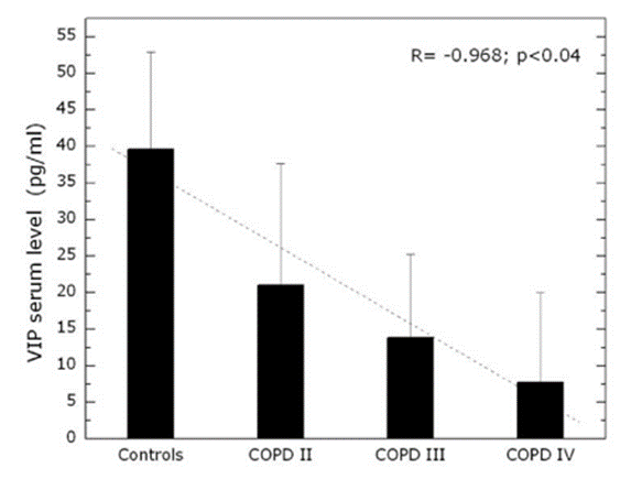
Figure 1: VIP serum concentration as determined by RIA of healthy controls and COPD patients. Healthy controls n=48, COPD (GOLD II) n=12, COPD (GOLD III) n=16, COPD (GOLD IV) n=14.
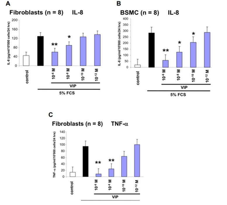
BSMC and fibroblasts obtained from COPD patients were stimulated with fetal calf serum (5%) following, the release of TNF and IL-8 was
determined. Serum induced a significant release of both cytokines (Figure 2). This effect was counterbalanced by VIP as a function of concentration (IC50, TNF 8.5 x 10⁻⁹ M and that for IL-8 2.6 x 10⁻⁹ M).
BSMC (n = 8) TNF
Bronchial smooth muscle cells did not produce significant levels of TNF within 24 hours.
Figure 2: Inhibitory effect of VIP on 5?S-stimulated IL-8 and TNF secretion by human lung BSMC and fibroblasts (A) IL-8 in BSMC (B) in fibroblasts (C) TNF in fibroblasts** indicates p Less-than sign 0.001, * indicates p Less-than sign 0.05 student’s paired t-test compared to 5?S. Bars represent the mean ± S.E.M. of three independent experiments performed in four individual primary human BSMC lines or fibroblasts. BSMC did not secrete TNF under the described cell culture conditions within 24 hours.
Bronchial biopsies were used for examining the mRNA transcription and protein expression of the cytokines before and following the inhalation of VIP. The biopsies were taken from 6 patients at baseline and after 3 months of VIP inhalation (50 μg, 4 x daily). Compared to 3 healthy controls RT-PCR results of biopsies obtained from COPD patients showed a increased transcription of IL-8 and TNF before therapy at baseline. In contrast, as depicted in Figure 3, the transcription of both genes was down-regulated in the tissue samples from COPD patients following treatment with VIP.
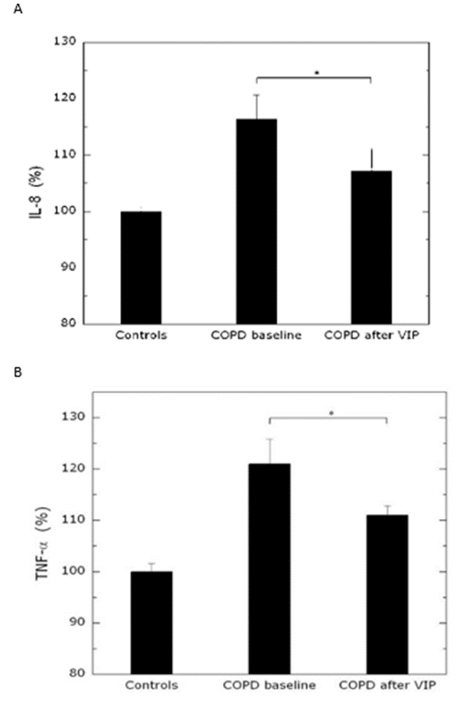
Figure 3: Transcription of IL-8 and TNF genes in bronchial biopsies before and after inhalation of VIP in COPD in vivo. Quantitative analysis of gene expression of IL-8 (A) and TNF (B) in human lungs. Total RNA was isolated from lung biopsies collected from patients before and after 3 months VIP treatment and from control subjects; mRNA levels of 18s were used for normalization. Data are expressed as mean±SD (n=6). P Less-than sign 0.01 in treated versus untreated patients.
The expression of the two genes in biopsies of lung tissue by immuno-histochemistry demonstrated a similar pattern. While before the inhalation of VIP the expression of the two genes was more pronounced in tissue sections of COPD patients, following treatment the signal declined (Figure 4).
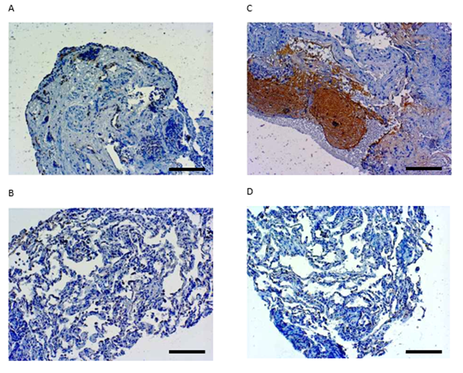
Figure 4: Expression of IL-8 and TNF in bronchial biopsies before and after inhalation of VIP in COPD. Immunohistochemical staining of IL-8 (A and B) and TNF (C and D) in lung tissue from COPD patients before (A and C) and after (B and D) 3 months VIP therapy. Bar ~ 200µm.
TNF activated the DNA binding of the three transcription factors AP-1, Stat3 and NF-κB with individual kinetics as assessed by DNA mobility shift assay, and which was confirmed by the transfer of the respective proteins from the cytosol into the nucleus by immuno-blot analysis (Figure 5). When pre-incubated with VIP (10-6 M - 10-12 M) for 30 minutes, the DNA binding of AP-1 and Stat3 was significantly reduced in
a dose-dependent manner (Figure 5A, 5.C) while that of NF-κB was only marginally reduced (Figure 5E). A similar reducing effect of VIP was observed on the protein translocation of the respective transcription factors as shown for AP-1 in figure 5B, for Stat3 in figure 5D and for NF-κB in figure 5F. Equal protein loading was confirmed by α-tubulin (loading).
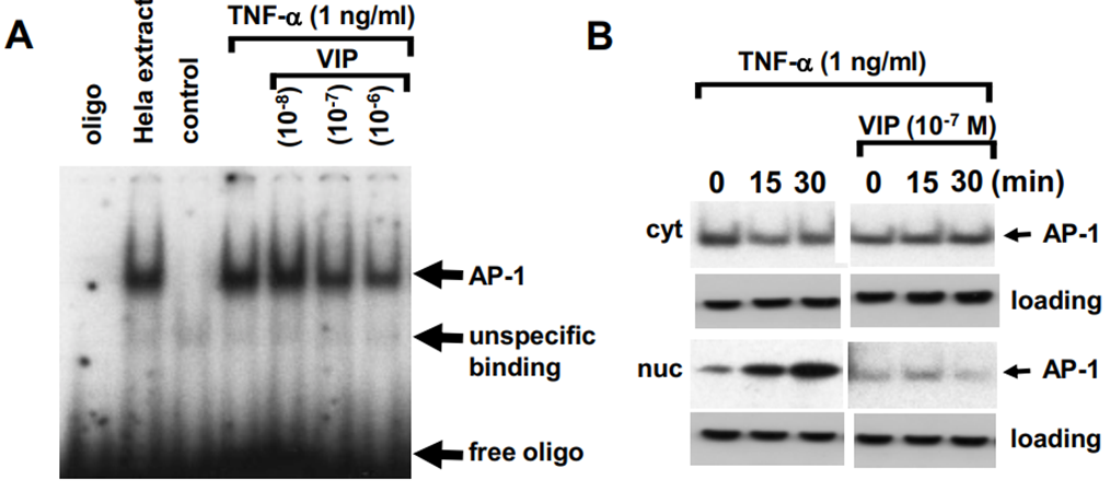
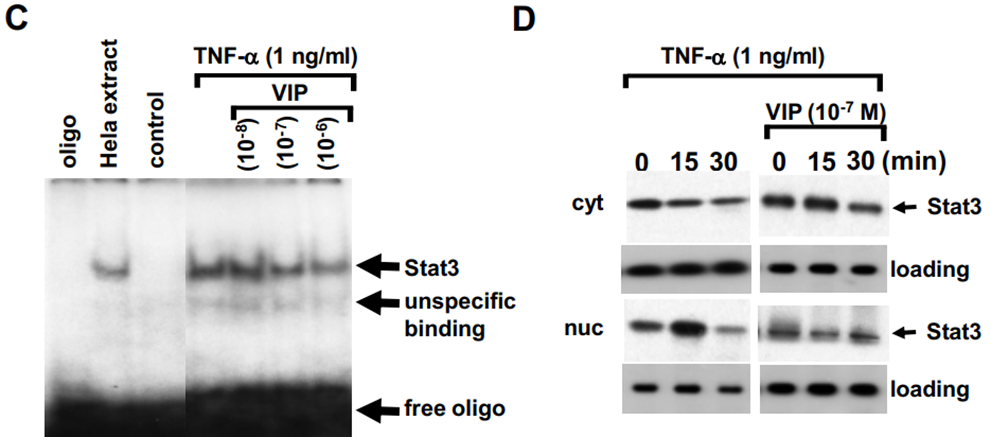
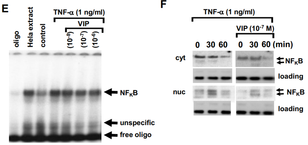
Figure 5: The effect of VIP on TNF induced transcription factor activity in primary BSMC and fibroblasts. A) A representative EMSA of the dose-dependent inhibition of TNF stimulated AP-1 to DNA binding by increasing concentrations of VIP. Similar results were obtained in two additional cell lines. B) A representative immuno-blot of the kinetic of TNF activated AP-1 translocation from the cytosol (cyt) into the nucleus (nuc) and its inhibition by VIP. Similar results were obtained in a second cell line. C) a representative EMSA of the dose- dependent inhibition of TNF stimulated Stat3 to DNA binding by increasing concentrations of VIP. Similar results were obtained in two additional cell lines. D) A representative immuno- blot of the kinetic of TNF activated Stat3 nuclear translocation and its inhibition by VIP. Similar results were obtained in a second cell line. E) a representative EMSA of the effect of VIP on TNF stimulated NF-κB to DNA binding. Similar results were obtained in two additional cell lines. F) A representative immuno-blot of the effect of VIP on TNF activated NF-κB nuclear translocation. Similar results were obtained in a second cell line.
A total of 34 participants were randomized out of 85 candidates who completed initial out-patient screening. The most common reason for exclusion was noncompliance and refusal of invasive diagnostic procedures. During the study, 4 subjects were withdrawn because of noncompliance with the study procedures: 2 in the VIP and 2 in the placebo group (Figure 6). Baseline characteristics of all 34 patients are
provided in table 1. All COPD patients had moderate to severe reduced pulmonary function with FEV1 before treatment of 1.33±0.70 l in the placebo group and 1.42±0.67 l in the VIP group, and the baseline lung function was comparable between both groups. More than 60% of the participants reported daily cough, more than 15% of the participants were smokers with a daily consummation of over 15 cigarettes, and 100% were former or current smokers.
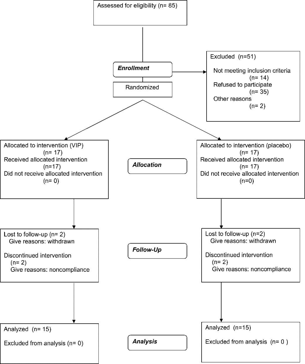
Figure 6: showing the flow of participants through each stage of the randomized trial.
The changes in post-bronchodilator FEV1 over time differed significantly (P Less-than sign 0,01) between the placebo and the VIP treatment group (Figure 7A and 7B). The placebo group showed a decline in FEVi with a slope of -0.03 l (from 1.36±0.86 to 1.34±0.85) after 3 months, while VIP increased
the FEV1 by 0.1 l (from 1.50 Plus–minus sign 0.68 to 1.60 Plus–minus sign 0.76) from baseline (P Less-than sign 0,01). Thus, the overall effect of VIP accounts for 0.13 l as compared to placebo (P Less-than sign 0.01). The post bronchodilator IVC increased by 0.155 l in the VIP group (Figure 7C) vs. baseline (P Less-than sign 0.05) and by 0.041 l in the placebo group (Figure 7D).
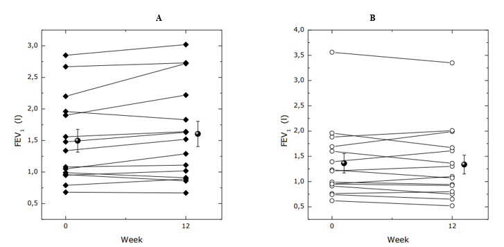
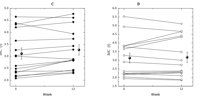
Figure 7: Forced Expiratory Volume in one Second (FEV1) ± SEM at selected time points (before and after 3 months treatment) by VIP (■) in group (A) and placebo (o) in group (B) (P Less-than sign 0,01). The numbers at each time point refer to data derived from VIP (■) or placebo (o) treatment group. Median change in Inspiratory Volume Capacity after 3 months treatment as compared to the value at randomization (Day 0) in the placebo (o) (D) or VIP group (■) (C).
As shown in table 1 (line 25), neither group of study patients had pulmonary hypertension. There was no significant change in pulmonary hemodynamic after treatment with VIP. (Data not shown)
| Pos. | Parameter | Placebo Group (N=17) | VIP Group (N=17) | Difference1 |
| 1 | Age (yr) | 60.8±6,7 | 57.4±4,6 | n.s. |
| 2 | Male sex no.(%) | 10 (65%) | 8 (48%) | n.s. |
| 3 | White race no.(%) | 17 (100%) | 17 (100%) | n.s. |
| 4 | PrebronchodilatorFEV1(litres) | 1,33±0,70 | 1,42±0,67 | n.s. |
| 5 | PrebronchodilatorFEV1(%predicted) | 43,1±19,20 | 45,3±17,80 | n.s. |
| 6 | FEV1:IVC ratio | 42,6±12,10 | 47,1±13,30 | n.s. |
| 7 | Cough - no.(%) | 15( 88,2%) | 11 (64,7%) | n.s. |
| 8 | Sputum production - no.(%) | 12 (71%) | 11 (64,7%) | n.s. |
| 9 | Wheezing - no.(%) | 8 (47,1%) | 8 (47,1%) | n.s. |
| 10 | Smokedinpast3mo - no.(%) | 6 (35%) | 5 (29%) | n.s. |
| 11 | Totalcigarettesmokingpack-yr | 58,0± 39,6 | 52,4±37,8 | n.s. |
| 12 | Regular medications - no.(%) | 16 (94%) | 16 (94%) | n.s. |
| 13 | Inhaled beta-adrenergic agonists - no. (%) | 16 (94%) | 16 (94%) | n.s. |
| 14 | Inhaled anti-cholinergic drug - no. (%) | 16 (94%) | 16 (94%) | n.s. |
| 15 | Oral beta-adrenergic agonists - no. (%) | 0 (0%) | 0 (0%) | n.s. |
| 16 | Theophilline - no.(%) | 13 (76%) | 10 (59%) | n.s. |
| 17 | Inhaled glucocorticosteroids - no.(%) | 14 (82%) | 15 (88%) | n.s. |
| 18 | Use of oxygen at home - no. (%) | 2 (12%) | 4 (23%) | n.s. |
| 19 | Other illness - no.(%) | 17 (100%) | 17 (100%) | n.s. |
| 20 | Diabetes mellitus - no.(%) | 0 (0%) | 1 (6%) | n.s. |
| 21 | History of ulcer - no.(%) | 1 (6%) | 2 (12%) | n.s. |
| 22 | Hypertension - no.(%) | 10 (59%) | 8 (47%) | n.s. |
| 23 | Disabling heart disease - no.(%) | 1 (6%) | 1 (6%) | n.s. |
| 24 | Disabling arthritis - no.(%) | 0 (0%) | 0 (0%) | n.s. |
| 25 | Mean pulmonary artery pressure (mmHg) | 18.8±6,7 | 16.9±4,6 | n.s. |
| 26 | Cardiac output (L/min) | 5,4±0,6 | 5,7±1,3 | n.s. |
| 27 | Pulmonary capillary wedge pressure (mmHg) | 9,1±3,9 | 7,2±2,9 | n.s. |
| 29 | Time from presentation to randomization days | 20.6±4,71 | 22.6±4,71 | n.s. |
* Data are either “means ± SD” or “counts and (percent)”.
t Significance=P Less-than sign 0.05 for differences among groups by statistical analysis
(Students T-Test for continuous variables and by the chi-square test for categorical variables
Table 1: Base-line characteristics of the patients
The six minutes walking distance increased by 24.1 m from 443.1 ± 90.8 to 467.2 ± 93.8 m after 12 weeks VIP treatment (P Less-than sign 0.05), and insignificantly decreased by 8.6 meters from 444.5 ± 115.5 to 436.0 ± 145.2 m after placebo (Figure 8A and 8B).
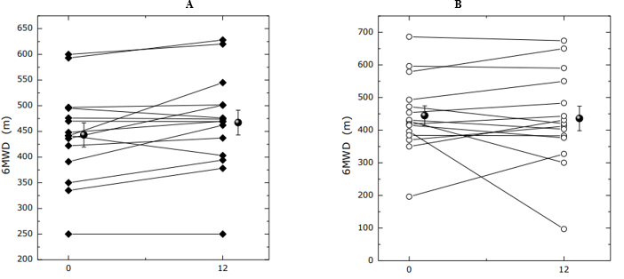
Figure 8: Change in Six-Minutes Walking Distance from baseline to day 90 in the VIP (■, A) group, and Placebo (o, B) group. P Less-than sign 0,05 for the comparison between VIP and placebo.
Borg dyspnea index, a primary variable, showed improvements (i.e., decreased score) with VIP (Figure 9). The changes from base-line in Borg dyspnea scale before 6MWT were -0,2 U for VIP (A), and +0,2 U for placebo (P Less-than sign 0,05) and -1,1 U for VIP, and 0,7 U for placebo after 6MWT (P Less-than sign 0,05).
Health-related quality of life assessed by SF-36, a primary variable (i.e., decreased score) and SGRQ showed improvements with VIP (Table 2). The changes from baseline in SF-36 - PHS were 4.08 for VIP and 0.20 for placebo (P Less-than sign 0.05). No significant changes were found in the mental health dimension in either group. In SGRQ the changes from baseline were -5.25 (from 46.52±4.06 to 41.27±4.35) for VIP and 1.72 for placebo (from 46.95±4.03 to 47.23±4.70, P Less-than sign 0.05).
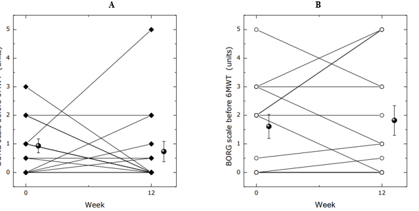
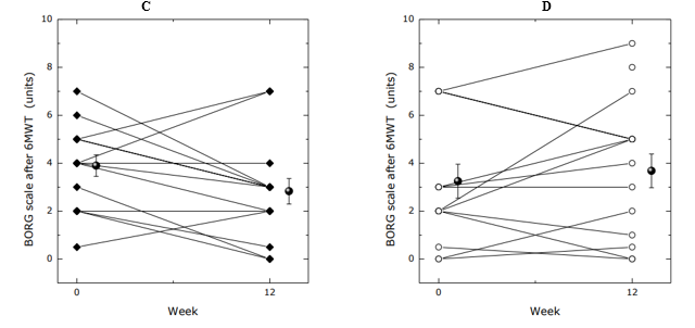
Figure 9: BORG dyspnea Score before (A and B) and after (C and D) 6MWT -exercise. Total scores from baseline to day 90 for the VIP (■) and placebo (o) groups. P Less-than sign 0,05 for comparison between VIP and placebo.
| SF - 36 and SGRQ | VIP | Placebo |
| PHS at baseline | 41.15±2.26 | 38.62±2.12 |
| PHS after treatment (week 12) | 45.23±2.07 (p=0,05) | 38.82±2.49 (p=0,898) |
| MHS at baseline | 44.45±2.41 | 44.89±2.53 |
| MHS after treatment (week 12) | 47.92±2.18 (p=0,366) | 45.64±3.18 (p=0,796) |
| SGRQ at baseline | 46.52±4.06 | 46.95±4.03 |
| SGRQ after treatment (week 12) | 41.27±4.35 | 47.23±4.70 |
* Data are either “means ± SD” or “counts and (percent)”.
t Significance=P Less-than sign 0.05 for differences among groups by statistical analysis
(Students T-Test for continuous variables and by the chi-square test for categorical variables
Table 2: Quality of life according to the short 36-item questionnaire - SF-36 and St. Georges respiratory questionnaire - SGRQ.
VIP is released through inflammatory processes as evidenced by the finding that the serum VIP levels were significantly higher in COPD patients with acute exacerbations compared to stable COPD [27].
We found an inverse relationship between the serum concentration of VIP and the progression of COPD. VIP inhibited the release of IL-8 and TNF in parallel with the inhibition of transcription and expression of the two genes and downregulated the transcription factors AP-1 Stat3 and NF-κB. The inhalation of VIP resulted in a significant improvement of lung function parameters of COPD patients (GOLD l-IV), a better quality of life and exercise capacity and in lesser exacerbations without relevant side effects. How can the efficacy of VIP in COPD be interpreted?
Firstly, an inverse relationship between the level of expression of VIP receptor proteins and of the level of circulating VIP was previously observed in pulmonary arterial hypertension [37]. Similarly, VIP serum levels in COPD patients were significantly decreased versus normal subjects suggesting a lack of VIP contributing to the pathogenesis of COPD. Elevated levels of several pro-inflammatory cytokines and other mediators have been demonstrated in the lungs of patients with COPD [8,17]. For example, IL-8, adhesion molecule (ICAM-1), and TNF are present in inducted sputum [22], and their levels increased further during exacerbation [51]. The levels of TNF, IL-8 and metalloproteinase-9 (MMP-9) are elevated in bronchoalveolar lavage fluid from chronic smokers compared to non-smokers [24]. Blood levels of TNF, as well as of the soluble receptors for these cytokines, are two to three times higher in subjects with COPD than in the non-COPD population. Importantly, these levels correlate directly with the severity of airflow impairment [47, 13]. A recently performed study on gene expression profiles using serial analysis of gene expression and microarray analysis in lung biopsies of GOLD II smokers revealed a significantly increased expression of a number of genes encoding transcription factors or related proteins [31, 32]. Analogously, measurements of IL-8 and TNF mRNAs in bronchial biopsies of the COPD patients of our trial using real-time PCR demonstrated lower transcription after VIP treatment. Among its various immuno-modulatory properties, VIP was reported to down-regulate the biosynthesis of CXC- and CC-chemokines such as IL-8, ICAM-1, MMP-9, and TNF [11, 49]. Moreover, VIP inhibits the binding and activation of the transcription factor NF-κB to corresponding promoter sequences of genes encoding mediators of inflammation, such as early growth factors 2/3 [11]. Another transcription factor which has been implicated in COPD is Stat3 [15, 38] and the observation that VIP inhibits TNF induced Stat3 activation in cells, as shown in this study, isolated from COPD patients indicates that VIP has a broad anti-inflammatory action. In mouse as well as in human fibroblasts cigarette smoke, the prevalent cause of COPD, activated AP-1 which was involved in the regulation of redox system-dependent pro-inflammatory factors such as heme oxygenase [2, 24]. Furthermore, it was suggested that the inhibition of NF-κB and AP-1 could circumvent steroid insensitivity in chronic inflammatory diseases [1]. Since steroids are not very efficient in the therapy of COPD the inhibition of NF-κB and AP-1 would explain the significant clinical improvement in COPD which we report here.
The molecular basis for the observed reduction of acute exacerbations following VIP treatment is unclear. Although various immuno-modulatory properties may be involved, experimental evidence suggests that VIP promotes Th-2 type and reduces inflammatory Th-1 type responses [10] that may lead to reduced susceptibility against infections.
Secondly, the fact that none of the patients included in this study exhibited pulmonary hypertension excludes the known vasodilatory effect of VIP as relevant effective mechanism. VIP has been shown to stimulate the dilatation of bronchial rings in vitro (Greenberg et al. 1987). Moreover, VIP inhalation resulted in increased FEV1 in asthmatics [33]. Similarly, in our trial the daily dose of 200 pg VIP significantly increased post-bronchodilator FEVi and that of IVC throughout the 12-week treatment period (P<0>
Thirdly, chronic inflammation is generally regarded as a central mechanism in the pathogenesis of COPD [17]. Analogously, VIP has been shown to inhibit the inflammatory processes associated with COPD, and bronchial biopsy specimens of COPD patients (GOLD 0) exhibited increased expression of VPAC1 and VPAC2 receptors [28]. Furthermore, mitochondrial dysfunction and the effects of severely reduced availability of the protein deacetylase sirtuin-1 (SIRT1) are evident in COPD patients. The reduced SIRT1 expression within cells is a result of severe oxidative stress. The effects of VIP in pneumoplegic solution were tested in isolated rat lungs. Rat lungs were isolated, flushed and stored for 24 hours in presence or absence of VIP in the restoration solution. Electron microscopy showed that lungs stored in VIP-containing solutions had significantly more normal shaped mitochondria, less mitochondrial edema, less distortion of mitochondrial cristae, thinner basal lamina, and less aggregation of nuclear chromatin than control solutions.
Treatment with VIP also reduced histopathological severity of colitis and cell death markers in a murine model, leading to partial recovery of inhibited mitochondrial respiratory complexes, altered mitochondrial membrane potential and lowered ATP generation.
Mice lacking VIP (VIP-KO mice) gene develop right ventricular hypertrophy, thickened pulmonary artery, perivascular inflammatory cell infiltrates in the lung, and exhibit airway hyperresponsiveness to the cholinergic agonist methacholine. Treatment of these mice with exogenous VIP attenuates both, the vascular remodeling and right ventricular remodeling. [55, 56, 57, 58, 61, 62, 63, 64].
The fact that the treatment of COPD is limited to only few therapeutic options indicates the need for research and product development of additional alternatives. The unique ability of VIP to improve cardiopulmonary circulation and lung function in COPD, and quality of life, and 6 MWD without relevant side effects suggests that it may become a new therapeutic option. Future studies on VIP and VIP analogues are therefore necessary to reveal its full potential for clinical use.
Our study is the first to report that the inhalation of VIP has clear immunoregulatory effects in COPD patients. VIP inhalation might be an attractive, new immunoregulatory treatment strategy without systemic immunosuppression and negative side effects. Inhaled VIP can act alone, or in combination with any other approved COPD treatment option, with excellent safety profile. Further multicentric controlled studies are needed to evaluate the clinical efficiency of this new treatment option in COPD.
Author’s Contributions
Wilhelm Mosgoeller, preclinical project design, histology, immunhistochemistry, Ventzislav Petkov, clinical study conceptualization, principal clinical investigator Bernhard Burian, patient recruitment and investigation, clinical study rollout Michael Roth, in vitro molecular laboratory studies, immunoblotting Christopher Lambers, in vitro cell isolation, sample administration Lutz-Henning Block, data review, manuscript writing, senior PI, Claudia Vonbank, clinical patient investigation and data administration
We are indebted to all participating physicians of the Department of Pulmonary Medicine of the Medical University of Vienna to their help in realizing this study.
Registration number for the clinical study:
ClinicalTrials.gov Identifier: NCT00464932
Scientific Knowledge on the Subject:
Molecular and clinical effects of the neuropeptide VIP in COPD.
What This Study Adds to the Field:
A new aspect of the role of Vasoactive Intestinal Peptide (VIP) in Chronic Obstructive Pulmonary Disease (COPD) and its therapeutical potential, when inhaled.
Dear Editorial Team, Clinical Medical Reviews and Reports. My experience with the journal was highly positive. The peer-review process was rigorous, constructive, and completed in a timely manner. The reviewers provided valuable comments that helped improve the quality and clarity of our manuscript. The editorial office was professional, responsive, and supportive throughout all stages of the publication process. Communication was clear and efficient, and any questions were addressed promptly. Overall, I found the journal to maintain high scientific standards and an excellent publication workflow. I would be pleased to consider submitting future work to this journal. Best wishes from, Elena Popa.

It was my pleasure to submit my testimonial concerning the Reviewer Board of our Scientific Journal “Brain and Neurological Disorders”. The Reviewers focused on some modifications and their contribution was helpful. The ladies of our Editorial Office were also supported my efforts. It was my honor to have such a co-operation and I am looking forward for more collaboration.

Dear Grace Pierce, Editorial Coordinator of Journal of Clinical Research and Reports, Thank you for the speedy and efficient peer review process. I appreciate the fact that your peer reviewers do not take months to respond like with some other journals. I would also like to thank the editorial office for responding quickly to my questions. It is an excellent journal. I plan to submit more manuscripts in the future. Best wishes from, Robert W. McGee

Dear Grace Pierce, Editorial Coordinator of Journal of Clinical Research and Reports, Working with you and your team on our recent publication in JCRR has been a truly wonderful and enjoyable experience. The responses were prompt, and the reviewers were patient, constructive, and highly professional. One reviewer in particular gave me the feeling that a professor was carefully reading and commenting on my coursework, which was deeply touching. The entire process was straightforward and hassle‑free, with no tedious online forms to complete. I highly recommend this journal. Best wishes from, DR Aibing Rao, Head of R&D

I Appreciate the Opportunity to Share my Experience with the Journal of Clinical Research and Reports. The peer review process was timely and constructive, and the feedback provided helped improve the quality of our manuscript. The editorial office was professional, responsive, and supportive throughout the process, ensuring smooth communication and efficient handling of the submission. Overall, it was a positive experience collaborating with your team.

Dear Mercy Grace, Editorial Coordinator of Obstetrics Gynecology and Reproductive Sciences, We would like to express our gratitude for your help at all stages of publishing and editing the article. The editors of the magazine answer all the necessary questions and help at every stage. We will definitely continue to cooperate and publish other works in the Obstetrics Gynecology and Reproductive Sciences! Best wishes from, Alla Konstantinovna Politova,
