
Review Article | DOI: https://doi.org/10.31579/2768-2757/025
*Corresponding Author: Aamir Jalal Al Mosawi, Advisor in Pediatrics and Pediatric Psychiatry Children Teaching Hospital of Baghdad Medical City, Iraq.
Citation: Aamir Jalal A, M. (2021). Chest CT-Scan Findings of an Iraqi Patient with Symptomatic Covid-19 Disease. Journal of Clinical Surgery and Research. 2(4); DOI: 10.31579/2768-2757/025
Copyright: ©2021 Aamir Jalal Al Mosawi, This is an open-access article distributed under the terms of the Creative Commons Attribution License, which permits unrestricted use, distribution, and reproduction in any medium, provided the original author and source are credited.
Received: 27 July 2021 | Accepted: 23 August 2021 | Published: 31 August 2021
Keywords: chest ct-scan; covid-19; Iraqi patient
Background: On the first of June, 2021, 1,201,352 cases of covid-19 were reported by the Iraqi Ministry of Health, and 16375 patients died because of the disease. The aim of this paper is to describe chest CT-scan findings of an Iraqi patient who was observed early during June, 2021, and had symptomatic covid-19, but he didn’t need hospitalization.
Patients and methods: A forty-year old school teacher developed covid-19 disease with fever, fatigue, anorexia, and cough. The patient recovered after about two weeks.
Results: Chest CT-scan performed during first week of illness showed:
(1) Multiple bilateral ground glass opacities.
(2) Atelectatic bands.
(3) Thickening of the interlobular septa.
(4) Vascular thickening.
Conclusion: The chest CT-scan findings in this Iraqi patient was rather typical of covid-19 disease and included the most commonly reported abnormality of ground-glass shadows.
On the first of June, 2021, 1,201,352 cases of covid-19 were reported by the Iraqi Ministry of Health, and 16375 patients died because of the disease [1]. A significant number of covid-19 patients who develop pneumonia were found to have normal chest radiographs. However, Yoon et al (2020) emphasized that most Korea patients with covid-19 pneumonia had abnormalities on chest CT-scan mostly including ground-glass opacities with bilateral patchy, confluent or nodular shadows [2].
The aim of this paper is to describe chest CT-scan findings of an Iraqi patient who was observed early during June, 2021, and had symptomatic covid-19, but he didn’t need hospitalization.
A forty-year old school teacher developed covid-19 disease with fever, fatigue, anorexia, and cough. The patient recovered after about two weeks.
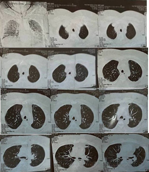
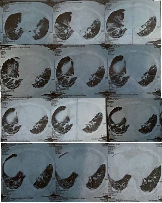
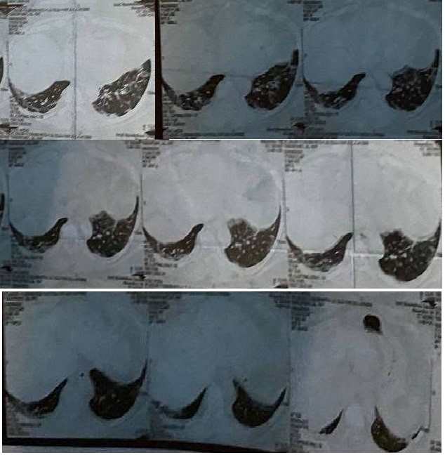
Chest CT-scan performed during first week of illness (Figure-1) showed:
(1) Multiple bilateral ground glass opacities.
(2) Atelectatic bands.
(3) Thickening of the interlobular septa.
(4) Vascular thickening.
There was no pleural effusion or thickening.
Conclusion: The chest CT-scan findings in this Iraqi patient was rather typical of covid-19 disease and included the most commonly reported abnormality of ground-glass shadows.
Xiang et al (2020) reported the CT-scan findings of fifty three patients (31 males, 22 females; mean age, 53 years) who had Covid-19 pneumonia. They observed the occurrence of ground-glass opacity with consolidation in 24 patients (45.3%) and pure ground-glass opacity in 28 patients (52.8%). Crazy-paving occurred in 14 patients (26.4%), bronchiectasis occurred in 12 patients (22.6%), atelectasis occurred in 7 patients (13.2%), parenchymal bands occurred in 6 patients (11.3%), air bronchogram occurred in 6 patients (11.3%), and interlobular thickening occurred in 5 (9.4%).
Xiang et al suggested that most Covid-19 patients who develop pneumonia had abnormalities detectable on chest CT-scan on the time of presentation [3].
4-Kong et al (2020) reported the CT-scan findings of twenty-two patients hospitalized with Covid-19 disease during the period from January 17, 2020 to February 15, 2020. On presentation nineteen patients had fever and eight patients had cough. Chest CT-scan showed ground-glass opacities in 18 patients, lung consolidation in 7 patients, interlobular septal thickening in five 5 patients, and fibrosis-like stripes in 4 patients.
Wang et al (2021) reported the CT-scan findings of 693 covid-19 patients, including 13 children (51% males and 49
Dear Editorial Team, Clinical Medical Reviews and Reports. My experience with the journal was highly positive. The peer-review process was rigorous, constructive, and completed in a timely manner. The reviewers provided valuable comments that helped improve the quality and clarity of our manuscript. The editorial office was professional, responsive, and supportive throughout all stages of the publication process. Communication was clear and efficient, and any questions were addressed promptly. Overall, I found the journal to maintain high scientific standards and an excellent publication workflow. I would be pleased to consider submitting future work to this journal. Best wishes from, Elena Popa.

It was my pleasure to submit my testimonial concerning the Reviewer Board of our Scientific Journal “Brain and Neurological Disorders”. The Reviewers focused on some modifications and their contribution was helpful. The ladies of our Editorial Office were also supported my efforts. It was my honor to have such a co-operation and I am looking forward for more collaboration.

Dear Grace Pierce, Editorial Coordinator of Journal of Clinical Research and Reports, Thank you for the speedy and efficient peer review process. I appreciate the fact that your peer reviewers do not take months to respond like with some other journals. I would also like to thank the editorial office for responding quickly to my questions. It is an excellent journal. I plan to submit more manuscripts in the future. Best wishes from, Robert W. McGee

Dear Grace Pierce, Editorial Coordinator of Journal of Clinical Research and Reports, Working with you and your team on our recent publication in JCRR has been a truly wonderful and enjoyable experience. The responses were prompt, and the reviewers were patient, constructive, and highly professional. One reviewer in particular gave me the feeling that a professor was carefully reading and commenting on my coursework, which was deeply touching. The entire process was straightforward and hassle‑free, with no tedious online forms to complete. I highly recommend this journal. Best wishes from, DR Aibing Rao, Head of R&D

I Appreciate the Opportunity to Share my Experience with the Journal of Clinical Research and Reports. The peer review process was timely and constructive, and the feedback provided helped improve the quality of our manuscript. The editorial office was professional, responsive, and supportive throughout the process, ensuring smooth communication and efficient handling of the submission. Overall, it was a positive experience collaborating with your team.

Dear Mercy Grace, Editorial Coordinator of Obstetrics Gynecology and Reproductive Sciences, We would like to express our gratitude for your help at all stages of publishing and editing the article. The editors of the magazine answer all the necessary questions and help at every stage. We will definitely continue to cooperate and publish other works in the Obstetrics Gynecology and Reproductive Sciences! Best wishes from, Alla Konstantinovna Politova,
