AUCTORES
Globalize your Research
Research Article | DOI: https://doi.org/10.31579/2637-8892/021
Department of Psycheatry, Adam International Hospital, Egypt.
*Corresponding Author: Anbis El Hakim,Department of Psycheatry, Adam International Hospital, Egypt.
Citation: Anbis El Hakim, Charactor of Modality and Resident Level in Pedicle Screw Accuracy and Neurosurgical Education. J. Psychology and Mental Health Care 2(1); DOI: 10.31579/2637-8892/021
Copyright: © This is an open-access article distributed under the terms of the Creative Commons Attribution License, which permits unrestricted use, distribution, and reproduction in any medium, provided the original author and source are credited.
Received: 30 January 2018 | Accepted: 20 February 2018 | Published: 25 March 2018
Keywords: pedicle screw; resident level; navigation and accuracy
Objective: Evolving pressure on surgical education necessitates safe and efficient learning of techniques. We evaluated the effect of training year using anatomic, percutaneous fluoroscopy guided and computer navigated techniques on the accuracy of pedicle screw placement to attempt to determine if different modalities may be better suited for different levels of training.
Methods: All instrumented thoracic and lumbar cases performed at Detroit Medical Center by the Neurosurgery Service between August 2012 and June 2013 were included.Cases had hardware verified by post-operative CT. Hardware placement was graded according to Mirza SK et al., grade 0 (within pedicle), grade 1 (< 2 mm breach), grade 2 (> 2 mm breach) , and grade 3 (extrapedicular). Pedicle screws were reviewed independently by a resident and attending surgeon. Rates of pedicle breach, EBL, length of case, pedicle size and pedicle starting point were all reviewed. Pedicles were analyzed on PACS system in axial views, using sagittal views to identify the correct level.
Results: A total of 306 pedicle screws were evaluated in 36 patients. The overall rate of accurate pedicle screw placement among residents defined as Grade 0 or 1 placement was 86.8%.Fluoroscopically placed screws had significantly less breaches than anatomic screws 11% vs 20% (p = 0.03). Fluoroscopic cases had significantly less medial breeches (20%) than anatomic (50%) (p < 0.05) and computer assisted cases (73%) (p < 0.05). EBL values for fluoroscopic, anatomic and Body Tom cases were 425 cc, 720 cc, and 816 cc respectively. Resident level was found to be inversely proportional to breech rate (R squared 0.45). We did not see any clear difference in breach rate for resident level in different modalities.
Conclusion: Supervised neurosurgical residents can place pedicle screws within published rates of acceptable breach. Interestingly our study revealed an inverse relationship between resident experience and pedicle screw accuracy. Fluoroscopic placement of pedicle screws compared to computer assisted and anatomic techniques results in lower medial breach rate and may be better suited for junior level residents.
Introduction
Surgical trainees today are training in an era when education, milestones, and quality improvement must be balanced with restricted work hours.Given these demands programs continue to look for safe and objective ways to measure resident performance, efficiently train residents and ensure patient safety. Unfortunately few objective measures of surgical competency outside of a simulator setting exist.Commonly simulated procedures in which accuracy can be measured include ventriculostomy and pedicle screw placement.Scott, et al. [1] states “Simulation-based training has gained significant momentum and will be a requirement for residencies in the near future”. However even with today’s’ modern simulators there still exists a difference between virtual surgery and intra operative experience. Pedicle screw placement we feel is a neurosurgical technique that has an objective measure of surgical performance, which can be easily documented: the breach rate. Optimizing surgical training requires maximizing the learning potential of different surgical techniques within a set period of time, all while documenting proficiency.Given that different residents and different resident levels may learn techniques through different methods we sought to determine whether one of the three methods we currently use for pedicle screw placement was better than the other for our different levels of surgical trainees.We hypothesized that image based modalities would allow more accurate screw placement for junior level residents.The goals of this study were to determine whether modality affects pedicle screw placement accuracy, if accuracy of a modality is affected by resident training level and finally to quantify our own resident experience with pedicle screws.
All instrumented thoracic and lumbar cases performed at Detroit Medical Center by the Neurosurgery Service between August 2012 and June 2013 were included.Cases had hardware verified by post-operative CT.Hardware placement was graded according to Mirza SK, et al [2], grade 0 (within pedicle), grade 1 (< 2> 2 mm breach) , and grade 3 (extrapedicular). Pedicle screws were reviewed independently by a resident and attending surgeon.Rates of pedicle breach, EBL, length of case, pedicle size and pedicle starting point were all reviewed.Pedicles were analyzed on PACS system in axial views, using sagittal views to identify the correct level. All screws were placed with the patient in the prone position. Charts and statistics were generated using Microsoft Excel.T tests were utilized for statistical significance where appropriate. As we were expecting screws placed with fluoroscopy or computer navigation to have lower breach rates than anatomic screws one Tailed Tests were used.Two tailed T tests were used for the remainder of comparisons.Pedicle screw starting point was determined by drawing a line down the center of the pedicle into the vertebral body on axial views. If the entry point of the screw was medial or lateral to this it was considered a medial or lateral starting point.Grade 2 and 3 breaches were utilized for calculating the breach rate because in the upper thoracic spine were the pedicles can be smaller than 5 mm, the smallest percutaneous screw is 5 mm in diameter, thus a small degree of breach is inevitable. Furthermore medial breaches less than 4 mm have been shown to be within a “safe zone” as there is typically 2 mm epidural space and 2 mm subarachnoid space [3,4]. However this has been refuted by other studies [5].
4.1 Percutaneous Fluoroscopically Guided Screw Placement Technique
Midline skin incision utilized. Subcutaneous tissue undermined laterally, fascia opened over the level of pathology and decompression completed.Percutaneous screws placed through the fascia under direct AP and lateral fluoroscopy.Jamshidi needles start lateral and superior to pedicle at desired level and are docked under AP fluoroscopy on superior lateral corner of pedicle.The Jamshidi is then advanced using still shots of AP fluoroscopy until the medial border of pedicle is reached.Lateral fluoroscopy is then used to ensure the Jamshidi is past the posterior wall of vertebral body.K wires are then placed, the Jamshidi needle removed and the screw placed over the K wire.
4.2 Anatomic Screw Placement Technique
Midline incision is utilized, paravertebral muscles are dissected down to lamina and out to the transverse processes.The correct level is verified with fluoroscopy.AM 8 burr is used to decorticate at well known anatomic entry points for thoracic and lumbar spines.Curved pedicle finder is utilized to gain access to posterior vertebral body through pedicle by initially point the tip laterally and coming medially at around 20 mm. Ball probe is used to determine if a breach was present. Self tapping screws are then We identified 36 patients with post operative imaging after posterior instrumentation in Lumbar and Thoracic spine.This included 212 resident screws and 94 attending placed screws.Patient demographics included 23 women and 13 men the average age was 53 (20-73). Case breakdown was as follows: 17 tumors, 7 motor vehicle related traumas, 2 osteoporotic related fractures, 7deformity cases (including scoliosis, butterfly, hemi vertebra), and 3 other cases (epidural abscesses, repeat cases for disc herniation and lumbar stenosis). Pedicle diameters were measured for Upper Thoracic (T1-6) (5.7 mm average), Lower Thoracic (T7-12) (6.7 mm average) and Lumbar Spine (L1-5) (10mm average).
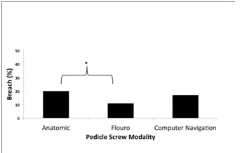
Fluoroscopically placed screws had significantly less breaches than anatomic screws 11% vs. 20% (p = 0.03) (Figure 1). Each modality had a breach rate between 11-20% breach rates. Overall resident breach rate was 13.2% for Grade 2/3 breaches. Overall grade 3 breach rate was 5 % (16 screws). Ten of these screws were resident placed, and only 3 were medial breaches.
inserted, ensuring same trajectory as pedicle finder.
4.3 Computer Navigated Screw Placement Technique
Body Tom (Neurologica, Intra Operative 32 slice CT scanner) registration was as follows: The reference arc was attached to spinous process 1-2 levels above planned instrumentation.Images were then acquired; typically 3-4 levels can be visualized with one spin.Once registration was completed screws were inserted using planned trajectory dictated by image guidance.
We identified 36 patients with post operative imaging after posterior instrumentation in Lumbar and Thoracic spine.This included 212 resident screws and 94 attending placed screws.Patient demographics included 23 women and 13 men the average age was 53 (20-73). Case breakdown was as follows: 17 tumors, 7 motor vehicle related traumas, 2 osteoporotic related fractures, 7deformity cases (including scoliosis, butterfly, hemi vertebra), and 3 other cases (epidural abscesses, repeat cases for disc herniation and lumbar stenosis). Pedicle diameters were measured for Upper Thoracic (T1-6) (5.7 mm average), Lower Thoracic (T7-12) (6.7 mm average) and Lumbar Spine (L1-5) (10mm average).
Fluoroscopically placed screws had significantly less breaches than anatomic screws 11% vs. 20% (p = 0.03) (Figure 1). Each modality had a breach rate between 11-20% breach rates. Overall resident breach rate was 13.2% for Grade 2/3 breaches. Overall grade 3 breach rate was 5 % (16 screws). Ten of these screws were resident placed, and only 3 were medial breaches.

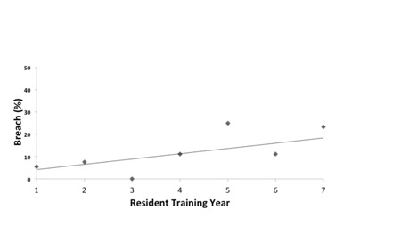
Comparing breach rates by resident level an inverse trend between training experience and breach rate was observed. Among residents, the PGY 7 resident placed 47 screws with a grade 0/1 placement of 76%, compared to the PGY2 resident who placed 53 screws with a grade 0/1 placement of 92%. There is an inverse correlation between PGY year and pedicle placement accuracy (R squared 0.45) (Figure 2).

There was an inverse trend between resident level and overall pedicle screw breach rate. Increasing resident level resulted in increased breach rate when compared to junior level residents. R² = 0.4548
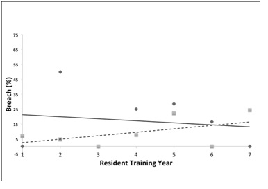
Analyzing individual modalities by resident year we found two different trends of performance. Anatomic screws trended towards improved accuracy as the PGY level increased, while fluoroscopic screws showed an inverse relationship between year and performance (Figure 3).

Different modalities have different trends among residents for breach rates. Fluoroscopically placed screws (Squares with dotted line) are more accurately placed by junior level residents while anatomic placement (Diamonds with solid line) tends to have better senior level placement.
There was no significant difference in medial or lateral breach by resident level. However, fluoroscopy guided percutaneous screw placement resulted in significantly less medial breaches (20%) than anatomic (50%) and navigation placed screws (73%) (p < 0> 9 mm had a breach rate of only 6%. (Figure 5)
No patients had to be taken back to the operating room for hardware replacement.There was a trend for supervision by spine fellowship trained attendings to have lower resident and attending rates of breaches overall, but due to low number of screws placed by non fellowship spine trained attendings this was not statistically significant.Fluoroscopy guided percutaneous screws trended towards having lower EBL and shorter case times however these finding did not reach significance.
Across surgical specialties it seems documenting surgical performance by training level and specialty has become more routine. Recent studies have shown early PGY general surgery residents performing hernia surgery take more time, have higher EBL’s, and have more recurrences of inguinal hernias than their older counterparts [6]. Another study looking across all surgical specialties noted an association between increased attending supervision and decreased morbidity and mortality [7]. Reflecting on our study and reviewing previously published data on neurosurgical trainees it becomes apparent that literature supports supervised neurosurgical trainees may have better outcomes surgically and respond differently to fatigue than published results of other training specialties. A study comparing post call neurosurgical trainees to general surgical trainees reported a distinct difference in post call performance.This study suggested that not all specialties trainees’ react the same to fatigue and neurosurgical trainees performance is affected less by fatigue [8]. Literature suggests supervised neurosurgical trainees performance in spine and intracranial aneurysm surgery results in comparable outcomes to attending level intervention [9-11]. Taken together this creates a case for neurosurgical in-vivo supervised training.
We have identified three studies related to resident level and screw placement accuracy.Only one of these studies was in vivo [10], one simulated [12] and one cadaveric [13].None of these studies incorporated navigational screws.To our knowledge there has been no comparison of modalities by resident level.
While our data is still limited it appears that fluoroscopic (MIS) screw placement may benefit the junior level resident the most. We have shown that supervised neurosurgical residents can use all three modalities with breach rates between 11-20% with no operative take backs. Our results are within available published rates of breach. Literature reported rates of pedicle breach have been documented as high as 40%, however commonly quoted rates are 10-25%, with upper thoracic and deformity cases having higher rates of pedicle breach [14-16]. Combined low breach rates in the hands of junior residents and low medial breach rate make fluoroscopic placement the ideal technique for junior level residents.Fluoroscopic screws provide the resident with necessary tactile feedback, trajectory and radiographic anatomy that will hopefully lay the foundation for future pedicle screw placement techniques. In addition, advocates of ex-vivo surgical training cite increased OR costs as deterrent to in-vivo training due to novice surgeons increased operating times [17].However, fluoroscopic screw placement by residents in our study trended towards having faster OR finishes times than either of the other techniques and may represent a strategy for safe, efficient, and cost effective junior level training
Interestingly the inverse relationship found in fluoroscopic screw placement between resident level and accuracy may be related to attending comfort with senior level residents and a decreased level, albeit still supervised interaction. Wang, et al. [10] came to similar conclusion with their findings that PGY 2/3 had lower breach rates compared to PGY 6 residents in thoracic spine with anatomic screws (13% vs. 19%). However in contrast to the Wang, et al. [10] study our experience with anatomic screws trended towards improvement in breach rate with increasing resident level. We feel fluoroscopy set up and end plate alignment may be another explanation for the increased breach rate observed with senior level residents as they likely have more of a direct role in AP and Lateral alignment.
Our data supports a centered or lateral starting point for pedicle screw placement as this was shown to have the least number of medial breaches.In addition junior residents may benefit from starting their screw placement in the low thoracic or lumbar spine as the pedicle widths are typically larger.
Both our study and the Wang, et al. [10] study demonstrates acceptable resident breach rates during training. Furthermore, literature shows novice residents who initially had high breach rates were able to reach literature accepted rates by the 4th cadaver in one study of anatomic screws[13], highlighting the importance of repetition.Thus a combination of fluoroscopic and anatomic cadaveric training may provide junior residents with a successful training regimen.
A recent meta-analysis reported that navigated and non navigated screws could be placed with 95.2% and 90.3
Out study demonstrates supervised neurosurgical residents can place pedicle screws within published rates of acceptable breach. Furthermore our data supports intra operative training of residents as safe and effective. Fluoroscopic placement of pedicle screws had lower overall and medial breach rates compared to computer assisted and anatomic techniques. Fluoroscopically guided screws may provide the needed tactile and trajectory feedback to facilitate early PGY trainees in accurate, safe, and efficient screw placement, while setting the foundation for future techniques.