
Case Report | DOI: https://doi.org/10.31579/2692-9406/129
1Central Radiology Institute, Kepler University Hospital, Medical Faculty of the Johannes Kepler University, Linz, Austria.
2Institute of Neuroradiology, Kepler University Hospital, Medical Faculty of the Johannes Kepler University, Linz, Austria.
3Medical Faculty of the Friedrich-Alexander-University of Erlangen-Nürnberg, Erlangen, Germany.
*Corresponding Author: Michael Hofko, Central Radiology Institute, Kepler University Hospital, Medical Faculty of the Johannes Kepler University, Linz, Austria.
Citation: Michael Hofko, Michael Sonnberger and Franz A. Fellner, (2022), Cerebral Dural Arterio-Venous Fistula – Part I: Virtual Anatomy and Pathoanatomy in Ct And Mr Imaging. J. Biomedical Research and Clinical Reviews, 7(4); Doi: 10.31579/2692-9406/129.
Copyright: © 2022, Michael Hofko, This is an open-access article distributed under the terms of the Creative Commons Attribution License, which permits unrestricted use, distribution, and reproduction in any medium, provided the original author and source are credited.
Received: 05 August 2022 | Accepted: 23 September 2022 | Published: 23 December 2022
Keywords: virtual anatomy; virtual pathoanatomy; cerebral dural arterio-venous fistula; computed tomography (ct); magnetic resonance (mr) imaging; cinematic rendering (cr)
Case report of a 60 years old male patient, at first his clinical symptoms were interpreted as a cerebrovascular stroke. Imaging work up with CT, MRI and DSA of the cerebral arteries and veins was performed. Final diagnosis revealed an occipital dural arteriovenous fistula classified Cognard Typ IV.
Dural arteriovenous fistula (DAVF) is a type of AVM in which there is a communication between dural arteries and cerebral venous sinuses. These lesions constitute 10–15% of all cerebral AVMs and most of them seemed to be acquired, only some are congenital. There is a female to male ratio of 2:1 and most of them are diagnosed in the fifth and sixth decade. The distinguishing feature between DAVF and cerebral AVM is the fact that there is no parenchymal nidus and there is a dural arterial supply [1].
We demonstrate a case of a 60-years old man, who came to our hospital with a clinical suspected diagnosis of a cerebrovascular stroke. Imaging work up consisted of CT and MRI scan, furthermore a DSA of the cerebrovascular arteries/veins was carried out.
We present a case of a 60-years old male patient, who was transferred to the neurology department with violent headache, dysesthesia of the right upper and lower limb and unconsciousness for an hour one day before with retrograde amnesia now. The neurologists suspected an ischemic stroke.
The patient´s history was largely unremarkable, mild arterial hypertonus was well treated with an ACE-Inhibitor. The patient denied any previous neurological diseases.
At first, a CT scan of the brain was performed to exclude intracerebral hemorrhage. This showed pronounced, slightly hyperdense tubular structures (Figure 1a) in the left temporal lobe and a hypodense area located in the left parietal and frontal lobe (Figure 1b).
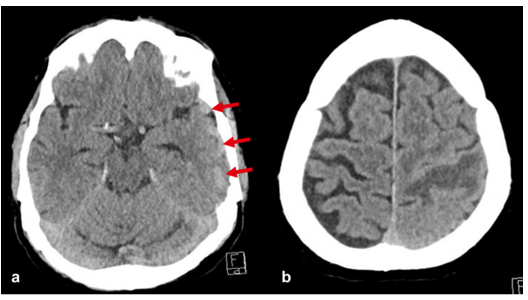
Figure 1: a) Slightly hyperdense tubular structures in the left temporal lobe (arrows). b) Hypodense area in the left central area.
We then immediately started a contrasted enhanced MR examination including standard imaging sequences, arterial and venous 3 D time-of-flight (TOF) MRA, susceptibility-weighted imaging (SWI), and contrast-enhanced T1-weighted 3D gradient-echo.
Standard MR imaging sequences before i.v. administration of contrast agent (FLAIR, T2-weighted spin-echo, T1-weighted spin-echo) revealed areas with late subacute brain hemorrhage in the left central region and the left frontal lobe (Figure 2).

Figure 2: Late subacute hemorrhage in the left central area and the left frontal lobe (arrows). a) T2-weighted turbo spin-echo, b) T1-weighted spin-echo before i.v. contrast agent administration.
Susceptibility weighted imaging showed multiple suspect vessels in the vicinity of the central hemorrhage (Figure 3) in the left hemisphere.
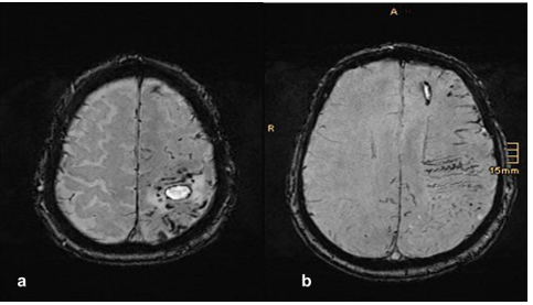
Figure 3: Susceptibility weighted imaging. Multiple suspect vessels are found in the area surrounding the central hemorrhage.
The arterial and the venous TOF-MRA, reconstructed with maximum-intensity projections and Cinematic Rendering [3,4] showed a suspicious vascular formation, probably of arterial origin, in the left occipital region and then strongly dilated veins (Figure 4).
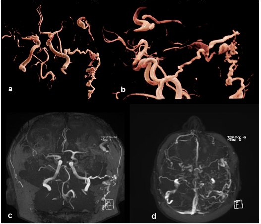
Figure 4: Suspicious arterial formation on the left occipital with subsequent dilated veins. a), b) arterial TOF-MRA reconstructed with Cinematic Rendering, b) arterial TOF-MRA reconstructed with maximum-intensity-projection, d) venous TOF-MRA reconstructed with maximum-intensity-projection.
The dilated veins in the left hemisphere were best seen in the contrast-enhanced T1-weighted 3D gradient-echo sequence (Figure 5).
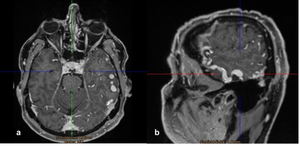
Figure 5: The contrast-enhanced T1-weighted 3D gradient-echo sequence best shows the multiple congested and tortuous veins in the left hemisphere.
Based on these MR findings, we suspected a vascular malformation, most likely in the sense of a dural arterio-venous fistula. Therefore, the next step was to perform an intra-arterial digital subtraction angiography to confirm this suspicion.
The intra-arterial digital subtraction angiography then verified a strong
arterio-venous fistula with feeders from the occipital artery and a petrous branch of the middle meningeal artery. There was a fistula at the inflow into the superficial temporal vein with retrograde filling in cortical veins and as well as a clearly dilated draining vein (Figure 6). Therefore, final diagnosis was occipital dural arterio-venous fistula on the left side, Cognard grade IV.
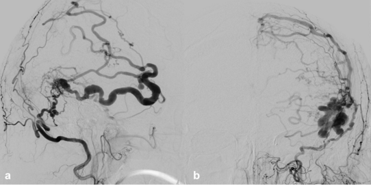
Figure 6: Intra-arterial digital subtraction angiography of the dural arterio-venous fistula Cognard grad IV. a) lateral view, b) ap view.
Dural arterio-venous fistulas are arteriovenous shunts, supplied by a dural artery to a dural venous channel, mostly located near a major venous sinus. These fistulas are divided into 5 grades:
| I | Normal antegrade flow into dural sinus
|
| II | a. Retrograde flow into sinus(es) b. Retrograde filling of cortical vein(s) c. Retrograde drainage into sinus(es) and cortical veins |
| III | Direct drainage into cortical veins without venous ectasia |
| IV | Direct drainage into cortical veins with venous ectasia >5 mm and 3x larger than diameter of draining vein |
| V | Drainage to spinal perimedullary veins |
Most of them seem to be acquired (i.e. trauma, surgical, chronic infection or sinus thrombosis), some of them are congenital. In the pediatric population, most of them are associated with venous anomalies.
Dural arterio-venous fistulas are diagnosed in all age groups, mainly in the fifth and sixth decades of life. There is a higher incidence in female patients (female to male ratio 2:1), estimated incidence is 0,17 case in 100000 population and they represent 10 – 15 % of cerebral vascular malformations [2].
Disclosure
All co-authors do not report conflicts of interest.
Dear Editorial Team, Clinical Medical Reviews and Reports. My experience with the journal was highly positive. The peer-review process was rigorous, constructive, and completed in a timely manner. The reviewers provided valuable comments that helped improve the quality and clarity of our manuscript. The editorial office was professional, responsive, and supportive throughout all stages of the publication process. Communication was clear and efficient, and any questions were addressed promptly. Overall, I found the journal to maintain high scientific standards and an excellent publication workflow. I would be pleased to consider submitting future work to this journal. Best wishes from, Elena Popa.

It was my pleasure to submit my testimonial concerning the Reviewer Board of our Scientific Journal “Brain and Neurological Disorders”. The Reviewers focused on some modifications and their contribution was helpful. The ladies of our Editorial Office were also supported my efforts. It was my honor to have such a co-operation and I am looking forward for more collaboration.

Dear Grace Pierce, Editorial Coordinator of Journal of Clinical Research and Reports, Thank you for the speedy and efficient peer review process. I appreciate the fact that your peer reviewers do not take months to respond like with some other journals. I would also like to thank the editorial office for responding quickly to my questions. It is an excellent journal. I plan to submit more manuscripts in the future. Best wishes from, Robert W. McGee

Dear Grace Pierce, Editorial Coordinator of Journal of Clinical Research and Reports, Working with you and your team on our recent publication in JCRR has been a truly wonderful and enjoyable experience. The responses were prompt, and the reviewers were patient, constructive, and highly professional. One reviewer in particular gave me the feeling that a professor was carefully reading and commenting on my coursework, which was deeply touching. The entire process was straightforward and hassle‑free, with no tedious online forms to complete. I highly recommend this journal. Best wishes from, DR Aibing Rao, Head of R&D

I Appreciate the Opportunity to Share my Experience with the Journal of Clinical Research and Reports. The peer review process was timely and constructive, and the feedback provided helped improve the quality of our manuscript. The editorial office was professional, responsive, and supportive throughout the process, ensuring smooth communication and efficient handling of the submission. Overall, it was a positive experience collaborating with your team.

Dear Mercy Grace, Editorial Coordinator of Obstetrics Gynecology and Reproductive Sciences, We would like to express our gratitude for your help at all stages of publishing and editing the article. The editors of the magazine answer all the necessary questions and help at every stage. We will definitely continue to cooperate and publish other works in the Obstetrics Gynecology and Reproductive Sciences! Best wishes from, Alla Konstantinovna Politova,
