
Research Article | DOI: https://doi.org/10.31579/2641-0419/362
Japan Association of Science Specialists.
*Corresponding Author: Shoshi Inooka, Japan Association of Science Specialists.
Citation: Shoshi Inooka (2024), Cell ring-like objects created in agar cultures of synthetic DNA (Streptomyces) crown cells with monolaurin, J Clinical Cardiology and Cardiovascular Interventions, 7(7); DOI: 10.31579/2641-0419/362
Copyright: © 2024, Shoshi Inooka. This is an open access article distributed under the Creative Commons Attribution License, which permits unrestricted use, distribution, and reproduction in any medium, provided the original work is properly cited.
Received: 29 January 2024 | Accepted: 01 July 2024 | Published: 08 July 2024
Keywords: synthetic DNA (Streptomyces) crown cells; agar plate cultures; Sphingosine-DNA; cells; cell ring-like objects; monolaurin
Synthesized DNA crown cells (artificial cells), which can proliferate within egg white in vivo, can be prepared in vitro using sphingosine (Sph)-DNA-adenosine-monolaurin compounds. Synthesized DNA crown cells form assemblies that proliferate in the presence of monolaurin and can be observed in agar cultures. In previous experiments on synthetic DNA (E. coli/Human placenta/Ascidian) crown cells, the proliferation of objects similar in appearance to thalli, double-cells, morning glory and daffodil inflorescences was observed on agar plates.
In the present experiments using synthetic DNA (Streptomyces) crown cells and monolaurin, various objects and examples of cell proliferation were observed on the agar plates. Here, the microscopic appearance of cell ring-like objects is described.
Artificial cells potentially useful for the study of a wide variety of problems in the life science. Until recently, most attempts in generate artificial cells have resulted in only vesicle like cells that package several cellular components such as DNA and enzymes. Such vesicles can do many things that cells can do, including transcribe RNA, translate proteins, and generates ATP, recently, division and replication of DNA have been observed in such vesicles.
Until the first report by the present author in 2012 (1), fully functional (self replicating) cells had not been produced. Moreover, in 2016 (2), the principal methods to prepare generated artificial cells which can be multiply within egg white and cultivated in vitro were reported. In 2016, artificial cells referred to as DNA crown cells were reported by the present author, (3). The cells derive their name from the facts that their exterior consists of DNA.
DNA crown cells were synthesized using four common commercial compounds; Sphigosine (Sph), DNA, adenosine and monolaurin using methods developed based on the author’s experience. of my research. Briefly, Sph, which is the core of sphingolipids, was synthesized using serine and metabolized immediately to form ceramides (4). Non-metabolized Sph (free Sph) exsits within cells in small amount. Free Sph within cells increased as a result of the catabolism of sphingomyelin which consisted of cell membrane and occured with cell injury,
Also, cell injury can increase the amount of of DNA within cells. The chance of binding to DNA of Sph can result in the formation. This free transfer of Sph from cell to cell (5, 6) can cause cell injury (7), because Sph was cytotoxic. Sph-DNA particles were found to be capable of binding to various cell components and cultivable particles were formed.(7, 8).
The method to prepare DNA crown cells is as follows. 1) Sph and DNA were mixture. 2) adenosine was added to the mixtures. 3) monolaurin is then added and results in the formation of synthetic DNA crown cells 4) DNA crown cells were formed from synthetic DNA crown cells within egg white.
Thus, synthetic DNA crown cells can be prepared using Sph-DNA and adenosine-monolaurin (A-M) compounds, and these can develop into DNA crown cells within egg white or in vitro cultures (9, 10).
Such DNA crown cells formed assemblies or cells that proliferated in cultures with monolaurin in test tubes (11-13) and the generated cells could also be cultivated on agar plates (14,15). In previous studies (16) , various types of objects, as well as cell proliferation, were observed when monolaurin-treated synthetic DNA (E. coli/Human placenta/Ascidian) crown cells were cultivated with or without egg white on agar plates and examined microscopically.
The present experiments examined whether such objects were formed when synthetic DNA (Streptomyces) crown cells were cultured with monolaurin on agar plates. Numerous kinds of objects and cell proliferation were observed. Here, the microscopic appearance of objects that are similar in appearance to cell rings are described.
Materials
The materials used were the same as those employed in previous studies (9, 10): Sph (Tokyo Kasei, Japan), DNA (From Streptomyces), adenosine (Sigma-Aldrich; Wako, Japan), monolaurin (Tokyo Kasei), and adenosine-monolaurin (A-M), a compound synthesized from a mixture of adenosine and monolaurin (9, 10). Monolaurin solutions were prepared to a final concentration of 0.1 M in distilled water. Agar plates were prepared using standard agar medium (SMA) (AS ONE, Japan).
Methods
Preparation of synthetic DNA (Streptomyces) crown cells (9, 10).
Step.1 180 µL of Sph (10 mM) and 50 µL of DNA (0.05 µg/µL) were combined, and the mixture was heated and cooled twice.
Step 2. A-M solution (100 µL) was added and the mixture was incubated at 37°C for 15 min.
Step3. 30 µL of monolaurin solution was added and the mixture was incubated at 37°C for another 5 min.
Step. 4 The resulting suspension was used as the synthetic DNA (Streptomyces) crown cells. The culture of monolaurin with DNA (Streptomyces) crown cells on agar plates was performed as follows:
Microscopic observations
Objects on plates were observed directly under a light microscope and with the naked eye.
Figure 1 shows a photograph of an agar plate (No. 1) of synthetic DNA (Streptomyces) crown cells after 7 days of monolaurin culture. Circular brownish objects (arrow), visible with the naked eye, were observed in the center of the Petri dish, which had a diameter of 8.0 cm.
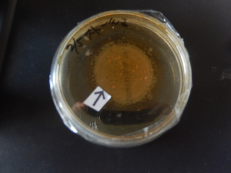
Figure 1. Photograph of an agar plate (No. 1) containing synthetic DNA (Streptomyces) crown cells after 7 days of monolaurin culture. Brownish objects (arrow) can be seen in the center of the Petri dish, which has a diameter of 8.0 cm.
Figure 2 shows a photograph of another agar plate (No. 3) containing synthetic DNA (Streptomyces) crown cells after 7 days of monolaurin culture. Cloudy, dot-like or round objects (arrow), visible with the naked eye, were observed on the Petri dish, which had a diameter of 8.0 cm.
Figure 3 shows the microscopic appearance of synthetic DNA (Streptomyces) crown cells shown in Fig. 1 after 5 days of culture with monolaurin. A ring-like object consisting of numerous cell-like objects was observed (arrow a). An independent cell (approximately 7–8 mm, arrow b) was also observed, and large objects were observed within the ring (arrow c). The approximate size in diameter of the ring-like object (arrow a) was 700–720 mm.
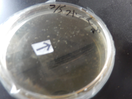
Figure 2. Photograph of an agar plate (No. 3) containing synthetic DNA (Streptomyces) crown cells after 7 days of monolaurin culture. Dot-like or round (arrow) objects can be seen in the Petri dish, which has a diameter of 8.0 cm.
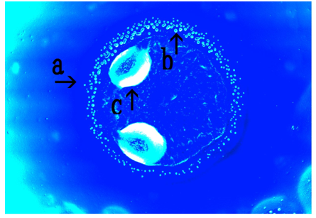
Figure 3. Micrograph of the synthetic DNA (Streptomyces) crown cells shown in Figure. 1 after 5 days of monolaurin culture.
A ring-like object (arrow a) and a cell are shown (arrow b). Objects can be observed within the ring (arrow c). The approximate diameter of the ring-like object (arrow a) is 700–720 mm. The cell (arrow b) measures approximately 7–8 mm.
Figure 4 shows the microscopic appearance of synthetic DNA (Streptomyces) crown cells shown in Fig. 1 after 5 days of culture with monolaurin. In addition, an object consisting of numerous cell-like objects was observed (arrow a). The cell-like objects (arrow c) appeared as though they were flowing and as being aggregated. These cell-like objects (arrow b) were approximately 7–8 mm in diameter
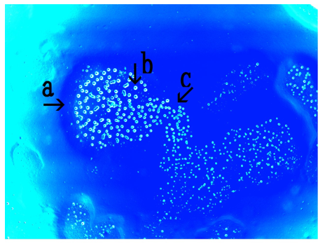
Figure 4. Microscopic appearance of the synthetic DNA (Streptomyces) crown cells shown in Figure. 1 after 5 days of monolaurin culture. An object is shown (arrow a). Cell-like objects (arrow c) are shown. The diameter of the object (arrow b) is approximately 7–8 mm.
Figure 5 shows the microscopic appearance of synthetic DNA (Streptomyces) crown cells shown in Fig. 2 after 7 days of culture with monolaurin. Cell ring-like objects (arrow a), which appeared to be composed of numerous cell-like objects (arrow b) were observed. The objects (arrow a) were approximately 540–600 mm in diameter.
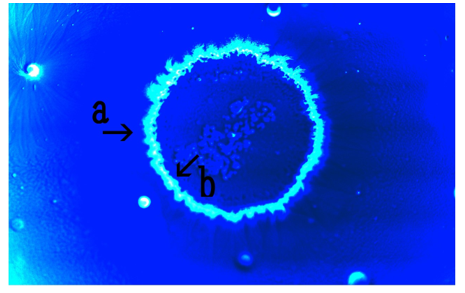
Figure 5. Microscopic appearance of synthetic DNA (Streptomyces) crown cells shown in Fig. 2 after 7 days of monolaurin culture.
Ring-like objects (arrow a) constructed of many cell-like objects (arrow b) are shown. The diameter of the object (arrow a) is approximately 540–600 mm.
Figure 6 shows the microscopic appearance of synthetic DNA (Streptomyces) crown cells shown in Figure. 2 after 7 days of culture with monolaurin. Objects similar in appearance to a divided ring, consisting of numerous cell-like objects were observed (arrows a). The objects, which had an approximate diameter of 500–520 mm (arrow a), appear empty (arrow c).
The single cell-like object (arrow c) measured approximately 18–20 mm.
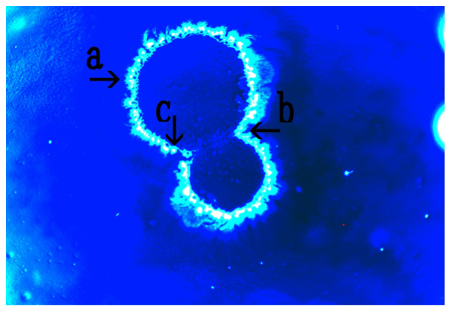
Figure 6. Microscopic appearance of synthetic DNA (Streptomyces) crown cells shown in Figure. 2 after 7 days of monolaurin culture.
An object like divided ring which consists of numerous cell-like objects (arrow b) is shown (arrow a). The diameter of the object is approximately 500–520 mm (arrow a). The size of the object (arrow c) is approximately 18–20 mm.
Figure 7 shows the microscopic appearance of synthetic DNA (Streptomyces) crown cells at 7 days after the addition of monolaurin (Plate was not shown).
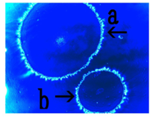
Figure 7: Microscopic appearance of synthetic DNA (Streptomyces) crown cells after 7 days of monolaurin culture.
Two objects are shown (arrows a and b). The diameter of the objects are approximately 440–460 mm (arrow a) and 280–290 mm (arrow b), respectively.
Two ring-like objects consisting of numerous cell-like objects were observed (arrows a and b); the approximate diameter of these objects was 440–460 mm (arrow a) and 280–290 mm (arrow b), respectively.
Figure 8 shows the microscopic appearance of synthetic DNA (Streptomyces) crown cells in egg white cultures for 7 days after the treatment of monolaurin (The method was not described). Objects consisting of numerous bamboo broom-like objects (arrows b and c) were observed (arrow a). Objects similar in appearance to single cells (approximate size 5–8 mm) were also observed (arrow d). These objects measured approximately 6.1–7.4 mm in diameter (arrow a)
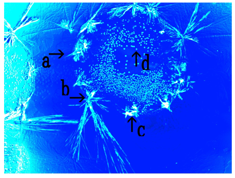
Figure 8. Microscopic appearance of synthetic DNA (Streptomyces) crown cells after 7 days of culture in monolaurin and egg white.
An object consisting of many bamboo broom-like objects (arrows b and c) is shown (arrow a). A single cell-like object measuring approximately 5–8 mm is shown (arrow d).
An object measuring approximately 6.1–7.4 mm in diameter (arrow a) is shown.
In previous experiments using synthetic DNA (E. coli/Human placenta/Ascidian) crown cells, objects similar in appearance to thalli, double cells, morning glory and daffodils were observed on agar plates (16,17, 18). The present experiments examined whether similar objects were observed in agar cultures of synthetic DNA (Streptomyces) crown cells in monolaurin.
Objects, visible with naked eye were observed on all plates (three plates); however, the appearance of the objects differed between plates. The reasons for these differences were not clear, because there is no evidence of the mechanisms underlying object formation. However, it was certain that they formed as a result of associations between the synthetic DNA crown cells and their related components, including monolaurin and egg white.
In this case, the formation of the assemblies in the different cultures of crown cells occurred immediately after the samples were prepared, as described previously. Therefore, ring-like objects (Figure. 1) may form as a result of the association of not cells, but assemblies with monolaurin, which then subsequently increase in size.
Ring-like objects (Figure. 3, arrow a) possibly consisting of numerous cell-like objects were observed (Figure. 4, arrows b and c). There was no evidence of the mechanisms underlying the formation of ring-like objects.
However, round objects that proliferated in the culture were often observed in cultures of synthetic DNA crown cells in monolaurin. Hence, the proliferated cells form rings and may form ring-like objects (Figure. 3, arrow a). Here, these objects are referred to as proliferated cell rings (pr-cell rings).
Large objects were observed within the ring-like objects; however, the identity of these objects is unclear (Figure. 3 arrow c). It is possible that
cell ring-like objects may enclose a kind of assembly composed of Sph/DNA: adenosine-monolaurin. Also, another type of cell ring-like object that consists of numerous cell-like objects (Figure. 5, arrow b) was observed (Figure. 5, arrow a). There were differences between both types of cell ring-like objects.
Cell-like objects (Figure. 5 b) that were constructed of ring-like objects (Figure. 5 a) were often observed in cells that were prepared as cell samples. Though there is no evidence, these findings imply that the objects are not constructed of proliferated cells, but original cells. Here, to distinguish between these two ring-like objects, they are named original cell ring (or-cell ring) and proliferated cells (pr-cell ring). Or-cell rings may be formed by binding of original synthetic DNA (Streptomyces) crown cells to form rings.
The ring may grow and divide (Figure. 6, arrow a) to form a new ring (Fig. 7 arrows a and b), implying that or-cell rings multiply and form new ring-like objects. However, this remains hypothetical.
Thus, two types of ring-like objects are introduced. One type consists of objects that are comprised of proliferated cells, the other type consists of original cells. On the other hand, various assemblies and/or objects of various sizes and shapes were formed when synthetic DNA crown cells were mixed with monolaurin or egg white. Here, although it is not described, another type of ring-like object was observed (Figure. 8). The outside was comprised of objects that were similar in appearance to bamboo brooms. Such objects were often observed in the mixtures of monolaurin-treated synthetic DNA (Streptomyces) crown cells cultured with egg white. Large spaces could be observed with the naked eye (Figs. 1 and 2), and numerous kinds of objects or the numerous features of cell proliferation could be observed within these spaces. Not all aspects of cell-proliferation could be described here.
Most synthetic DNA crown cells formed objects and proliferated. However, the formation of such ring-like objects appears to be rare, as these characteristics were not observed in cultures of other synthetic DNA (E. coli/Human placenta/Ascidian) crown cells. It was not clarified in the present study whether this type of ring formation is characteristic to synthetic DNA (Streptomyces) crown cells.
As described previously, it is important to clarify whether the objects being observed are living. To date, including this study, it has been demonstrated that characteristic objects were formed in four kinds of synthetic DNA (E. coli/Human placenta/Ascidian) crown cells.
Future research will clarify whether synthetic DNA (tumor cells) crown cells form characteristic objects. In addition, experiments will be conducted to clarify whether living cells were contained in the objects observed with the naked eye (Figure. 1 and 2).
These objects have been named as follows: Pro-cell ring Bacteria (Streptomyces) SDCCM (p CR) and Or-cell ring Bacteria (Streptomyces) SDCCM (o-CR). Here, Streptomyces in brackets is the name of the DNA source, SDCC refers to synthetic DNA (crown cells), and M refers to monolaurin. If cells were treated with both monolaurin and egg white, then they are named SDCCME.
Unlimited types of DNA crown cells can be prepared and cultivated (1922).
These cells could be used in a variety of practical fields, including industry, life science, medical science, and may contribute to the welfare of human beings.
The experiments described in this Special Discussion aim to clarify whether such cells can be applied to practical fields, as this is currently unclear. Potential applications in the field of clinical medicine are considered, including specific applications to humans, neurite elongation and intestinal bacteria.
Potential application to humans (23)
There were many chance of binding to DNA with Sph within nucleus or exosome.
In the nucleus, Sph may be derived from shingomyerin in the nucleus membrane and this Sph bind to DNA within nucleus.
In addition, many substances such as protein, DNA, and m-RNA exist in the exosome, and there is a possibility of these molecules bindings to DNA bound to Sph from the membrane. Sph-DNA particles may be released from nucleus and with or without exosome. These conditions could be favorable for the development of DNA crown cells in certain locations, such as where specific lipids (as monolaurin) are present.
Potential applications in neurite elongation
When Sph-DNA particles are cultivated with cells, tube like objects was formed. (7). Objects in synthetic DNA crown cells also became elongated and form long, thin fiber-like tubes (13). Synthetic DNA crown cells have be shown to cause elongation of Bacillus subtilis (24).
These findings indicate that components of DNA crown cells have the abilities to elongate part of organisms, forming objects similar to axons.
Potential application to intestinal bacteria.
The gut flora of animals and human comprises thousand of different kinds of bacteria, many of which have not been identified. Similarity, a wide variety of bacteria exist on Earth, suggesting that many kinds of new bacteria are produced in nature. Previous studies, as well as the present experiments (16-18), suggested that new organisms that are similar to bacteria, which as synthetic DNA crown cells, can arise when culture conditions are favorable. These new organisms can inhabit enviorments such as the gut or other habitats on Earth. New findings in this field could therefore benefit both human and the plant we inhabit.
Dear Editorial Team, Clinical Medical Reviews and Reports. My experience with the journal was highly positive. The peer-review process was rigorous, constructive, and completed in a timely manner. The reviewers provided valuable comments that helped improve the quality and clarity of our manuscript. The editorial office was professional, responsive, and supportive throughout all stages of the publication process. Communication was clear and efficient, and any questions were addressed promptly. Overall, I found the journal to maintain high scientific standards and an excellent publication workflow. I would be pleased to consider submitting future work to this journal. Best wishes from, Elena Popa.

It was my pleasure to submit my testimonial concerning the Reviewer Board of our Scientific Journal “Brain and Neurological Disorders”. The Reviewers focused on some modifications and their contribution was helpful. The ladies of our Editorial Office were also supported my efforts. It was my honor to have such a co-operation and I am looking forward for more collaboration.

Dear Grace Pierce, Editorial Coordinator of Journal of Clinical Research and Reports, Thank you for the speedy and efficient peer review process. I appreciate the fact that your peer reviewers do not take months to respond like with some other journals. I would also like to thank the editorial office for responding quickly to my questions. It is an excellent journal. I plan to submit more manuscripts in the future. Best wishes from, Robert W. McGee

Dear Grace Pierce, Editorial Coordinator of Journal of Clinical Research and Reports, Working with you and your team on our recent publication in JCRR has been a truly wonderful and enjoyable experience. The responses were prompt, and the reviewers were patient, constructive, and highly professional. One reviewer in particular gave me the feeling that a professor was carefully reading and commenting on my coursework, which was deeply touching. The entire process was straightforward and hassle‑free, with no tedious online forms to complete. I highly recommend this journal. Best wishes from, DR Aibing Rao, Head of R&D

I Appreciate the Opportunity to Share my Experience with the Journal of Clinical Research and Reports. The peer review process was timely and constructive, and the feedback provided helped improve the quality of our manuscript. The editorial office was professional, responsive, and supportive throughout the process, ensuring smooth communication and efficient handling of the submission. Overall, it was a positive experience collaborating with your team.

Dear Mercy Grace, Editorial Coordinator of Obstetrics Gynecology and Reproductive Sciences, We would like to express our gratitude for your help at all stages of publishing and editing the article. The editors of the magazine answer all the necessary questions and help at every stage. We will definitely continue to cooperate and publish other works in the Obstetrics Gynecology and Reproductive Sciences! Best wishes from, Alla Konstantinovna Politova,
