
Case report | DOI: https://doi.org/10.31579/2578-8965/011
Siraj Hospital,4th Nizampura,Vanjarpatti Naka,Bhiwandi,Thane,Maharashtra state
*Corresponding Author: Maheshgir S.Gosavi, Siraj Hospital,4th Nizampura,Vanjarpatti Naka, Bhiwandi, Thane, MH, India.
Citation: Maheshgir S.Gosavi, Tubal Twin Ectopic Gestation Report of Three Different Cases .J. Obstetrics Gynecology and Reproductive Sciences, 2(2). doi.10.31579/2578-8965/011
Copyright: © S.Gosavi, Tubal Twin Ectopic Gestation Report of Three Different Cases .J. Obstetrics Gynecology and Reproductive Sciences, doi.10.31579/2578-8965/011
Received: 30 May 2018 | Accepted: 18 June 2018 | Published: 25 June 2018
Keywords: spontaneous, tubal twin ectopic, transvaginal usg
Gestation outside the uterine cavity in which the implantation occurs in any tissue other than the endometrium is referred as ectopic pregnancy. The most place for occurring ectopic pregnancy (97% of cases) is the fallopian tubes including ampulla (55%), isthmus (25%), and fimbria (17%), and in 3% of patients ectopic pregnancy occurs in the abdominal cavity, ovary, or cervix.[1]The tubal twin ectopic pregnancy is a rare condition, and the first unilateral tubal twin was reported by De Ott in 1891, and the first live twin tubal ectopic pregnancy was reported in 1944 [2].A live tubal twin ectopic pregnancy is a very rare condition and among >100 reports of tubal twin pregnancies, till now, only 8 cases were live [3]. Early diagnosis and treatment of women with tubal twin ectopic pregnancy is very important and may decrease the risk of tubal rupture.. I present three cases of tubal twin ectopic gestation. In the first case, spontaneous unilateral live tubal twin ectopic gestation .The second and third cases spontaneous ruptured twin ectopic gestation .All three cases were successfully managed and there was no history of assisted reproductive techninique fertilization or pelvic inflammatory disease .
Objective : The above case report was to describe the ultrasonography findings of tubal twin ectopic gestation , to describe various surgical techniques in management of the condition.
Gestation outside the uterine cavity in which the implantation occurs in any tissue other than the endometrium is referred as ectopic pregnancy The most place for occurring ectopic pregnancy (97% of cases) is the fallopian tubes including ampulla (55%), isthmus (25%), and fimbria (17%), and in 3% of patients ectopic pregnancy occurs in the abdominal cavity, ovary, or cervix.[1] The tubal twin ectopic pregnancy is a rare condition, and the first unilateral tubal twin was reported by De Ott in 1891, and the first live twin tubal ectopic pregnancy was reported in 1944 [2].A live tubal twin ectopic pregnancy is a very rare condition and among >100 reports of tubal twin pregnancies, till now, only 8 cases were live.[3] Early diagnosis and treatment of women with tubal twin ectopic pregnancy is very important and may decrease the risk of tubal rupture.. The present report describes a successful management of spontaneous twin tubal ectopic pregnancies with no history of assisted reproductive technique fertilization or pelvic inflammatory disease. Objective of the above case report was to describe the ultrasonography findings of tubal twin ectopic gestation, to describe various surgical techniques in management of the condition
Abbreviations: USG -- Ultrasonography EP – Ectopic Pregnancy
Case Presentation:
A 35 year old female G2P1L1A0, came to casualty at midnight with pain in abdomen since three days with 8 weeks of amenorrhea and bleeding per vaginum since morning. She had history of two spontaneous abortions. Upon examination, her vitals were stable. Abdomen examination was unremarkable .Upon per vaginum examination the cervical movement was tender, the uterus was bulky and soft, and bogginess and tenderness were felt in the right adnexa. However, her urine pregnancy test was positive.
Ultrasonography with doppler study showed a uterus of 8.3x 5.4x 4.2 cms with endometrial thickness of 10 mm and there was evidence of large heterogeneous lesion in the right adnexa of a size 7.1x 5.1 cms, with sac like structure within which two foetal poles measuring 1.4 cm = 7 weeks and five days .Fetal cardiac activity was appreciated .Mild free fluid was seen in the abdomen. An impression of live twin ectopic pregnancy in the right adnexa was made with mild free fluid in an abdomen. A pseudosac like structure was noticed in endometrial cavity .Both ovaries were visualised separately from the right adnexal mass.
In view of the Twin live ectopic pregnancy she was immediately operated. Intraoperatively, the ampullary part of the right fallopian tube was found showed a sac like structure. A right salphingotomy was done. She was discharged on the 7 th day. On cut section of the sac there was evidence of two embryos Pathological evaluation of the surgical specimen showed a diamniotic monochorionic twin’s pregnancy within the fallopian tube and measurement of the twin feti estimated their gestational age at 7 weeks.

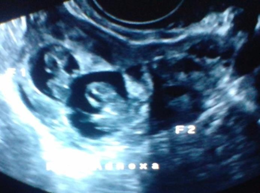


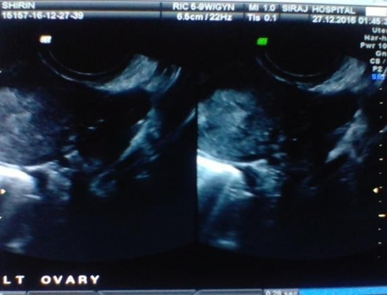
Figure1:Shows Right adnexal sac with Twin live ectopic gestation of approximately 7wks 5 days .No intrauterine pregnancy .Both ovaries are separately visualised.
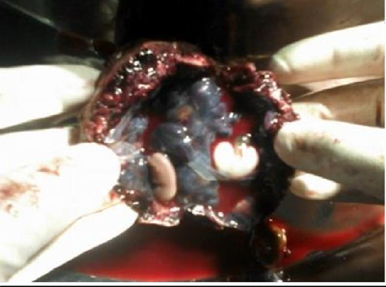
Figure 2: Pathological evaluation of the surgical specimen showed a diamniotic monochorionic twins pregnancy
A 30 year old female, G2L1A0 came to casualty with severe pain in abdomen since two days with 6 weeks of amenorrhea and bleeding per vaginum since morning .She had one living issues with full term normal deliveries.. Upon examination, her vitals were stable. Abdomen examination was unremarkable. However, her urine pregnancy test was positive .Spontaneous pregnancy there was no history of induction of ovulation by drugs or artificial reproductive techniques
The patient was taken for ultrasonography
Transvaginal Sonography showed a heterogeneous mass in right adnexa measuring approximately 7.3 x 4.6 cm The mass showed irregular Gestational sac measuring approximately 0.3 cm corresponding to 5 weeks and another irregular sac measuring 0.5 cm corresponding to 5weeks 2 days Right ovary was visualised separately from this mass .
Left ovary was visualised and appears normal .Moderate hemoperitoneum was seen .Uterus was bulky however there was no intrauterine gestational.
The diagnosis of right adnexal ruptured ectopicgestation was made .Preliminary investigations were done and patient was taken up for laparotomy in view of right adnexal mass and hemoperitoneum.
Per operatively gross morphology was: Right adnexal complex mass with moderate hemoperitoneum. Right salpinghostomy was performed. Patient’s post-operative period was uneventful
Histopathology report came out to be right adnexal two gestational sacs were noted
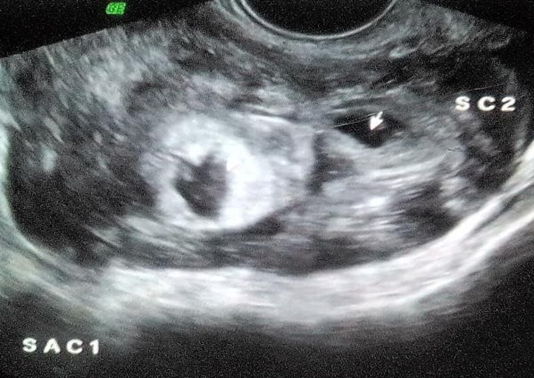
Figure 3: Trans vaginal Sonography shows Right adnexa heterogenous mass with two irregular sacs and hemoperitoneum. Left ovary is visualised
A 36 year old female, G3L2A0 came to casualty with severe pain in abdomen since three to four days with 6 weeks of amenorrhea and bleeding per vaginum since morning. Upon examination, her vitals were stable. Abdomen examination was unremarkable .Upon per vaginum examination the cervical movement was tender, the uterus was bulky and soft, and bogginess and tenderness were felt in the both adnexae. However, her urine pregnancy test was positive.
Spontaneous pregnancy there was no history of induction of ovulation by drugs or artificial reproductive techniques
The patient was taken for ultra sonography
Transvaginal Sonography showed a heterogeneous mass in right adnexa with irregular Gestational sac measuring approximately 1.0 cm corresponding to 5 weeks 5 days Right ovary was visualised separately from this mass.
Left adnexa irregular Gestational sac measuring approximately 0.8cm corresponding to 5 weeks 4days. Left ovary was visualised separately from this mess .Mild to Moderate hemoperitoneum was seen .Uterus was bulky however there was no intrauterine gestational sac.
The diagnosis of Bilateral adnexal ruptured ectopic gestation was made .Preliminary investigations were done and patient was taken up for laparotomy in view of bilateral adnexal masses and hemoperitoneum.
Per operatively gross morphology was: Bilateral adnexal complex masses with moderate hemoperitoneum. Bilateral salpingotomy was performed. Patient’s post-operative period was uneventful
Histopathology report came out to be gestational sacs in bilateral adnexa.
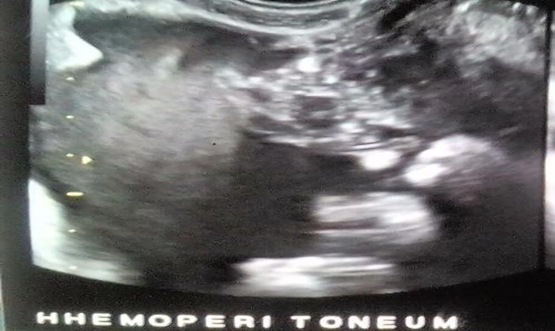

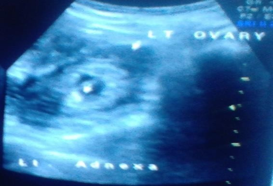
Figure 4: Trans vaginal Sonography shows both adnexae heterogeneous mass with two irregular sacs and hemoperitoneum. Both ovaries are visualised
It is found that the incidence of ectopic pregnancy has risen fourfold compare with the rates of 1970 in the United States (from 0.5% of all pregnancies in 1970 to 2% in 1992),[4] however, unilateral tubal twin ectopic pregnancy are still rare and until now about 100 cases have been reported in the literature.[5] Moreover, fetal cardiac activity has been reported in about <10>3]
The twin pregnancy is more prevalent among patients with a family history of multiple pregnancies.[6] The incidence of ectopic pregnancy is around 1-2% of all pregnancies[7] and the incidence of spontaneous twin pregnancy is 1:90.[8] However, live unilateral tubal twin ectopic pregnancy is a very rare condition and occurs in about 1:125,000 of pregnancies.[9] Several risk factors for tubal ectopic pregnancy were identified including active and passive cigarette/tobacco smoking, tubal damage as a result of surgery or infection (particularly Chlamydia trachomatis), and in vitro fertilization.[7] Furthermore, some authors indicated that the number of prior deliveries, ectopic pregnancy , and spontaneous or induced abortions were strongly associated with occurrence of ectopic pregnancy.[10,11. No such risk factors were present in our patients .It has been demonstrated that the history of ectopic pregnancy leads to an increased recurrence rate of about 10% and 25% for one and two/more previous ectopic pregnancy, respectively.[12]
A history of pelvic pain along with amenorrhea and vaginal bleeding are found in 45% of ectopic pregnancies [2] and probability of ectopic pregnancy in a patient with only abdominal pain and vaginal bleeding is 39%. The likelihood of ectopic pregnancy rises to 54% if the patient has other risk factors, including history of tubal surgery, previous ectopic pregnancy, or pelvic inflammatory disease. [1]
In addition, ultrasound evaluations have facilitated the early EP diagnosis which may lead to a reduction in maternal mortality and morbidity. Also, use of β-hCG assay, especially serial measurements, may improve these evaluations. Studies demonstrated that a β-hCG value of above 1500 mIU/ml corresponds to an approximately 91.5
A spontaneous twin tubal pregnancy can occur in patients who have no known predisposing factor. Early diagnosis has made this disorder amenable to appropriate treatment. The high-resolution transvaginal sonography is very helpful in the diagnosis of this condition. Twin-ectopic gestations are extremely rare but there aretreatment options. These have typically been classified as either conservative or surgical.
Dear Editorial Team, Clinical Medical Reviews and Reports. My experience with the journal was highly positive. The peer-review process was rigorous, constructive, and completed in a timely manner. The reviewers provided valuable comments that helped improve the quality and clarity of our manuscript. The editorial office was professional, responsive, and supportive throughout all stages of the publication process. Communication was clear and efficient, and any questions were addressed promptly. Overall, I found the journal to maintain high scientific standards and an excellent publication workflow. I would be pleased to consider submitting future work to this journal. Best wishes from, Elena Popa.

It was my pleasure to submit my testimonial concerning the Reviewer Board of our Scientific Journal “Brain and Neurological Disorders”. The Reviewers focused on some modifications and their contribution was helpful. The ladies of our Editorial Office were also supported my efforts. It was my honor to have such a co-operation and I am looking forward for more collaboration.

Dear Grace Pierce, Editorial Coordinator of Journal of Clinical Research and Reports, Thank you for the speedy and efficient peer review process. I appreciate the fact that your peer reviewers do not take months to respond like with some other journals. I would also like to thank the editorial office for responding quickly to my questions. It is an excellent journal. I plan to submit more manuscripts in the future. Best wishes from, Robert W. McGee

Dear Grace Pierce, Editorial Coordinator of Journal of Clinical Research and Reports, Working with you and your team on our recent publication in JCRR has been a truly wonderful and enjoyable experience. The responses were prompt, and the reviewers were patient, constructive, and highly professional. One reviewer in particular gave me the feeling that a professor was carefully reading and commenting on my coursework, which was deeply touching. The entire process was straightforward and hassle‑free, with no tedious online forms to complete. I highly recommend this journal. Best wishes from, DR Aibing Rao, Head of R&D

I Appreciate the Opportunity to Share my Experience with the Journal of Clinical Research and Reports. The peer review process was timely and constructive, and the feedback provided helped improve the quality of our manuscript. The editorial office was professional, responsive, and supportive throughout the process, ensuring smooth communication and efficient handling of the submission. Overall, it was a positive experience collaborating with your team.

Dear Mercy Grace, Editorial Coordinator of Obstetrics Gynecology and Reproductive Sciences, We would like to express our gratitude for your help at all stages of publishing and editing the article. The editors of the magazine answer all the necessary questions and help at every stage. We will definitely continue to cooperate and publish other works in the Obstetrics Gynecology and Reproductive Sciences! Best wishes from, Alla Konstantinovna Politova,
