
Case report | DOI: https://doi.org/10.31579/2578-8868/271
¹ Instituto de Assistência Médica ao Servidor Público Estadual, Sao Paulo, Sao Paulo, Brazil.
² Universidade Federal de São Paulo, Sao Paulo, Sao Paulo, Brazil.
*Corresponding Author: Nilton Amorim de Souza , Instituto de Assistência Médica ao Servidor Público Estadual, Sao Paulo, Sao Paulo, Brazil.
Citation: Ambiel Magalhães LZ, De Souza NA (2023), Case Report Recurrent Painful Ophthalmoplegic Neuropathy, J. Neuroscience and Neurological Surgery, 13(4); DOI:10.31579/2578-8868/271
Copyright: © 2023, Luciana Zelante Ambiel Magalhães. This is an open-access article distributed under the terms of The Creative Commons Attribution License, which permits unrestricted use, distribution, and reproduction in any medium, provided the original author and source are credited
Received: 19 June 2023 | Accepted: 24 August 2023 | Published: 01 September 2023
Keywords: recurrent painful ophthalmoplegic neuropathy ; ophthalmoplegic migraine ; headache ; ophthalmoplegia
This is a case report of recurrent painful ophthalmoplegic neuropathy (RPON), a rare disorder characterized by recurrent unilateral headaches and paralysis of the ocular cranial nerves. The condition was previously referred to as ophthalmoplegic migraine but was renamed due to potential misinterpretation of its underlying mechanisms. The case involves a 16-year-old female patient treated at the Hospital do Servidor Público Estadual de São Paulo. She experienced episodes of headache, periorbital pain, and cranial nerve paresis. Neurological examinations and imaging tests ruled out other causes. After diagnosis of RPON was considered, the patient received corticosteroid treatment. This article case report discusses the diagnostic criteria, clinical features, atypical aspects of the case, diagnostic challenges, treatment options, and the unresolved pathophysiology of RPON. Since RPON is a rare syndrome, research relies on observational studies and case reports like the one presented to enhance understanding and treatment approaches.
Recurrent painful ophthalmoplegic neuropathy (RPON) is a rare disorder described as recurrent bouts of unilateral headache associated to ipsilateral ocular cranial nerve paralysis. [1]
RPON was previously known as ophthalmoplegic migraine, it was renamed in 2013 because the use of the term “migraine” could associate RPON with an inaccurate pathophysiology, which may lead to inappropriate treatment strategies. [1–3].
The pathophysiology of RPON remains controversial and a better understanding of its mechanisms is still necessary to establish the most appropriate treatment, but it may be considered an idiopathic inflammatory neuropathy and usually responds to corticosteroid treatment. [4,5]
RPON usually presents in childhood, but there are reports of cases starting in adults, a systematic review of 84 cases of the so-called Ophthalmoplegic Migraine considered that two thirds of the affected patients were women and one third men. The median age of the first crisis was 8 years old and varied between 7 months and 50 years old. Most patients had third cranial nerve palsy (83%), followed by the sixth nerve palsy (20%) and, finally, the fourth cranial nerve palsy, affected in only 2% of patients, 6% of patients had involvement of multiple cranial nerves. [5]
The aim of the article is to present a case report of RPON, discuss the diagnostic criteria, clinical features, atypical aspects of the case, diagnostic challenges, treatment options, and the unresolved pathophysiology of RPON.
This case is a 16-year-old female patient with a history of polycystic ovary syndrome and migraine since the age of 14 years. She was admitted at the Hospital do Servidor Público Estadual de São Paulo (HSPE) in October 2018 with a complaint of diplopia and a different pattern of her usual headache, which was described as periorbital pain on the left, initiated the day before. Neurological examination revealed paresis of the left sixth cranial nerve.
Brain magnetic resonance (MRI) and magnetic resonance angiography were normal. The following laboratory tests were performed to rule out other causes: rheumatoid factor, antiRo, antiLa, erythrocyte sedimentation rate, glucose, immunoglobulin A and immunoglobulin G, C-reactive protein, complement C3, alphaglycoprotein, renal, hepatic and thyroid functions, the results were all normal. Negative serologies for syphilis, HIV, hepatitis B and C. Serology for cytomegalovirus and varicella zoster with negative IgM and positive IgG. The cerebrospinal fluid (CSF) examination was normal
The patient was discharged with complete resolution of headache and persistence of the sixth cranial nerve paresis. The diagnostic hypothesis was idiopathic sixth cranial nerve palsy. About four months after discharge, the abduction of her left eye was just slightly impaired and the holocranial headache was less frequent and less intense.
In May 2021, the patient had a second episode of periorbital headache starting the day before and diplopia starting on the same day of evaluation. The ophthalmoparesis of the sixth cranial nerve on the left had worsened. Brain and orbits MRI were performed, no abnormalities were observed. This time, the hypothesis of RPON was considered and the patient received prednisone 30 mg/day for 20 days with recommendation to gradually reduce the dose after this period and, to treat migraine episodes, the dose of topiramate was increased from 100 mg/day to 150 mg/day. [6-7]
About one month later, the patient reported improvement in the diplopia and the paresis had already remitted up to the intensity that affected her before the second episode (Figures 1, 2).
In October 2021 the patient maintained regular use of topiramate at a dose of 150 mg per day, even though, she reported that in the last three months she had twelve episodes of headache characterized as pulsatile, frontal or holocranial, intensity 8/10, without association with nausea and vomiting, with worsening pain on movement and associated with photophobia and phonophobia. The association of topiramate 150 mg/day with propranolol 40 mg/day was proposed for better control of migraine episodes. [6-7]
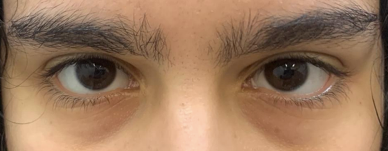
Figure 1: Primary gaze with mild esotropia of the left eye
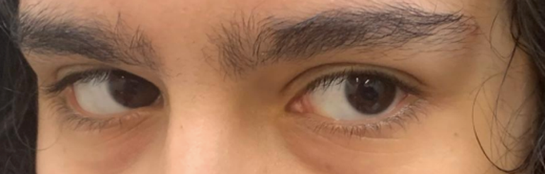
Figure 2: Conjugated gaze to the left with mild paresis of the VI cranial nerve at left
The diagnostic criteria for RPON are:
A. At least two attacks fulfilling criterion B;
B. Both of the following: Unilateral headache AND ipsilateral paresis of one, two, or all three ocular motor nerves;
C. Orbital, parasellar, or posterior fossa lesions has been excluded by appropriate investigation;
D. It cannot be better explained by another diagnosis of The International Classification of Headache Disorders 3rd edition (ICHD-3). [3]
The patient had two episodes of headache with ophthalmoparesis, fulfilling criteria A and B. The neuroimaging exams performed excluded lesions, vascular alterations or neoplasia that could cause ophthalmoparesis, fulfilling criterion C.
In a systematic review of 84 patients diagnosed with RPON, the clinical features observed were: most patients affected were female, the interval between the onset of headache and the observation of ophthalmoparesis had a median of 1.6 days, the headache remained for less time than ophthalmoparesis, and the headache that appears during the RPON outbreak is usually unilateral, on the same side of the ophthalmoparesis and has the maximum intensity in the peri- or retro-orbital region. The patient reported presented a difference of one day between the onset of the headache and the ophthalmoparesis, the headache of the RPON attack was periorbital and on the same side as the ophthalmoparesis and ceased before the VI cranial nerve paresis. [8,9]
The patient had some atypical features such as: the affected cranial nerve was the VI, usually, it is affected in only 20% of all cases, it is known that the third cranial nerve is the most frequently affected, in addition, she maintained the neurological deficit of the first episode for years, which occurs in only 30% of cases. [10].
The contrast-enhanced MRI scan of the brain did not show any remarkable features, a review of published cases of RPON demonstrates that cranial MRI performed in the acute phase may demonstrate increased enhancement and enlargement of the affected nerves with frequencies ranging from 25% to 81
In conclusion, RPON is a rare syndrome, this makes difficult to conduct experimental studies to determine the real effectiveness of the treatments used, and there is still much to be clarified about the pathophysiology of the disease, making researchers rely on the analysis of observational studies and reports of case like the one presented.
Dear Editorial Team, Clinical Medical Reviews and Reports. My experience with the journal was highly positive. The peer-review process was rigorous, constructive, and completed in a timely manner. The reviewers provided valuable comments that helped improve the quality and clarity of our manuscript. The editorial office was professional, responsive, and supportive throughout all stages of the publication process. Communication was clear and efficient, and any questions were addressed promptly. Overall, I found the journal to maintain high scientific standards and an excellent publication workflow. I would be pleased to consider submitting future work to this journal. Best wishes from, Elena Popa.

It was my pleasure to submit my testimonial concerning the Reviewer Board of our Scientific Journal “Brain and Neurological Disorders”. The Reviewers focused on some modifications and their contribution was helpful. The ladies of our Editorial Office were also supported my efforts. It was my honor to have such a co-operation and I am looking forward for more collaboration.

Dear Grace Pierce, Editorial Coordinator of Journal of Clinical Research and Reports, Thank you for the speedy and efficient peer review process. I appreciate the fact that your peer reviewers do not take months to respond like with some other journals. I would also like to thank the editorial office for responding quickly to my questions. It is an excellent journal. I plan to submit more manuscripts in the future. Best wishes from, Robert W. McGee

Dear Grace Pierce, Editorial Coordinator of Journal of Clinical Research and Reports, Working with you and your team on our recent publication in JCRR has been a truly wonderful and enjoyable experience. The responses were prompt, and the reviewers were patient, constructive, and highly professional. One reviewer in particular gave me the feeling that a professor was carefully reading and commenting on my coursework, which was deeply touching. The entire process was straightforward and hassle‑free, with no tedious online forms to complete. I highly recommend this journal. Best wishes from, DR Aibing Rao, Head of R&D

I Appreciate the Opportunity to Share my Experience with the Journal of Clinical Research and Reports. The peer review process was timely and constructive, and the feedback provided helped improve the quality of our manuscript. The editorial office was professional, responsive, and supportive throughout the process, ensuring smooth communication and efficient handling of the submission. Overall, it was a positive experience collaborating with your team.

Dear Mercy Grace, Editorial Coordinator of Obstetrics Gynecology and Reproductive Sciences, We would like to express our gratitude for your help at all stages of publishing and editing the article. The editors of the magazine answer all the necessary questions and help at every stage. We will definitely continue to cooperate and publish other works in the Obstetrics Gynecology and Reproductive Sciences! Best wishes from, Alla Konstantinovna Politova,
