
Review Article | DOI: https://doi.org/10.31579/2690-8794/075
Energy Systems Engineering Department, Engineering Faculty, Cinarcik Road 5th km, Central Campus, Yalova University, 77200, Yalova, Turkey
*Corresponding Author: Cemil Koyunoğlu, Energy Systems Engineering Department, Engineering Faculty, Cinarcik Road 5th km, Central Campus, Yalova University, 77200, Yalova, Turkey
Citation: Koyunoğlu C. (2021) Cancer Cell Growth: - A Mini-Review Part-4: First threat (atherosclerotic lesions). Clinical Medical Reviews and Reports 3(4); DOI: 10.31579/2690-8794/075
Copyright: © 2021 Cemil Koyunoğlu, This is an open access article distributed under the Creative Commons Attribution License, which permits unrestricted use, distribution, and reproduction in any medium, provided the original work is properly cited.
Received: 02 March 2021 | Accepted: 15 March 2021 | Published: 20 March 2021
Keywords: after proliferation; mimics of a cancer cell; ancient times behavior; first threat; atherosclerotic lesions
The first 3 issues about cancer cells have been tried to explain -in the author papers- how the cells grow by using more energy in the tissue or organ compared to other cells, with the justification of scientific sources. The 4th series of articles will include detailed determinations about the initial threat of a cell growing in an organ or tissue to the organ or tissue by multiplying more than other cells. When a cell proliferates to a sufficient number of cells as mentioned in previous chapters, it begins to make its first threat. It fulfills this cell threat by expressing more than 100 disease forms within the organ. This is a phenomenon that occurs in all living cells. This was even seen in the most primitive invertebrates such as Mollusca and Arthropoda. Examinations of dinosaurs living 100 million years ago revealed the presence of neoplasms. Traces of the above constriction have been found in the cell work done on a 100,000-year-old human fossil. The most important sign of the first threat is phenotypic and genetic change. It creates the impression that it is threatened by a bacterial colony relative to other cells. These cells MIMIC the survival behavior and resistance of unicellular organisms that lived in the same ancient times.
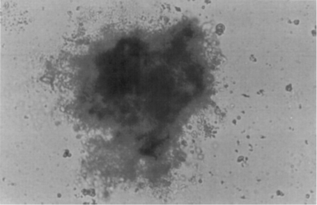
However, since their historical imitations are not enough (see Figures 1 and 2), they try to mimic the effects of environmental factors (chemical, biological and physical carcinogenic substances) that create a real response. Thus, the body's defense mechanism becomes unable to separate this uncontrolled creature from inanimate elements. This phase is the most critical. The cell has performed this behavior as a requirement of the defense mechanism it claims. These lesion effects are given below. This study reveals the threat to the reader as a result of the development of a cancer cell, apart from other cells, in relation to previously published topics. The first threat (atherosclerotic lesions) has been brought to the knowledge and attention of relevant researchers and scientists for this purpose.
Atherosclerotic lesions
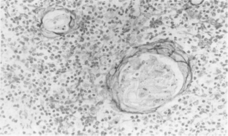
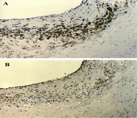
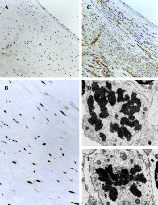
Detection of increased cell proliferation is not sufficient in atherosclerotic lesions. However, evidence of cell proliferation levels in different types of human great arteries is still lacking. A study has proven that carotid and coronary arteries make up almost one-third to almost 50% of the total cell count in lipofibrosis plaques in hematogenous cells. The basis of the impulses "I am the majority" and "will attack me" that I mentioned in the introduction is that the cell that I described in the first 3 sections grows using more energy than other cells. He was one like other cells. However, the symptom of the cell containing the first threat behavior, for example in the aorta, is 15% of the other cell behaviors. This has experimentally proved that the community formed by single-cell growth, which I suggested in the first 3 chapters, is based on the parent cell, not like other cells. Also, the findings showed that evidence of threat occurs less frequently in the arteries than in the coronary and carotid regions. With the detection of tumor group cells that act dependent on the main cell, they form the tumor in a bell-shaped structure (see figure 3(a) and (b)), and the signs of the threat they give to the organ or tissue (see Figure 4 (a, b, c, d, e)) can be listed as follows [2-5].
This sequence was determined in the carotid and coronary arteries. For example, the maximum number of resident cells was determined for item 4. 0.7 times more proliferation between a healthy carotid artery and a carotid artery with proliferation related to the first threat, while this ratio is 0.9 in the coronary artery, this ratio changes in the carotid artery and coronary artery cells with tumor growth, respectively, 11 and 11, It attracts attention with 2 values. The authors commented that this study is the first experimental study in this field [2].
These findings are important. The first threat of cancer cells can be determined by methods such as imaging techniques. Besides, the mother cell, mentioned in the introduction, imitating physical and chemical substances is another research subject that should be considered.
Dear Editorial Team, Clinical Medical Reviews and Reports. My experience with the journal was highly positive. The peer-review process was rigorous, constructive, and completed in a timely manner. The reviewers provided valuable comments that helped improve the quality and clarity of our manuscript. The editorial office was professional, responsive, and supportive throughout all stages of the publication process. Communication was clear and efficient, and any questions were addressed promptly. Overall, I found the journal to maintain high scientific standards and an excellent publication workflow. I would be pleased to consider submitting future work to this journal. Best wishes from, Elena Popa.

It was my pleasure to submit my testimonial concerning the Reviewer Board of our Scientific Journal “Brain and Neurological Disorders”. The Reviewers focused on some modifications and their contribution was helpful. The ladies of our Editorial Office were also supported my efforts. It was my honor to have such a co-operation and I am looking forward for more collaboration.

Dear Grace Pierce, Editorial Coordinator of Journal of Clinical Research and Reports, Thank you for the speedy and efficient peer review process. I appreciate the fact that your peer reviewers do not take months to respond like with some other journals. I would also like to thank the editorial office for responding quickly to my questions. It is an excellent journal. I plan to submit more manuscripts in the future. Best wishes from, Robert W. McGee

Dear Grace Pierce, Editorial Coordinator of Journal of Clinical Research and Reports, Working with you and your team on our recent publication in JCRR has been a truly wonderful and enjoyable experience. The responses were prompt, and the reviewers were patient, constructive, and highly professional. One reviewer in particular gave me the feeling that a professor was carefully reading and commenting on my coursework, which was deeply touching. The entire process was straightforward and hassle‑free, with no tedious online forms to complete. I highly recommend this journal. Best wishes from, DR Aibing Rao, Head of R&D

I Appreciate the Opportunity to Share my Experience with the Journal of Clinical Research and Reports. The peer review process was timely and constructive, and the feedback provided helped improve the quality of our manuscript. The editorial office was professional, responsive, and supportive throughout the process, ensuring smooth communication and efficient handling of the submission. Overall, it was a positive experience collaborating with your team.

Dear Mercy Grace, Editorial Coordinator of Obstetrics Gynecology and Reproductive Sciences, We would like to express our gratitude for your help at all stages of publishing and editing the article. The editors of the magazine answer all the necessary questions and help at every stage. We will definitely continue to cooperate and publish other works in the Obstetrics Gynecology and Reproductive Sciences! Best wishes from, Alla Konstantinovna Politova,
