

Research Article | DOI: https://doi.org/10.31579/2690-8808/335
1School of Advanced Technologies in Medicine, Tehran University of Medical Sciences, Tehran, Iran.
2Physics and Medical Engineering, Tehran University of Medical Sciences, Tehran, Iran.
*Corresponding Author: Fatemeh Mahdavi, School of Advanced Technologies in Medicine, Tehran University of Medical Sciences, Tehran, Iran.
Citation: Fatemeh Mahdavi, Hosseini Dokht AS, (2026), Calculation of Absorbed Dose in Skin, Adipose, Cortical Bone and Soft Tissue in Skin Cancer Radiotherapy Methods: Monte Carlo Study, J, Clinical Case Reports and Studies, 7(8); DOI:10.31579/2690-8808/335
Copyright: ©, 2026, Fatemeh Mahdavi. This is an open access article distributed under the Creative Commons Attribution License, which permits unrestricted use, distribution, and reproduction in any medium, provided the original work is properly cited.
Received: 07 July 2026 | Accepted: 24 July 2026 | Published: 20 August 2026
Keywords: brachytherapy; radiation therapy; skin; cortical bones; Iridium 192; Monte Carlo simulation
Purpose: The aim of this project was to evaluate the absorbed dose on the cortical bone during radiation therapy of the skin with EBT, IR-HDRS, and EEBRT treatment techniques. These evaluations are performed using the Monte Carlo simulation technique. The absorbed dose at the cortical bone is evaluated as a function of the radiation source, bone thickness, and bone depth in the soft tissue.
Materials and Methods: Using the Monte Carlo simulator, the Leipzig applicator is simulated with an Iridium 192 source as well as a heterogeneous phantom that includes skin, adipose, cortical bone, and soft tissue. In this research, simulation has been performed using MCNPX code version 2.6. In these simulations, the monoenergetic photon beams of 10 keV, 15 keV, 20 keV, 30 keV, 50 keV, and 70 keV for covering the energy range of eBT, 380 keV photons for IR-HDRS with Leipzig applicator, and 4 MeV and 6 MeV monoenergetic electron beam for EEBRT were modeled.
Results: This study shows that, for the treatment of skin cancer in conditions where the bone is immediately under the skin, in the treatment method 380 keV photons for IR-HDRS with Leipzig applicator, the amount of absorbed dose into the bone relative to the skin compared to other methods is the lowest value and the absorbed dose on the surface of the skin, which includes the target volume (tumor) is the highest of all methods, which can provide a more ideal treatment than eBT and EEBRT methods.
The prevalence of skin cancer worldwide is growing steadily and has been diagnosed as the most common tumor. Non-melanoma skin cancer (NMSC) is the most common type of skin cancer. Although NMSC has a low mortality rate, its prevalence is still increasing, significantly affecting the quality of life. Basal cell carcinoma (BCC) and squamous cell carcinoma (SCC) of the skin usually occur in sun-exposed areas, where the face is one of the most dangerous places and accounts for 95% of cases. Surgery in these areas (nose, ears, eyelids, lips) may cause abnormalities or require complex plastic surgery techniques under general anesthesia.
Radiation therapy has been used to treat NMSC for more than 40 years, and different techniques include superficial X-rays, orthovoltage X-rays, megavoltage photons, electron beam irradiation, and HDR brachytherapy (BT).
There are many radiation therapy techniques that can be used to treat skin cancers. External beam radiotherapy (EBRT) uses partial or photon radiation from linear accelerators. In contrast, brachytherapy uses the energy of photons or particles from radioactive isotopes located in the tumor, interstitial brachytherapy (BT interstitial) or near the tumor, superficial brachytherapy (BT superficial).
Brachytherapy (BT) in specific locations is a valuable tool of accurate irradiation within the tumor mass. Brachytherapy is a safe method for high-risk organs due to the rapid drop in dose outside the applicator axis.
Complications are acceptable, and treatment costs are low. In some tumors (skin lesions on the scalp, near the eyes, or nose), brachytherapy significantly reduces the dose to surrounding healthy tissue or organ at risk. Brachytherapy gives the least dose to the surrounding healthy tissues. In almost all cases, treatment is possible on an outpatient basis.
Brachytherapy with the radioactive isotope Ir-192 (source) on small skin cancer lesions, using Leipzig surface applicators is one of the new developments in the field of skin cancer radiotherapy. [3]
Continuous technological advances in brachytherapy in recent years allow us to use appropriate programs prepared from radioactive sources (usually Ir-192) using techniques (micro after loading systems). Under the control of a computer system by treatment planning systems, a single source enters and is targeted at the target area based on the planned treatment area. The source moves along the axis at a certain distance, which is approximately equal to the size of the tumor. General principles of treatment planning include: tumor spread with the applicator, applicator stabilization (interstitial or superficial), accurate and appropriate prescribed dose that covers the tumor with sufficient margin.
Due to the fact that skin cancers are more in areas exposed to sunlight, such as: (nose, ears, eyelids, lips) In these areas, the bone is located under the skin, which is of special importance in calculating the absorbed dose in the bone. Because the bone dose should not exceed the allowable limit.
Using the Monte Carlo simulation method, the Leipzig applicator is simulated with an Iridium 192 source as well as a heterogeneous phantom that includes skin, adipose tissue, bone, and soft tissue.
In these simulations, the monoenergetic photon beams of 10 keV, 15 keV, 20 keV, 30 keV, 50 keV, and 70 keV for covering the energy range of eBT, 380 keV photons for IR-HDRS with using Leipzig applicator, and 4 MeV and 6 MeV monoenergetic electron beam for EEBRT were used.
Generally, for external electron beam therapy, a protocol with about 40Gy to 60Gy total dose is delivered at the rate of 2 Gy to 3 Gy per fraction within four to six weeks. However, for brachytherapy, skin treatment by IR-HDRs and eBT, the patient receives 6 to 8 fractions for a total dose ranging from 30 Gy to 40 Gy in two weeks.
Using the absorption dose calculation by the Monte Carlo code can prove which energy and which method delivers the lowest dose to the bone and creates an optimal treatment.
In this research, the main component is the Leipzig applicator. The simulation was performed using MCNPX code version 2.6.
In these simulations, the monoenergetic photon beams of 10 keV, 15 keV, 20 keV, 30 keV, 50 keV, and 70 keV for covering the energy range of eBT, 380 keV photons for IR-HDRS with Leipzig applicator, and 4 MeV and 6 MeV monoenergetic electron beam for EEBRT were modeled.
The point source of radiation with the applicator is made of tungsten with an opening angle of 75 degrees and 1.5 cm distance from the source to the skin surface. The radiation field with a diameter of 2 cm, is assumed to be circular and parallel.
The field size on the surface of the skin is 2 cm, so the length of the phantom under the applicator can be considered 2 cm. The geometry of the cone is 2 cm and 0.3 cm in radius. There is a cylinder at the top of the applicator. The applicator is made of a cylindrical joint and a cone.
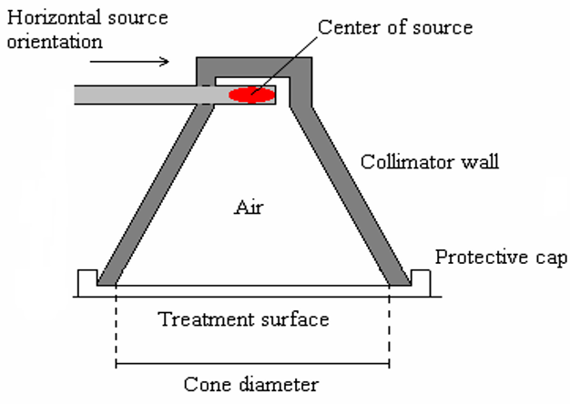
Figure 1: Schematic image of the Leipzig applicator
The phantom makeup and dimensions chosen here are exactly the same as treating the skin in areas such as the scalp, forehead, knees, hands, feet, ears, and back along the spine or upper ribs. Photon or electron beams are perpendicular to the surface of the skin. For the treatment of skin cancer, the diameter of the field is 2 cm.
Dimensions of the inhomogeneous phantom are first considered as 0.3 cm thickness of skin, then 0.2 cm of adipose and 0.5 cm of cortical bone, after bone 14 cm of soft tissue.
Each piece in the phantom is divided into thicknesses of 1 mm with a diameter of 1 mm, which are small enough to record dose changes along the central axis of the radiation beam.
| organs | Thickness | Density |
| Skin | 0.3 cm | 1.09 gr/cm3 |
| Adipose | 0.2 cm | 0.95 gr/cm3 |
| Cortical bone | 0.5 cm | 1.9 gr/cm3 |
| Soft tissue | 14 cm | 1.7 gr/cm3 |
Table 1: Features of simulated heterogeneous phantoms.
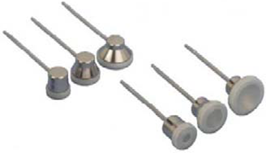
Figure 2: Leipzig applicators in 6 different types, three models for horizontal positioning of the source and three models for vertical positioning of the radioactive source. [7]
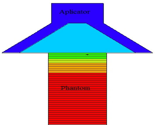
Figure 3: Schematic representation of heterogeneous phantom geometry and Leipzig applicator simulated with Monte Carlo code in this research. The phantom has dimensions of 15 cm × 15cm× 15 cm cubes, which includes layers of skin, adipose, cortical bone, and soft tissue.
Here, in order to be accurate in the calculations, the phantom method should be divided into smaller parts (meshing) according to the code method to give a better result. In this research, the F6 tally is used to estimate the deposited energy. (F6 (MeV / g / source-particle)). In this study, calculations have been performed for the number of 107 particles in history to reduce the error rate.
To compare the effects of dose on the bone for the low-energy photon techniques eBT, IR-HDRS, and EEBRT, two different definitions have been used, defined as "normal dose":

Where D represents the dose at a depth of "r" in the homogeneous or heterogeneous phantom and Dmax represents the maximum dose in the phantom.
The simulation results in the following diagrams are the ratio of the absorbed dose in the heterogeneous phantom for the monoenergetic photon beams of 10 keV, 15 keV, 20 keV, 30 keV, 50 keV, and 70 keV for covering the energy range of eBT, 380 keV photons for IR-HDRS with Leipzig applicator, and 4 MeV and 6 MeV monoenergetic electron beam for EEBRT have been identified.
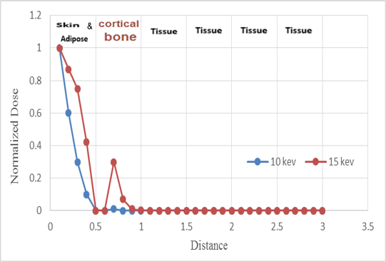
Figure 4: Comparison of absorbed dose in heterogeneous phantoms with 10 and 15 keV photons
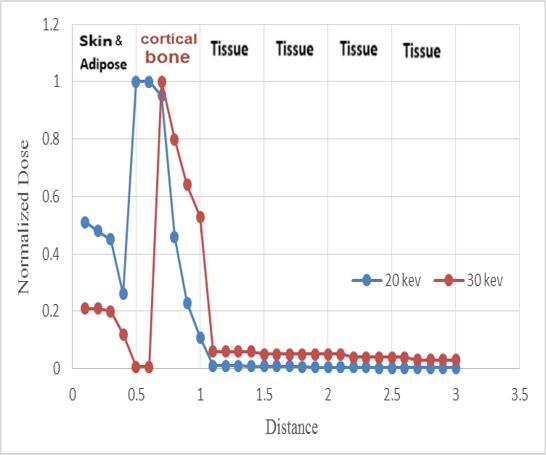
Figure 5: Comparison of absorbed dose in heterogeneous phantoms with 20 and 30 keV photons
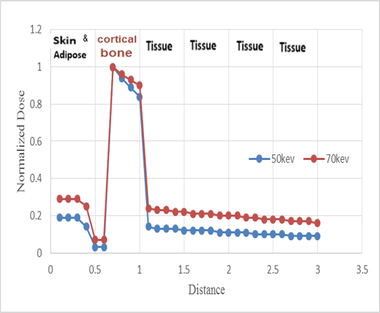
Figure 6: Comparison of absorbed dose in heterogeneous phantoms with 50 and 70 keV photons
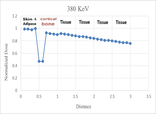
Figure 7: Absorbed dose in heterogeneous phantoms with 380 keV photons for IR-HDRS
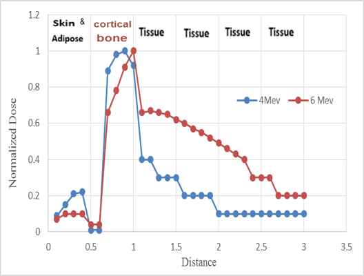
Figure 8: Comparison of absorbed dose in heterogeneous phantoms with 4 MeV and 6 MeV monoenergetic electron beam for EEBRT
The results obtained for the MCNP simulated dose distribution as a function of distance (g / cm2) for the treatment of skin cancer are assumed to be heterogeneous phantom conditions for beams with the energies listed the bone thickness of 0.5 cm. These results show that:
For 50 keV photons, the eBT method, the amount of absorbed dose on the skin surface is 3.24 times the amount of absorbed dose in adipose. The absorbed dose at the cortical bone is 6.4 times the absorbed dose at the skin surface.
For 70 keV photons, the eBT method, the amount of absorbed dose on the skin surface is 2.66 times the amount of absorbed dose in adipose. The absorbed dose at the cortical bone is 4.4 times the absorbed dose at the skin surface.
For 380 keV photons, the IR-HDRS method, the amount of absorbed dose on the skin surface is 2.01 times the amount of absorbed dose in adipose. The absorbed dose at the cortical bone is 1.4 times the absorbed dose at the skin surface.
For 4 MeV EEBRT photons, the absorbed dose by the skin is 1.92 times the absorbed dose by the adipose. The absorbed dose at the cortical bone is 8.1 times the absorbed dose at the skin surface.
For 6 MeV EEBRT photons, the absorbed dose by the skin is 1.62 times the absorbed dose by the adipose. The absorbed dose at the cortical bone is 8.6 times the absorbed dose at the skin surface.
These large differences in the amount of absorbed doses at the skin surface and adipose in 50 Kev and 70 KeV photons are due to changes in chemical composition and density of different tissues, especially at low energy levels where the photoelectric reaction occurs.
The large differences in the amount of absorbed dose at the skin and cortical bone for the 4 MeV EEBRT and 6 MeV photons are due to multiple scatterings in the bone area and differences in mass stopping power. After bone thickness, there is a slight increase in the absorbed dose into the soft tissue immediately due to the increase in electron scattering.
Figure (8) clearly shows that there is a large difference between the absorbed dose in the soft tissue and the absorbed dose at the cortical bone between the 4 MeV and 6 MeV electron beams (EEBRT) compared to the 50 keV and 70 keV photons. However, for both of these methods, the absorbed dose at the cortical bone is still significantly higher than the skin.
For 380 keV IR-HDRS, the absorbed dose at the cortical bone is smaller than the absorbed dose at the skin, adipose, and soft tissues due to the Compton interaction range, which is independent of the atomic number of materials and depends on the electron density, which is approximately All textures are the same.
In radiation therapy for skin cancers such as the scalp, forehead, knees, hands, feet, ears, and back along the spine or upper ribs where the lower bones are adjacent to the target, the choice of the radiation source is very important. According to the results of this study for these treatments, the use of low energy photons from eBT sources is desirable due to the minimal need to protect the treatment room. In addition, this technique does not involve any radioactive sources, and it is easier to meet the requirements in terms of regulatory requirements. The clinical benefits of this system have been discussed by various researchers, and the dosimetric properties of eBT have been published in various scientific journals. [2]
The Monte Carlo code has the ability to estimate the error rate. In this study, the error rate for all energies is less than 5%, which indicates the appropriate and acceptable accuracy of the results.
The results of this study indicate that, for the treatment of skin cancer with adjacent bones, eBT and EEBRT techniques with 4 MeV and 6 MeV electron beams are not the most appropriate methods because the absorbed dose into the bone may be higher than 380keV IR-HDRS with applicator be in Leipzig.
In 380 keV IR-HDRS treatment method with Leipzig applicator, the absorbed dose in the bone relative to the skin is the lowest compared to other methods, and the absorbed dose on the skin surface, which includes the target volume (tumor), is the highest compared to all methods, that can provide the ideal treatment. Therefore, it can be concluded that HDRS brachytherapy treatment with Iridium 192 source with Leipzig applicator causes the least possible side effects for cortical bone.
Dear Editorial Team, Clinical Medical Reviews and Reports. My experience with the journal was highly positive. The peer-review process was rigorous, constructive, and completed in a timely manner. The reviewers provided valuable comments that helped improve the quality and clarity of our manuscript. The editorial office was professional, responsive, and supportive throughout all stages of the publication process. Communication was clear and efficient, and any questions were addressed promptly. Overall, I found the journal to maintain high scientific standards and an excellent publication workflow. I would be pleased to consider submitting future work to this journal. Best wishes from, Elena Popa.

It was my pleasure to submit my testimonial concerning the Reviewer Board of our Scientific Journal “Brain and Neurological Disorders”. The Reviewers focused on some modifications and their contribution was helpful. The ladies of our Editorial Office were also supported my efforts. It was my honor to have such a co-operation and I am looking forward for more collaboration.

Dear Grace Pierce, Editorial Coordinator of Journal of Clinical Research and Reports, Thank you for the speedy and efficient peer review process. I appreciate the fact that your peer reviewers do not take months to respond like with some other journals. I would also like to thank the editorial office for responding quickly to my questions. It is an excellent journal. I plan to submit more manuscripts in the future. Best wishes from, Robert W. McGee

Dear Grace Pierce, Editorial Coordinator of Journal of Clinical Research and Reports, Working with you and your team on our recent publication in JCRR has been a truly wonderful and enjoyable experience. The responses were prompt, and the reviewers were patient, constructive, and highly professional. One reviewer in particular gave me the feeling that a professor was carefully reading and commenting on my coursework, which was deeply touching. The entire process was straightforward and hassle‑free, with no tedious online forms to complete. I highly recommend this journal. Best wishes from, DR Aibing Rao, Head of R&D

I Appreciate the Opportunity to Share my Experience with the Journal of Clinical Research and Reports. The peer review process was timely and constructive, and the feedback provided helped improve the quality of our manuscript. The editorial office was professional, responsive, and supportive throughout the process, ensuring smooth communication and efficient handling of the submission. Overall, it was a positive experience collaborating with your team.

Dear Mercy Grace, Editorial Coordinator of Obstetrics Gynecology and Reproductive Sciences, We would like to express our gratitude for your help at all stages of publishing and editing the article. The editors of the magazine answer all the necessary questions and help at every stage. We will definitely continue to cooperate and publish other works in the Obstetrics Gynecology and Reproductive Sciences! Best wishes from, Alla Konstantinovna Politova,
