Blastomycosis Prostatitis and Blastomycosis Prostate Abscess: A Review and Update

Research Article | DOI: https://doi.org/10.31579/2767-7370/065

Blastomycosis Prostatitis and Blastomycosis Prostate Abscess: A Review and Update

  • Anthony Kodzo-Grey Venyo

North Manchester General Hospital, Department of Urology, Delaunays Road, Manchester, M8 5RB. United Kingdom

*Corresponding Author: Anthony Kodzo-Grey Venyo, North Manchester General Hospital, Department of Urology, Delaunays Road, Manchester, M8 5RB. United Kingdom

Citation: Grey Venyo A. K, (2023). Blastomycosis Prostatitis and Blastomycisis Prostate Abscess: A Review and Update. J New Medical Innovations and Research, 4(7); DOI:10.31579/2767-7370/065

Copyright: © 2023, Anthony Kodzo-Grey Venyo. This is an open access article distributed under the Creative Commons Attribution License, which permits unrestricted use, distribution, and reproduction in any medium, provided the original work is properly cited.

Received: 18 October 2023 | Accepted: 25 October 2023 | Published: 31 October 2023

Keywords: blastomycoses; blastomycis; prostatitis, prostate abscess; soil; fungus; yeast; asymptomatic; non-specific; smear, biopsy; aspiration; fever; urinary retention; ultrasound scan

Abstract

Blastomycosis is a globally accepted terminology that is used for a fungal infection which is also referred to as Gilchrist’s disease, which typically afflicts the lungs, but which in addition has the cpability of spreading to the brain, the stomach, the intestine, as well as the skin, where it does manifests as crusting, purple looking warty plaques that are associated with a roundish, bumpy edge as well as central depression. About half of individuals who are afflicted by Blastomycosis, remain asymptomatic and the remaing half do tend to develop symptoms which could include fever, cough, night sweats, muscle pains, loss of weight, chest pain as well as a sensation of feeling tired. The symptoms of Blastomycosis tend to manifest between three weeks and three months ensuing breathing inhalation of Blastomyces spores. It is known that in approximately 25% to 40% of cases of Blastomycosis, the infection could also traverse to other parts of the human body including the skin, bones, central nervous system and on rare occasions. Blastomycosis, could secondarily afflict the urogenital system, or the affliction could be on extremely rare occasions, afflict only the genitourinary tract system only without affecting any other part of the human body. It is known that even though blastomycosis tends to be very dangerous for individuals who have immunosuppression as multiple other co-morbidities. Many individuals who are afflicted by Blastomycosis are ummunocompetent. Bla stomyces dermatidis is encountred within the soil, as well as within decaying organic matter including wood and leaves. It is understood that outdoor activities including: toiletting the soil, hunting and camping within wooden areas do increas the risk for the development of Blastomycosis. Even though up to date there is no well-developed vaccine for the prevention of Blastomycosis, the risk for the development of Blastomycosis could be reduced by individuals not disturbing the soil. Blastomycosis has existed for millions of years; nevertheless, Blastomycosis, was first described by Thomas Caspar Glichrist in 1894. In view of this, Blastomycosis has sometimnes been called Gilchrist’s disease. The manifestations of Blastomycosis, do cover a wide range of symptoms that overlap with the manifestations of more common conditions and for this reason, Blastomycosis has been referred to as the great pretender. Many cases of Blastomycosis have tended to be asymptomatic or sub-clinical. The involvement of the prostate gland in the form of prostatitis or prostate abscess could be non-specific and this does entail symptoms of dysuria, perineal discomfort, supra-pubic discomfort, visible haematuria, retention of urine and other lower urinary tract symptoms. This non-specific manifestation of Blastomycosis of the prostate gland often tends to result in delay in the correct diagnosis due to the fact that clinicians tend to pursue more common diagnosis including benign prostatic hypertrophy, carcinoma of prostate and actute and chronic bacterial prostatitis. Diagnosis of Blastomycosis is best established by visualization of the distinct yeast within smears, cultures or direct tissue/biopsy specimens. Most clinicians had opinioned that even though acute Blastomycosis could be self-limitinmg they had recommended that treatment with antifungal medication for all cases with utilization of at least 6 months of itraconazole for mild to moderate infectionsand amphotericin for life treatening cases or involvemnent of the central nervous system. Fungal abscesses of the prostate gland or prostatitis are rare pathologies that tend to simulate alternative diagnoses and hence a high-index of suspicion is required to establish an accurate diagnosis and to provide correct treatment. Evethough genitourinary tract involvement of Blastomycosis usually ensues haematogeneous spread, pursuant to pulmonary infections, some patients might manifest with isolated urinary tract symptoms. In view of the fact some patients who have Blastomycoses infection of the prostate gland may manifest with isolated lower urinary tract symptoms, it is pertinent for health care professionals to have a high-index of suspicion with regard to individuals who dwell within or have travelled from Blastomycoses endemic areas that are found to have refactory lower urinary tract symptoms who are not responding to treatment with the usual treatments for lower urinary tract symptoms and bacterial prostatitis. When diagnosis of Blastomycosis prostatitis is confirmed, this should generally be treated for at least 6 months of itraconazole and ensued by regular clinical laboratory and radiology-image follow-up assessment. If a patient has Blastomycosis prostate abscess, the infection should be treated generally by radiology-image-guided complete aspiration / drainage of the abscess plus at lest 6 months treatment with itraconazole and regular clinical, radiology image guided, and laboratory test – follow-up assessments in order to confirm absence of recurrence as well to diagnose any future recurrence early. A high index of suspicion is required in order to diagnose Blastomycoses infection of the prostate gland in both the immunocompetent and immunosuppressed individuals globally especially in North and South America, Canada, Africa and India.    

Introduction

It has been iterated that Blastomycosis is an endemic infection which is caused by Blastomyces dermatitidis, and which is found primarily within the South-Eastern, South-Central, and Mid-West United States of America, with prevalence that has ranged from 0.5 to 4 cases per 100,000 per year. [1] [2] [3] It has been pointed out that Blastomycosis infection often initially manifests with non-specific systemic symptoms which include weight loss, fevers, and fatigue. [1] It has also been documented that majority of Blastomycosis infections do manifest with pulmonary presentations, with rare instances of extra-pulmonary involvement including the skin, central nervous system (CNS), bone, and the genitourinary (GU) system. In addition, it has been iterated that very few cases of blastomycosis infection with initial presentation isolated to the prostate had been reported in the global literature. [1] [4] Considering the fact that Blastomycosis has been reported to occur within some parts of United States of America, Canada, Africa and India and because of global travel of individuals for holidays as well as to settle within other areas of the world, it would be envisaged that Blastomycosis can be encountered anywhere in the world. In view of the rarity of Blastomycosis prostatitis and Blastomycosis prostatic abscess, it would be envisaged that both clinicians and patients globally would tend not to be familiar with the manifestations, diagnostic features, treatment, as outcome ensuing treatment of blastomycosis prostatitis and blastomycosis prostate abscess. There is therefore the need to review and summate experiences that had been reported by few clinicians that had reported cases of blastomycosis of the prostate in order to provide a birds eye view on blastomycosis of the prostate gland to enable both clinicians and patients all over the world to be conversant with the diagnostic features of the infection to help all individuals to have a high index of suspicion for the infection which would enable the establishment of a prompt diagnosis of the infection in order to also enable prompt and effective treatment of the infection. The ensuing article contains a review and update of the literature on Blastomyces prostatitis and Blastomyces prostate abscess which has been divided into two parts (A) Overview which contains General Overview of various aspects of Blastomyces and (B) Miscellaneous narrations and discussions from some case reports, case series and studies related to Blastomycoses prostatitis and blastomycoses prostatic abscess. 

AIMS:

To review and update the literature on Blastomyces and Blastomyces Prostatitis as well as Blastomyces prostate abscess. 

Methods:

Internet Data Bases were searched including: Google; Google Scholar; PUBMED, Yahoo and AOL. The search words that were used included: Blastomyces; Blastomyces of prostate; Blastomyces Prostatic abscess; Blastomycosis prostatitis; Blastomycosis Prostate Abscess; Blastomycosis prostatic abscess; Blastomycosis abscess of prostate gland. Forty-eight (48) references were identified which were used to write the article which has been divided into two parts: The ensuing article contains a review and update of the literature on Blastomyces prostatitis and Blastomyces prostate abscess which has been divided into two parts (A) Overview which contains General Overview of various aspects of Blastomyces and (B) Miscellaneous narrations and discussions from some case reports, case series and studies related to Blastomycoses prostatitis and blastomycoses prostatic abscess. 

RESULTS:

[A] OVERVIEW 


Definition / general statements [5] 

The ensuing summations had been made related to the definition and general aspects of Blastomyces: [5]

  • Taxonomy: [5]
    • Blastomyces belong to a Class of Eurotiomycetes of the order Onygenales and a family of Ajellomycetaceae
  • Common species of Blastomyces include the ensuing: [5]
    • Blastomyces dermatitidis: is the commonest cause of cause of blastomycosis
    • Blastomyces gilchristi: which is encountered within northern part of United States of America (U.S.A) and Canada
    • Blastomyces helicus: which tends to be found within western United States of America (U.S.A)
    • Blastomyces percursus: which is found within Africa

Essential features [5] 

  • Blastomyces is a dimorphic mold
  • Body temperature, uniformly sized, extracellular round yeasts with a thick, refractile, double contoured cell wall and broad-based budding
  • Blastomyces could afflict immunocompetent individuals and tends to manifest as a chronic granulomatous and suppurative disease of the lung or skin
  • Blastomyces tends to afflict immunocompromised individuals which tends to be disseminated disease that is associated with poor prognosis

Epidemiology

The ensuing summations related to the epidemiology of the infection had also been iterated: [5]

  • The infection that is caused by Blastomyces is referred to by terminology as blastomycosis which represents a chronic pyogranulomatous inflammatory disease
  • Blastomyces are soil organisms, which tend to be associated with moist areas near riverbeds
  • Blastomyces tend to be found within the Midwestern, south central and southeastern areas of the United States of America (U.S.A), particularly within areas encompassing the Ohio and Mississippi River valleys, the Great Lakes and the Saint Lawrence River
  • Blastomyces had been found present within Canada, with a small number of documented cases encountered within the continent of Africa as well as within India [6] [7] 

Sites 

Summations related to the sites of Blastomyces infection include: [5]

  • Blastomycosis does tend to affect the lungs and pulmonary blastomycosis could be encountered as an asymptomatic infection or it may entail afflictions with stated manifestation that tend to range from self-limited pulmonary infection to life threatening infections [8] 
  • Extrapulmonary blastomycosis:
  • Cutaneous blastomycosis does afflict the skin with the development of verrucous (wart-like) or ulcerative skin lesions
  • Osseous blastomycosis with the development of osteomyelitis together with contagious tissue abscesses and draining sinuses
  • Genitourinary tract blastomycosis which usually has tended to afflict males with the involvement of the prostate gland as well as the epididymis
  • Central nervous system blastomycosis with the development of: brain abscess, cranial abscess or epidural abscess and meningitis

Pathophysiology

The pathophysiology of blastomycosis had been summated as follows: [5]

  • Inhaled spores of Blastomyces transform into yeasts that resulting in the development of acute and chronic pulmonary (lung) inflammation
  • Cutaneous blastomycosis lesions could demonstrate pseudo-epitheliomatous hyperplasia of the epidermis
  • Dissemination of blastomycosis to extra-pulmonary sites of the body, especially to the skin, has been presumed to be by haematogenous spread [6] 

Clinical features [5] 

  • Blastomycosis does involves the lung in over 90% of cases; infection can range from asymptomatic self-limited infection (about 50% of cases) to severe diffuse pneumonia causing respiratory failure
  • Extrapulmonary dissemination occurs in approximately 25 - 50% of cases of blastomycosis [9] 

Laboratory tests 

Haematology blood tests

  • Routine haematology blood tests tend to be undertaken but the results would not be specific for the diagnosis of Blastomycosis but the results would express the general status of the patients in that the results generally would tend to be normal; however, the eosinophil count could be raised and the white blood cell count could be raised in scenarios of co-infections. 

Biochemistry blood tests

  • Routine biochemistry blood tests tend to be undertaken but the results would not be specific for the diagnosis of Blastomycosis but the results would express the general status of the patients in that the results generally would tend to be normal; however, the level of the eGFR, serum urea and electrolytes, as well as liver function tests would reflect the general state of the patient in that if there is urinary retention, there could be results that reflect renal function impairment as well as the serum prostate specific antigen (PSA) level may be raised as well as digital rectal examination finding of benign prostate gland, enlarged prostate gland, tender prostate gland or mass / nodularity of prostate gland which are all non-specific. 

Urinalysis, urine microscopy and culture 

  • The results of urinalysis, urine microscopy and urine culture may be normal most often but on rare occasions there would be a positive growth of Blastomyces. 

Microscopy Pathology Examination

Microscopy pathology examination of specimens containing Blastomyces does demonstrate the ensuing: [5]

  • Direct examination demonstrates large, thick-walled, yeast forms with single broad based budding cells (8 to 10 μm)
  • Slow-growing yeasts that transition to a fluffy white mycelium or glabrous, tan, nonsporulating colonies within 1 - 4 weeks
  • Microconidia which simulate Histoplasma capsulatum but macroconidia are not formed
  • Microscopy examination of growth demonstrates thick-walled yeast cells with broad based budding
  • Conversion to the yeast phase could occur on routine media incubated at 37 °C

Antibody detection tests

Blastomyces antibody detecting tests are available in a number of established laboratory centres but these would tend not to be available within small district centre laboratories. Some of the available tests for the identification of Blastomyces include the ensuing: [5]

  • Immunodiffusion utilizes purified B. dermatitidis A antigen and this is associated with relatively low sensitivity and specificity.
  • Enzyme immunoassays use BAD1 antigen, which is the most sensitive approach, that is associated with cross reactivity seen with histoplasmosis and other fungal infections. [7] 

Treatment

The ensuing iterations had been made regarding the treatment of blastomycosis: [5]

  • Acute pulmonary blastomycosis could be encountered as a mild and self- limited infection which does not require treatment in immunocompetent hosts
  • More severe cases of blastomycosis disease, particularly blastomycosis afflicting immunocompromised hosts, requires treatment with itraconazole with or without liposomal amphotericin B
  • The duration of treatment duration could range from 6 months to 1 year [10] [11] 

Microscopy (histology) description

The microscopy histopathology examination features of specimens of containing blastomycosis infecting agent include: [5] 

  • Microscopy histopathology examination of specimens containing Blastomyces infection does demonstrate Pyogranulomatous inflammation with inclusion of neutrophil infiltration admixed with epithelioid histiocytes as well as granulomatous inflammation
  • Cutaneous Blastomycosis infection lesions upon microscopy examination demonstrate pseudoepitheliomatous hyperplasia of the epidermis
  • During examination of lesions containing blastomycosis infection organisms the microscopy examinations would tend to demonstrate uniformly sized, refractile round yeast cells which may be observed at low power in H&E-stained tissue sections of the specimens. 
  • High power examination of specimen containing blastomycosis infection lesions may show cell contents within the refractile cell wall but often this material washes away during processing
  • Periodic acid-Schiff (PAS) and Gomori methenamine silver (GMS) stains do highlight organisms
  • Mucicarmine may be found to be weakly positive; and this is in contrast with a strongly positive staining that is found in Cryptococcus
  • Fontana-Masson stain for melanin in blastomycosis tends to be negative [7] 

Immunohistochemistry staining studies

Positive stains

It has been pointed out immunohistochemistry staining in cases of blastomycosis tends to demonstrate the ensuing positive staining findings: [5]

  • Periodic acid-Schiff (PAS) as well as Gomori methenamine silver (GMS) stains do highlight the organisms.
  • Mucicarmine may exhibit weakly positive staining which is in contrast with a strong positive staining for Cryptococcus

Molecular / cytogenetics description [5] 

  • It has been pointed out that Nucleic acid probe hybridization assays are commercially available (AccuProbe by Hologic, Inc.) for definitive identification on culture of the organism. 
  • It has also been iterated that additional laboratory developed PCR tests, sequencing and matrix assisted laser desorption / ionization time of flight (MALDI-TOF) mass spectrometry (MS) might enable identification of the organism

Differential diagnoses

Some of the documented differential diagnoses of blastomycosis had been documented to include the following: [5]

  • Acute illness:
  • Community acquired pneumonia is one of the differential diagnoses of blastomycosis pneumonia.
  • Viral, bacterial, atypical bacterial
  • Chronic illness:
  • Tuberculosis as well as infection with non-tuberculous mycobacteria:
  • Acid fast organisms
  • Histoplasmosis:
  • Small, uniform, narrow based budding yeasts; large tuberculate macroconidia
  • Coccidioidomycosis:
  • Thick-walled spherules with endospores in tissue
  • Sarcoidosis:
  • Confirmation of diagnosis is by means of diagnosis of exclusion after excluding all the infectious agents

[B] Miscellaneous Narrations and Discussions from Some Case Reports, Case Series, And Some Studies Related to Blastomocosis Prostatitis and Blastomycosis Prostate Abscess. 

Sloan et al. [1] reported a 59-year-old man who had recently been diagnosed as having prostatitis and an elevated serum prostate specific antigen level of (PSA of 9.7), but otherwise he did not have any significant past medical history, who had presented to the emergency department (ED) with urinary retention. A Foley urethral catheter was inserted and the results of her urine culture obtained was negative for urinary tract infection. The patient was discharged with instructions to continue taking tamsulosin, a six-week course of sulfamethoxazole-trimethoprim, and to consult with urology. He was seen in the urology clinic the following week, during which time his symptoms had improved. He underwent digital rectal examination which had revealed a slightly firm non-tender prostate gland. He was advised on management options for his enlarged prostate and his Foley urethral catheter was removed. He was able to void with a residual urine volume of 37mL. He was scheduled to have a repeat serum PSA test 3 months later. Many days subsequently, the patient presented to the Emergency Department after he had developed recurrent urinary retention, as well as night sweats and fevers of up to 102.7 °F. During his presentation, the patient had a temperature of 100.2 °F, but he was otherwise hemodynamically stable. He had a leucocytosis up to 15.5 × 109/L and mild anaemia (haemoglobin 12.8 g/dL). His renal function was at baseline (creatinine 1.0. mg/dL). His urinalysis revealed 3+ leukocyte esterase, negative nitrites, >182 WBCs, 1+ bacteria, and 19 RBCs. He had a Chest x-ray which revealed a vague opacity that was reported to be concerning for possible early pneumonia, but was otherwise unremarkable. He denied having any upper respiratory symptoms, including cough or shortness of breath. He had a CT scan of pelvis which demonstrated multiple low-density regions within his prostate gland which was reported to be suggestive of a possible abscess (see figure 1). The Emergency Department staff inserted a Foley urethral catheter, as well as obtained blood and urine cultures, and the patient was commenced on broad-spectrum antibiotics.

Fig. 1

Figure 1: Enlarged and heterogeneous prostate with low-density regions concerning for prostatic abscesses. Reproduced from: [1]

During his admission to hospital, the infectious disease team was consulted, at which time it had been revealed that the patient had participated in gardening, outdoor work, and he had recently returned from a trip to the Nevada desert. He was continued on ceftriaxone while fungal cultures and antigens to Coccidioides and Blastomyces had been collected. The patient continued to develop intermittent fevers and he had developed occasional nausea and vomiting. He was scheduled to undergo cystoscopy and transurethral resection of the prostate and prostate abscess (TURP). Nevertheless, on the day of his surgery, his fungal urine cultures returned positive for Blastomyces dermatitidis. His surgery was accordingly rescheduled and his antibiotics were discontinued with commencement of oral itraconazole. He had CT scan of his thorax which revealed many pulmonary nodules bilaterally. Forty-eight hours pursuant to commencement of his antifungal treatment, the patient continued to develop intermittent fevers and he had a persistent leucocytosis to 12.1 × 109/L. The decision was made to proceed with the undertaking of cystoscopy and TURP. His intra-operative assessment had demonstrated a normal-appearing urinary bladder and bi-lobar hyperplasia of his prostate gland. Resection of his prostate gland demonstrated friable prostatic tissue with multiple cavities that contained purulent fluid which were sent for pathology examination. The patient remained afebrile post-operatively and his leucocytosis had settled. His urethral catheter was removed and he voided without any problem. He was discharged home the next day with a 12-month course of itraconazole. Pathology examination of his resected prostate and prostatic purulent fluid showed that the final specimen was 6.7grams in weight with pathology demonstrating granulomatous prostatitis with many yeast forms that had features compatible with Blastomyces (see figure 2 and figure 3).

Fig. 2

Figure 2: Prostatic tissue demonstrating necrotizing granulomatous prostatitis with numerous yeast forms compatible with Blastomyces species (H&E, 40X magnification). Reproduced from: [1]

Fig. 3

Figure 3: Prostatic tissue demonstrating necrotizing granulomatous prostatitis with numerous yeast forms compatible with Blastomyces species (PAS-D Stain, 40X magnification).  Reproduced from [1].

It was also reported that the patient was reviewed during his follow-up assessment 1 month pursuant to his surgical operation and it was noted that he was doing well and his post-void residual urine volume was 2mL that had indicated complete emptying of the urinary bladder. He also had follow-up computed tomography (CT) scan of thorax, 3 months pursuant to his surgery which had shown significant improvement with illustration of decreasing size of his remaining pulmonary nodules. He was maintained on itraconazole for 12 months, and at the time of publication of the article, the patient had ceased taking his medications and upon his assessment it was confirmed that the patient was doing well and he was asymptomatic. The authors made the ensuing detailed educative summation: [1]

  • It has been iterated that Blastomyces dermatitidis is a fungal pathogen with thermal dimorphism, that grows in mycelial form at room temperature and yeast form at body temperature. [2] 
  • Primary Blastomyces infection usually emanates from inhalation of conidia spores, with primary immunity mediated by the innate immune system. 
  • Those spores which are able to evade natural host defences do convert to yeast phase, which is conjectured to confer a survival advantage and further increase virulence.
  • There is a wide spectrum of clinical manifestations of Blastomycosis including asymptomatic infections that tend to be identified upon screening, non-specific systemic complaints that include: weight loss, fatigue, and cough, or fulminant infections with respiratory failure and high fevers.[3] 
  • The lungs tend to be most commonly involved, with hematogenous spread that leads to involvement of extrapulmonary sites including skin, bone, adrenal glands, the CNS, and the GU system. [3] [12] 
  • The most commonly involved organs of the Genito-Urinary (GU) tract are the prostate gland and epididymis. [3] 
  • A retrospective study that was undertaken by Eickenberg et al. found GU involvement of 21% of patients who had blastomycosis. [13] 
  • Prostate gland involvement by Blastomyces infection could manifest with non-specific symptoms that tend to include the following: dysuria, perineal or suprapubic discomfort, haematuria, and urinary retention. [2] [4] [12] This often does tend to lead to a delay in the correct diagnosis, due to the fact that clinicians tend to pursue more common diagnoses such as benign prostatic hyperplasia and/or prostate cancer.
  • Diagnosis of Blastomyces prostatic abscess is best made by means of visualization of the distinct yeast within smears, cultures, or direct tissue specimens. [2] 
  • Whilst majority of cases of acute blastomycosis are considered to be self-limiting, most experts do agree on antifungal therapy for all cases with utilization of at least 6 months of itraconazole for mild to moderate infections, and amphotericin for life-threatening cases or with involvement of the CNS. 
  • While systemic treatment has tended to be successful in most cases, select patients, such as the one they had reported, may require operative intervention for adequate source control.
  • Sloan et al. [1] made the ensuing conclusions: 
  • Fungal prostatic abscesses are rare pathology findings that can simulate alternative diagnoses and in view of this diagnosis of fungal abscesses of the prostate gland, therefore require a high level of clinical suspicion to accurately diagnosis and correctly manage the abscesses. 
  • While GU involvement by Blastomyces infection usually emanates from hematogenous spread that ensue pulmonary infection, patients may manifest with isolated urinary complaints. In view of this, health care providers should maintain a high level of suspicion in individuals from Blastomyces endemic areas with refractory urinary complaints.

Wittman et al. [14] reported an otherwise healthy 25-year-old man who had manifested to the emergency department with fevers and left sided testis pain. He had urinalysis which was suspicious for infection and ultrasound scan of his scrotum and scrotal contents was undertaken, which demonstrated hyperaemia of his left testis, and left epididymis which was indicative of left epididymoorchitis. He was also found to have several nodular lesions of his skin upon his back, extremities, as well as scalp. One of these lesions was incised and sent for culture and syphilis RPR, which were negative. His sexually transmitted infection testing was also negative, including HIV. He was treated by means of intramuscular gentamicin and a ten-day course of doxycycline. He reattended the emergency department two weeks later with worsening scrotal pain and he had reported a 20-pound weight loss over the preceding month. His assessment workup revealed a leucocytosis of up to 14 × 109/L and a scrotal ultrasound scan which demonstrated progression of his epididymoorchitis with development of a 1.5 cm scrotal abscess. He had Computed tomography (CT) scan of his pelvis which showed a 6 cm prostate abscess as well as infection within his left sacro-iliacc joint. (see figure 4). He was admitted to the hospital and commenced upon broad-spectrum antibiotics. Concern was high for blastomycosis based upon the appearance of his cutaneous lesions, so a urine Blastomyces antigen test was obtained, which was positive. He was transitioned to intravenous fluconazole treatment. He had computed tomography (CT) scan of thorax and abdomen which showed innumerable pulmonary nodules in a miliary pattern that was adjudged to be consistent with the diagnosis of blastomycosis. He was taken to the operating theatre for scrotal exploration and drainage of the scrotal abscess. Culture of the abscess fluid grew out Blastomyces dermatitidis. The next day he was taken for insertion of a trans-gluteal drain by the Interventional Radiology team into his prostate with drainage of 65 mL of green purulent fluid. Culture of this also grew out Blastomyces dermatitidis. His trans-gluteal drain was removed 3 days subsequently following the recording of minimal drainage output. He was transitioned to oral itraconazole for a total of 12 months of treatment. He had a repeat urine Blastomyces antigen test which was negative 9 months after commencement of his treatment. 

Fig. 1

Figure 4: Complex low-density fluid collection with rim enhancement within the prostate concerning for abscess. Reproduced from: [14].

Wittman et al. [14] made the ensuing educative iterations:

  • Blastomycosis is a fungal infection which is caused by the fungus Blastomyces dermatitidis. 
  • Blastomycosis is endemic to the Mid-west, south-central, and south-eastern regions of the United States of America and it is found in moist soil in forests or river valleys.1 
  • Blastomycosis infection does occur via inhalation of the spores and typically infects healthy hosts, unlike most fungal infections. [12] 
  • Disseminated blastomycosis is a systemic fungal infection that is caused by Blastomyces dermatitidis, a dimorphic fungus. 
  • It had been explained that within the environment, Blastomyces exists as mycelia, but, at the temperature of the human body, it converts to a broad-based budding yeast. [2] 
  • Blastomyces infection typically starts with inhalation of its spores with primary immunity that is provided by the innate immune system. [1]
  • It has been iterated that blastomycosis most commonly presents with pulmonary and cutaneous signs and symptoms, even though less than 10% of cases of Blastomyces infections do manifest within the genitourinary system, most commonly within in the prostate gland. [15] 
  • It has I addition been stated that patients who are afflicted by Blastomyces infection, often do manifest with symptoms that include: dysuria, urinary bladder or perineal pain, haematuria, or even urinary retention. [1] 
  • Diagnosis of Blastomyces infection usually is not straightforward. 
  • It has been pointed out that whilst urine antigen tests for Blastomyces had been found to be quite sensitive, there is cross reactivity with other fungi which cause endemic mycoses, particularly Histoplasma capsulatum. [16] 
  • Cultures growing the fungi are usually confirmatory of the diagnosis of blastomycosis infections, as direct visualization of the organism in cytology or histopathology samples is usually the quickest and most common path to establish the diagnosis. [16] 
  • According to the Infectious Diseases Society of America, itraconazole is the preferred first-line of treatment for patients who have mild to moderate infection, precluding those patients who have central nervous system (CNS) involvement.
  • Oral itraconazole treatment has been recommended for a total of 6 months to 12 months. [16] 
  • Amphotericin B is usually reserved for those individuals who have life-threatening infection, central nervous system (CNS) involvement, or for immunocompromised patients. [16] 

Wittman et al. [14] made the ensuing conclusions: 

  • Genitourinary manifestations of blastomycosis are relatively rare. 
  • Clinicians should have a high index of suspicion for this fungus, especially within Blastomyces-endemic areas of the world and when symptoms are refractory to typical antimicrobial treatment.

[17] Neal and Nikolai [17] reported a healthy 51-year-old man who had manifested with a 1-month history of lower urinary tract irritative symptoms. He had urinalysis which was suggestive of infection, and the patient was treated with multiple antibiotics without relief of symptoms. A urology examination demonstrated abnormal induration of his prostate gland. He had biopsy of the prostate gland which upon pathology examination demonstrated features indicative of Blastomyces dermatitidis. He was treated with itraconazole for 6 months. He reported symptom improvement within 2 weeks of beginning treatment. Neal and Nikolai [17] concluded by providing an advice that within areas where Blastomyces dermatitidis is endemic, clinicians should be aware of the presence of this fungus and possible sites of infection.

Busato et al. [18] reported the case of blastomycosis manifesting as epididymitis and prostatitis. The diagnosis was suggested based upon pathology examination of a prostate biopsy specimen of a man who had manifested with lower urinary tract symptoms (LUTS) and that was confirmed by culture. Special staining was utilized to identify Paracoccidioides brasiliensis in the prostate tissue. Despite the treatment the patient died. Busato et al. [18] made the ensuing iterations: 

  • The South American blastomycosis is caused by Paracoccidioides brasiliensis. 
  • Even though pulmonary exposure is more frequent, the urinary tract involvement had been described in 45% of cases, being the prostatic site involved in 1.5%. 
  • Prostatic symptoms could be the first manifestations of the disease.

Inoshita et al. [19] reported upon 2 patients who had manifested with prostatic involvement as the first prominent clinical manifestation of systemic blastomycosis. The clinical symptoms of both patients had commenced with dysuria and urinary retention. In 1 patient skin his lesions had developed 4 weeks later and his initial chest x-ray findings were positive. The other patient became aware of skin lesions concurrently with his urinary tract symptoms and he had negative chest x-ray findings. Successful treatment of the patients consisted of amphotericin B in 1 case and ketoconazole in the other case. A review of the literature had revealed 8 well documented cases of blastomycosis with the initial manifesting symptom of prostatic involvement. A summary of the previously reported cases was presented and the importance of recognition of skin lesions in such patients was stressed by the authors.

Labastida et al. [4] reported A 70-year-old man, who was born and raised within a rural community (Cerralvo) in the north-eastern Mexican state of Nuevo León, who had stated in his medical interview that he had not had any pathology history of diabetes mellitus, high blood pressure, or any immunodeficiency. he stated that he was a farmer and he had denied having travelled to other regions of North America over the preceding recent years. His deficient hygiene habits were apparent, but his nutritional status was adequate. He attended the emergency room due to him having acute urinary retention and urinary bladder distention. and he had presented with having irritative and obstructive urinary symptoms over the preceding 6 months. During his clinical examination a Grade II-III prostate gland was found. A Foley urethral catheter was inserted and 1500cc of urine was drained. He was discharged with an appointment at the urology outpatient clinic. Ten days subsequently the patient was re-admitted to the emergency room manifesting with fever, pallor, and visible haematuria. His laboratory tests abnormal findings included neutrophilia upon complete blood count and a serum PSA of 8.3 ng. He underwent trans-rectal ultrasound scan which had shown a marked distention of his urinary bladder and a urinary bladder calculus. his prostate gland had shown a moderate increase in size measuring 4.76 cm x 5.3 cm x 4.4 cm and a volume of 59.4 cc, with heterogeneous echogenicity and borders with a lobular appearance. The initial clinical diagnosis was benign prostatic hypertrophy which was complicated by acute prostatitis; nevertheless, in view of the increase in his serum PSA level and the ultrasound characteristics, a trans-rectal biopsy (TRB) of his prostate gland was decided upon to exclude other prostate pathologies, mainly carcinoma. Utilizing the traditional haematoxylin and eosin technique, the histopathology TRB sections had demonstrated a predominantly lymphocytic inflammatory process, with scarce epithelioid histiocytes which had clumped together and which had formed granulomas; giant cells were also found. (see Figure 5 and figure 6). 

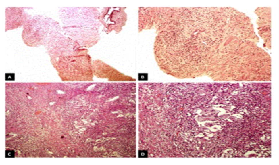
Figure 5: A. Microscopic photograph of transrectal biopsy (x5); in this image scattered inflammatory infiltrate in the stroma predominates. B. At a higher magnification (x10) granuloma with giant cells was identified. C. Histologic panoramic image (x5) of transurethral resection; an inflammatory process is evident in the stroma and surrounding the prostate glands. D. Close-up (x10) to observe the granulomas and giant cells. Reproduced from: [4]

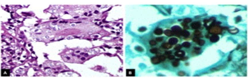
Figure 6: A. Close-up (x40) of multinucleated giant cells with yeast fungal structures in the interior. B. In this Grocott staining, the morphologic characteristics of fungal structures, which characteristically show an ovoid shape with a variable size of 8 to 15 microns and a wide base budding, are better appreciated. Reproduced from: [4]
Yeast fungal structures were identified within these giant cells and granulomatous formations and Grocott staining was undertaken in order to study the morphological characteristics of these fungal structures; they measured 8 microns to 15 microns in width and had shown broad-based budding. these findings had led to the diagnosis of granulomatous prostatitis which was associated with fungal structures that were consistent with the diagnosis of blastomycosis. The diagnosis was confirmed by the undertaking of molecular biology techniques. Total genomic DNA was extracted from a slice of paraffin-embedded tissue which then underwent PCR to amplify fungal ribosomal RNA sequences (ITS1-5.8S ribosomal RNA-ITS2) with the ITS4 (TCCTCCGCTTATTGATATGC) and ITS5 (GGAAGTAAAAGTCGTAACAAGG) consensus primer set [20] The amplification product of 666 base pairs (bp) were sequenced for both borders, assembled, and utilized as a hook in the Basic Local Alignment Search Tool (BLAST) of the NCBI. The results were 100% Ajellomyces dermatitidis (anamorph Blastomyces dermatitidis). Radiology imaging studies were ordered and the patient underwent a thorough clinical examination to exclude a systemic condition. Pulmonary involvement was intentionally evaluated and radiology imaging studies and examination had not identified no abnormalities. The patient commenced medical treatment with 400 mg daily of itraconazole orally for 6 months. After one week of his treatment the patient had demonstrated slight improvement, but due to the persistence of the obstructive symptoms, it was decided to undertake a transurethral resection of the prostate (TURP). Histological tissue sections from the TURP specimen had shown the same findings which had been previously described in the TRB, which had supported the diagnosis of granulomatous prostatitis due to prostatic blastomycosis. the TURP had resolved the obstructive urinary symptomatology and the patient was discharged from the hospital to continue his treatment as an outpatient for at least 6 months. He was asymptomatic at his last follow-up visit prior to publication of the article.

Discussions:


  • It has been iterated that granulomatous prostatitis is an unusual benign inflammatory process [21] 
  • Granulomatous prostatitis is a heterogeneous clinical entity that encompasses infectious, iatrogenic (post-surgery), or idiopathic (non-specific) lesions, malakoplakia, and other cases that are associated with systemic granulomatous disease, such as Wegener granulomatosis and allergy [22] 
  • Non-specific granulomatous prostatitis is the documented most common type [21] [23] and the most frequent aetiological factor for the development of non-specific granulomatous prostatitis is surgery (transurethral resection of prostate gland) or prostate needle biopsy. [24] [25] 
  • Clinical manifestation of granulomatous prostatitis may simulate that of benign prostatic hypertrophy or it might even be clinically indistinguishable from that of carcinoma of prostate [26], in view of the fact that it tends to be frequently associated with an abnormal digital rectal examination finding [27], an elevated serum prostate-specific antigen (PSA) results [8], or both. 
  • Infectious granulomatous prostatitis could be caused by Mycobacterium tuberculosis [29] [30] BCG instillation for transitional cell carcinoma of the bladder [22] [31] syphilis [32] malakoplakia, [33], [34] and various fungi [3]. 
  • Fungal granulomatous prostatitis is stated to be uncommon. [35] 
  • The fungal species which tend to most frequently cause prostatitis, include: candida, aspergilla, and cryptococcus. 
  • The patients that are at higher risk for developing fungal infections include the following: chronically ill, debilitated, and immune-compromised. 
  • Other factors related to the development of fungal infections do include: changing patterns of patient demographics which increase environmental exposure. [20] 
  • There are few cases of prostate afflictions reported as an initial manifestation of blastomycosis in the literature. 
  • The genitourinary system is a target for fungal infections including Candida, Cryptococcus and Aspergillums, Blastomyces, Coccidioides, and Histoplasma, which could be present and tend to be more aggressive within vulnerable patients. [36] 
  • Blastomyces dermatitidis is a dimorphic fungus which does predominate within North America and which is endemic to the Ohio and Mississippi River Valley regions, that is why this fungal infection is also known as North American Blastomycosis, Chicago disease, and Gilchrit's disease. [37] 
  • Blastomycosis usually tends to manifest as a systemic infection.
  • Blastomyces dermatitidis is acquired pursuant to inhaling the infectious conidia which is aerosolized as the mycelial form of the fungus growing as a saprophyte in soil, and hence, in most cases, the lungs are the primary focus of infection. 
  • Primary Blastomyces cutaneous infection is uncommon and is usually caused by accidental inoculation in clinical microbiologists and pathologists; and it has also been documented to occur pursuant to dog bites [38] 
  • It has been documented that there is no racial predilection for the development of Blastomyces dermatitidis; nevertheless, the male-to-female ratio is as high as 15 to 1. [39] 
  • Most cases of pulmonary blastomycosis are asymptomatic and self-limiting in immunocompetent patients, but they might spread from the lung to other organs by haematogenous means. 
  • Similar to majority of other fungal infections, it has been iterated that severely immunodeficient patients are at increased risk for the development of blastomycosis [40] 
  • The organs that are most commonly afflicted include: the skin, larynx, bones and joints, central nervous system, lymph nodes, heart, adrenal gland, and the genitourinary tract. [41]
  • Eickenberg et al. [13] had undertaken a retrospective study of 51 cases of systemic blastomycosis. Eleven patients that amounted to 21.5% of the patients had been found to have genitourinary involvement, with the prostate and epididymis being the commonest afflicted sites. 
  • Clinically, prostate involvement by Blastomyces manifests with dysuria, nocturia, urinary retention, perineal or suprapubic pain, haematuria, and hematospermia. 
  • Blastomyces prostatitis could also simulate bacterial prostatitis, benign hyperplasia, or carcinoma. 
  • An abnormal urology examination could demonstrate induration of the prostate gland [12] [42] 
  • There are only a few reported cases of blastomycosis of the prostate gland in the literature to their knowledge (less than 10): One case had manifested with localized prostate involvement, [43], two cases had involved the prostate and epididymis [12] [44] two cases had involved the prostate and skin [45] [46], and a final case of systemic dissemination was initially diagnosed by the undertaking of trans-rectal prostate biopsy [17] 
  • Within tissue sections typical yeast-like cells are morphologically distinctive. They are spherical, hyaline, 8-microns to 15-microns with regard to diameter, multinucleated, and have vacuolated cytoplasm and thick double contoured walls. 
  • The yeast-like cells do reproduce by budding, and buds are attached to their parent cells by broad bases. [41] 
  • The predominant inflammatory reaction within the lesions of acute blastomycosis is suppurative, with infiltration of neutrophils and abscesses. 
  • Fungal cells are usually extracellular, numerous, and scattered in the lesions or localized to abscesses. 
  • In chronic lesions, there is a mixed suppurative and granulomatous inflammatory reaction with classic granulomas that are composed of compact epithelioid histiocytes, giant cells, lymphocytes, as well as fibroblasts, however, caseation and calcification tend to be uncommon [38] 
  • Whenever fresh tissues are available, microbiology culture should be undertaken to complement histopathology studies. [47] 
  • In view of the fact the yeast forms of Blastomyces dermatitidis and Histoplasma capsulatum or duboissi both have thick walls and are similar in size and shape, they could be mistaken for each other in tissue sections. The latter; nevertheless, usually bud by a narrower base, whereas the diagnostic budding from Blastomyces dermatitidis is broad-based. [48] 

 Diagnostic confirmation via molecular biology was strength of their reported case. It did not require fresh tissue and was undertaken out from paraffin-embedded tissue. The methodology employed was based upon amplifying sequences that are common among fungi and customarily utilized for conducting phylogenetic studies. 

  • PCR utilizing ITS4 and ITS5 primers amplifies ribosomal sequences of a wide variety of fungi and the sequencing of the product enables species identification. This methodology is interesting due to the fact that it could aid in the diagnostic confirmation of pathologies in which fungi are microscopically visualized in biopsies, as occurred in their reported case.
  • Infectious granulomatous prostatitis does necessitate medical treatment. 
  • It has been stated that surgical management of granulomatous prostatitis leads to complications such as bladder neck contracture and requires repeated resection. [21] 
  • It had been iterated that the current treatment of choice for blastomycosis is itraconazole, except for patients who have central nervous system involvement and in cases of life-threatening infection in which amphotericin B should be used [42] and [17] 
  • With regard to their reported case, the urinary tract obstruction had resolved through TURP and itraconazole was also indicated; nevertheless, long-term disease progression could not be evaluated because patient follow-up was lost after 6 months. 

               Labastida et al. [4] made the ensuing conclusions: 

  • Fungal granulomatous prostatitis is a rare simulator of other prostatic pathologies.
  • Fungal granulomatous prostatitis is a rare condition and the diagnosis of prostatic blastomycosis does require a high level of clinical suspicion, which is why physicians should be aware of this entity, especially in patients who dwell within Blastomyces-endemic areas and who have a history of chronic urinary obstruction symptoms, and those diagnosed with chronic prostatitis.

Conclusions

  • Genitourinary tract manifestations of blastomycosis are relatively rare that tend to be sporadically reported in various areas of the world. 
  • Blastomycosis prostatitis and Blastomycosis prostate abscess are extremely rare clinical entities that have been sporadically reported globally even though they would tend to be encountered more often in North America, South America, Some Parts of Canada, Africa and India, because of global travel, it would be envisaged that Blastomycosis infections of the skin, lungs, prostate and other genitourinary tract organs could be encountered anywhere in the world. 
  • There is the possibility there has been under-reporting of cases of Blastomycoses prostatitis and Blastomyces prostate abscess due to the fact that the symptoms of prostate infections by Blastomycoses infections are non-specific and the symptoms simulate the non-specific symptoms of more commonly encountered prostate pathologies including: acute and chronic bacterial prostate infections, benign prostate hyperplasia, carcinoma of prostate and if bacterial culture or prostate biopsies are not taken for pathology examination, some cases may be misdiagnosed. 
  • Clinicians all over the world due to global travel should have a high index of suspicion for this fungus, especially within Blastomyces-endemic areas of the world and when the symptoms are refractory to typical antimicrobial treatment.

Conflict Of Interest – Nil 

Acknowledgements 

Acknowledgements to:

  • Urology Case Reports and Hindawi Publishing Limited for granting Copyright permission for reproduction of figures and contents of their Journal article under Copyright: 
  • Fungal Genomics & Biology for granting permission for reproduction of figures and contents of their journal article under copyright: Copyright: © 2015 Labastida LG, et al., This is an open-access article distributed under the terms of the Creative Commons Attribution License, which permits unrestricted use, distribution, and reproduction in any medium, provided the original author and source are credited.

References

Clearly Auctoresonline and particularly Psychology and Mental Health Care Journal is dedicated to improving health care services for individuals and populations. The editorial boards' ability to efficiently recognize and share the global importance of health literacy with a variety of stakeholders. Auctoresonline publishing platform can be used to facilitate of optimal client-based services and should be added to health care professionals' repertoire of evidence-based health care resources.

img

Virginia E. Koenig

Journal of Clinical Cardiology and Cardiovascular Intervention The submission and review process was adequate. However I think that the publication total value should have been enlightened in early fases. Thank you for all.

img

Delcio G Silva Junior

Journal of Women Health Care and Issues By the present mail, I want to say thank to you and tour colleagues for facilitating my published article. Specially thank you for the peer review process, support from the editorial office. I appreciate positively the quality of your journal.

img

Ziemlé Clément Méda

Journal of Clinical Research and Reports I would be very delighted to submit my testimonial regarding the reviewer board and the editorial office. The reviewer board were accurate and helpful regarding any modifications for my manuscript. And the editorial office were very helpful and supportive in contacting and monitoring with any update and offering help. It was my pleasure to contribute with your promising Journal and I am looking forward for more collaboration.

img

Mina Sherif Soliman Georgy

We would like to thank the Journal of Thoracic Disease and Cardiothoracic Surgery because of the services they provided us for our articles. The peer-review process was done in a very excellent time manner, and the opinions of the reviewers helped us to improve our manuscript further. The editorial office had an outstanding correspondence with us and guided us in many ways. During a hard time of the pandemic that is affecting every one of us tremendously, the editorial office helped us make everything easier for publishing scientific work. Hope for a more scientific relationship with your Journal.

img

Layla Shojaie

The peer-review process which consisted high quality queries on the paper. I did answer six reviewers’ questions and comments before the paper was accepted. The support from the editorial office is excellent.

img

Sing-yung Wu

Journal of Neuroscience and Neurological Surgery. I had the experience of publishing a research article recently. The whole process was simple from submission to publication. The reviewers made specific and valuable recommendations and corrections that improved the quality of my publication. I strongly recommend this Journal.

img

Orlando Villarreal

Dr. Katarzyna Byczkowska My testimonial covering: "The peer review process is quick and effective. The support from the editorial office is very professional and friendly. Quality of the Clinical Cardiology and Cardiovascular Interventions is scientific and publishes ground-breaking research on cardiology that is useful for other professionals in the field.

img

Katarzyna Byczkowska

Thank you most sincerely, with regard to the support you have given in relation to the reviewing process and the processing of my article entitled "Large Cell Neuroendocrine Carcinoma of The Prostate Gland: A Review and Update" for publication in your esteemed Journal, Journal of Cancer Research and Cellular Therapeutics". The editorial team has been very supportive.

img

Anthony Kodzo-Grey Venyo

Testimony of Journal of Clinical Otorhinolaryngology: work with your Reviews has been a educational and constructive experience. The editorial office were very helpful and supportive. It was a pleasure to contribute to your Journal.

img

Pedro Marques Gomes

Dr. Bernard Terkimbi Utoo, I am happy to publish my scientific work in Journal of Women Health Care and Issues (JWHCI). The manuscript submission was seamless and peer review process was top notch. I was amazed that 4 reviewers worked on the manuscript which made it a highly technical, standard and excellent quality paper. I appreciate the format and consideration for the APC as well as the speed of publication. It is my pleasure to continue with this scientific relationship with the esteem JWHCI.

img

Bernard Terkimbi Utoo

This is an acknowledgment for peer reviewers, editorial board of Journal of Clinical Research and Reports. They show a lot of consideration for us as publishers for our research article “Evaluation of the different factors associated with side effects of COVID-19 vaccination on medical students, Mutah university, Al-Karak, Jordan”, in a very professional and easy way. This journal is one of outstanding medical journal.

img

Prof Sherif W Mansour

Dear Hao Jiang, to Journal of Nutrition and Food Processing We greatly appreciate the efficient, professional and rapid processing of our paper by your team. If there is anything else we should do, please do not hesitate to let us know. On behalf of my co-authors, we would like to express our great appreciation to editor and reviewers.

img

Hao Jiang

As an author who has recently published in the journal "Brain and Neurological Disorders". I am delighted to provide a testimonial on the peer review process, editorial office support, and the overall quality of the journal. The peer review process at Brain and Neurological Disorders is rigorous and meticulous, ensuring that only high-quality, evidence-based research is published. The reviewers are experts in their fields, and their comments and suggestions were constructive and helped improve the quality of my manuscript. The review process was timely and efficient, with clear communication from the editorial office at each stage. The support from the editorial office was exceptional throughout the entire process. The editorial staff was responsive, professional, and always willing to help. They provided valuable guidance on formatting, structure, and ethical considerations, making the submission process seamless. Moreover, they kept me informed about the status of my manuscript and provided timely updates, which made the process less stressful. The journal Brain and Neurological Disorders is of the highest quality, with a strong focus on publishing cutting-edge research in the field of neurology. The articles published in this journal are well-researched, rigorously peer-reviewed, and written by experts in the field. The journal maintains high standards, ensuring that readers are provided with the most up-to-date and reliable information on brain and neurological disorders. In conclusion, I had a wonderful experience publishing in Brain and Neurological Disorders. The peer review process was thorough, the editorial office provided exceptional support, and the journal's quality is second to none. I would highly recommend this journal to any researcher working in the field of neurology and brain disorders.

img

Dr Shiming Tang

Dear Agrippa Hilda, Journal of Neuroscience and Neurological Surgery, Editorial Coordinator, I trust this message finds you well. I want to extend my appreciation for considering my article for publication in your esteemed journal. I am pleased to provide a testimonial regarding the peer review process and the support received from your editorial office. The peer review process for my paper was carried out in a highly professional and thorough manner. The feedback and comments provided by the authors were constructive and very useful in improving the quality of the manuscript. This rigorous assessment process undoubtedly contributes to the high standards maintained by your journal.

img

Raed Mualem

International Journal of Clinical Case Reports and Reviews. I strongly recommend to consider submitting your work to this high-quality journal. The support and availability of the Editorial staff is outstanding and the review process was both efficient and rigorous.

img

Andreas Filippaios

Thank you very much for publishing my Research Article titled “Comparing Treatment Outcome Of Allergic Rhinitis Patients After Using Fluticasone Nasal Spray And Nasal Douching" in the Journal of Clinical Otorhinolaryngology. As Medical Professionals we are immensely benefited from study of various informative Articles and Papers published in this high quality Journal. I look forward to enriching my knowledge by regular study of the Journal and contribute my future work in the field of ENT through the Journal for use by the medical fraternity. The support from the Editorial office was excellent and very prompt. I also welcome the comments received from the readers of my Research Article.

img

Dr Suramya Dhamija

Dear Erica Kelsey, Editorial Coordinator of Cancer Research and Cellular Therapeutics Our team is very satisfied with the processing of our paper by your journal. That was fast, efficient, rigorous, but without unnecessary complications. We appreciated the very short time between the submission of the paper and its publication on line on your site.

img

Bruno Chauffert

I am very glad to say that the peer review process is very successful and fast and support from the Editorial Office. Therefore, I would like to continue our scientific relationship for a long time. And I especially thank you for your kindly attention towards my article. Have a good day!

img

Baheci Selen

"We recently published an article entitled “Influence of beta-Cyclodextrins upon the Degradation of Carbofuran Derivatives under Alkaline Conditions" in the Journal of “Pesticides and Biofertilizers” to show that the cyclodextrins protect the carbamates increasing their half-life time in the presence of basic conditions This will be very helpful to understand carbofuran behaviour in the analytical, agro-environmental and food areas. We greatly appreciated the interaction with the editor and the editorial team; we were particularly well accompanied during the course of the revision process, since all various steps towards publication were short and without delay".

img

Jesus Simal-Gandara

I would like to express my gratitude towards you process of article review and submission. I found this to be very fair and expedient. Your follow up has been excellent. I have many publications in national and international journal and your process has been one of the best so far. Keep up the great work.

img

Douglas Miyazaki

We are grateful for this opportunity to provide a glowing recommendation to the Journal of Psychiatry and Psychotherapy. We found that the editorial team were very supportive, helpful, kept us abreast of timelines and over all very professional in nature. The peer review process was rigorous, efficient and constructive that really enhanced our article submission. The experience with this journal remains one of our best ever and we look forward to providing future submissions in the near future.

img

Dr Griffith

I am very pleased to serve as EBM of the journal, I hope many years of my experience in stem cells can help the journal from one way or another. As we know, stem cells hold great potential for regenerative medicine, which are mostly used to promote the repair response of diseased, dysfunctional or injured tissue using stem cells or their derivatives. I think Stem Cell Research and Therapeutics International is a great platform to publish and share the understanding towards the biology and translational or clinical application of stem cells.

img

Dr Tong Ming Liu

I would like to give my testimony in the support I have got by the peer review process and to support the editorial office where they were of asset to support young author like me to be encouraged to publish their work in your respected journal and globalize and share knowledge across the globe. I really give my great gratitude to your journal and the peer review including the editorial office.

img

Husain Taha Radhi

I am delighted to publish our manuscript entitled "A Perspective on Cocaine Induced Stroke - Its Mechanisms and Management" in the Journal of Neuroscience and Neurological Surgery. The peer review process, support from the editorial office, and quality of the journal are excellent. The manuscripts published are of high quality and of excellent scientific value. I recommend this journal very much to colleagues.

img

S Munshi

Dr.Tania Muñoz, My experience as researcher and author of a review article in The Journal Clinical Cardiology and Interventions has been very enriching and stimulating. The editorial team is excellent, performs its work with absolute responsibility and delivery. They are proactive, dynamic and receptive to all proposals. Supporting at all times the vast universe of authors who choose them as an option for publication. The team of review specialists, members of the editorial board, are brilliant professionals, with remarkable performance in medical research and scientific methodology. Together they form a frontline team that consolidates the JCCI as a magnificent option for the publication and review of high-level medical articles and broad collective interest. I am honored to be able to share my review article and open to receive all your comments.

img

Tania Munoz

“The peer review process of JPMHC is quick and effective. Authors are benefited by good and professional reviewers with huge experience in the field of psychology and mental health. The support from the editorial office is very professional. People to contact to are friendly and happy to help and assist any query authors might have. Quality of the Journal is scientific and publishes ground-breaking research on mental health that is useful for other professionals in the field”.

img

George Varvatsoulias

Dear editorial department: On behalf of our team, I hereby certify the reliability and superiority of the International Journal of Clinical Case Reports and Reviews in the peer review process, editorial support, and journal quality. Firstly, the peer review process of the International Journal of Clinical Case Reports and Reviews is rigorous, fair, transparent, fast, and of high quality. The editorial department invites experts from relevant fields as anonymous reviewers to review all submitted manuscripts. These experts have rich academic backgrounds and experience, and can accurately evaluate the academic quality, originality, and suitability of manuscripts. The editorial department is committed to ensuring the rigor of the peer review process, while also making every effort to ensure a fast review cycle to meet the needs of authors and the academic community. Secondly, the editorial team of the International Journal of Clinical Case Reports and Reviews is composed of a group of senior scholars and professionals with rich experience and professional knowledge in related fields. The editorial department is committed to assisting authors in improving their manuscripts, ensuring their academic accuracy, clarity, and completeness. Editors actively collaborate with authors, providing useful suggestions and feedback to promote the improvement and development of the manuscript. We believe that the support of the editorial department is one of the key factors in ensuring the quality of the journal. Finally, the International Journal of Clinical Case Reports and Reviews is renowned for its high- quality articles and strict academic standards. The editorial department is committed to publishing innovative and academically valuable research results to promote the development and progress of related fields. The International Journal of Clinical Case Reports and Reviews is reasonably priced and ensures excellent service and quality ratio, allowing authors to obtain high-level academic publishing opportunities in an affordable manner. I hereby solemnly declare that the International Journal of Clinical Case Reports and Reviews has a high level of credibility and superiority in terms of peer review process, editorial support, reasonable fees, and journal quality. Sincerely, Rui Tao.

img

Rui Tao

Clinical Cardiology and Cardiovascular Interventions I testity the covering of the peer review process, support from the editorial office, and quality of the journal.

img

Khurram Arshad

Clinical Cardiology and Cardiovascular Interventions, we deeply appreciate the interest shown in our work and its publication. It has been a true pleasure to collaborate with you. The peer review process, as well as the support provided by the editorial office, have been exceptional, and the quality of the journal is very high, which was a determining factor in our decision to publish with you.

img

Gomez Barriga Maria Dolores

The peer reviewers process is quick and effective, the supports from editorial office is excellent, the quality of journal is high. I would like to collabroate with Internatioanl journal of Clinical Case Reports and Reviews journal clinically in the future time.

img

Lin Shaw Chin

Clinical Cardiology and Cardiovascular Interventions, I would like to express my sincerest gratitude for the trust placed in our team for the publication in your journal. It has been a true pleasure to collaborate with you on this project. I am pleased to inform you that both the peer review process and the attention from the editorial coordination have been excellent. Your team has worked with dedication and professionalism to ensure that your publication meets the highest standards of quality. We are confident that this collaboration will result in mutual success, and we are eager to see the fruits of this shared effort.

img

Maria Dolores Gomez Barriga