
Case Report | DOI: https://doi.org/10.31579/2690-8808/190
Cardiovascular Center, Onze Lieve Vrouwziekenhuis (OLV), Aalst, Belgium
*Corresponding Author: Marc Vanderheyden, MD Cardiovascular Center, OLV Ziekenhuis, Moorselbaan 164, 9300 Aalst, Belgium.
Citation: Augustijn Mortele, Marta Belmonte, Eline Bogaerts, Marc Vanderheyden, (2024), Bilateral Coronary Artery Fistulas and Pulmonary Sequestration. An Unusual Combination of a Rare Disease: Case Report and Review of the Literature. J, Clinical Case Reports and Studies, 5(2); DOI:10.31579/2690-8808/190
Copyright: © 2024, Marc Vanderheyden. This is an open access article distributed under the Creative Commons Attribution License, which permits unrestricted use, distribution, and reproduction in any medium, provided the original work is properly cited.
Received: 19 March 2024 | Accepted: 08 April 2024 | Published: 19 April 2024
Keywords: coronary artery fistulas; pulmonary sequestration; coronary vessel anomalies; diagnosis
Coronary artery fistulas are a rare entity, especially when they origin from both the left and right coronary arteries. CAF may be asymptomatic but severe complications are possible. They are usually congenital but acquired forms have been described. The management of CAF remains undetermined and depends on various factors. Pulmonary sequestration is another rare congenital malformation. It is a non-functioning lung segment without continuity with the normal lung parenchyma.
In this case report an 83-year-old male with newly diagnosed idiopathic pulmonary fibrosis, history of ischemic cardiomyopathy and severe tricuspid regurgitation was admitted for syncope. Due to episodes of non-sustained ventricular tachycardia an invasive coronary angiography was ordered which showed 2 coronary artery fistulas. The two CAF, arising respectively from the right coronary artery and the left anterior descending artery, both supplied blood to vessels in a sequestrated segment of the lower lung lobe.
This is the first report of two coronary artery fistulas, arising from both left and right coronary artery, providing blood supply to a pulmonary sequester. Although this entity is extremely rare, knowledge of it can prevent inappropriate management and fatal results.
CAF: coronary artery fistula
CCA: conventional coronary angiography
CTA: coronary CT angiography
CPAF: coronary artery-to-pulmonary artery fistula
IPF: idiopathic lung fibrosis
PCI: percutaneous coronary intervention
LAD: left anterior descending artery
RCA: right coronary artery
LV: left ventricular
EF: ejection fraction
RV: right ventricular
TR: tricuspid regurgitation
ERO: effective regurgitant orifice
FAC: fractional area change
TAPSE: tricuspid annular plane systolic excursion
HRCT: high-resolution CT scan
FFR: Fractional flow reserve
TC: transcatheter closure
Coronary artery fistulas (CAF) were first described in 1865 [1] and are rare vascular anomalies characterized by abnormal connections between coronary arteries and structures such as the heart cavity, pulmonary circulation, coronary sinus, or vena cava. [2-4] Usually congenital, acquired forms have also been reported. [5,6] The clinical presentation of CAF varies based on the shunt's location, size, and associated conditions. [3,7]
Conventional diagnostic techniques like coronary angiography (CCA) provide limited insights into the 3D anatomy and tissue interactions. In contrast, coronary CT angiography (CTA) is a superior alternative. CTA offers improved visualization of the fistulas' origin, drainage, number, and can detect aneurysms, thrombi and intricate vascular details. [8] Routine CTA has revealed a prevalence of 0.9% for CAF, with coronary artery-to-pulmonary artery fistula (CPAF) being the most common, followed by coronary artery-to-bronchial artery fistula and coronary artery-to-cardiac chamber fistula. [5] They more frequently arise from the right coronary artery (50%) than the left coronary artery (42%), and in about 5% of cases, they originate from both arteries. [9] CAF may remain asymptomatic or cause symptoms such as chest pain or dyspnea related to a steal phenomenon. [4] In rare cases, complications may occur, including massive hemoptysis, pulmonary hypertension, congestive heart failure, or arterial aneurysm rupture. [4,6,7] CAF can co-occur with lung-related diseases such as bronchiectasis, pulmonary tuberculosis, cystic fibrosis, chronic bronchitis, and interstitial fibrosis. [3,5,7,10] In extremely rare cases, CAF may supply blood to a sequestrated lung segment. [11] Treatment of CAF varies, depending on size, symptoms, anatomical characteristics, concomitant disorders, and the patient's age. Management strategies range from a "watchful waiting" approach to antiplatelet therapy, antibiotics and surgical or percutaneous closure. [4,12] The purpose of this article is to review the literature on CAF, focusing on an exceptional case report.
An 83-year-old male non-smoker (with no known exposure to any occupational hazards) diagnosed with idiopathic lung fibrosis (IPF), treated with antifibrotic medication, and having undergone a proximal LAD PCI due to unstable angina, was referred for evaluation of recurrent syncopal episodes. These episodes occurred at rest, characterized by sudden loss of consciousness and quick recovery. They were unexplained and did not involve palpitations. Previous investigations, such as long-term holter monitoring, EEG, brain CT scan and carotid artery duplex were inconclusive. The patient's medication regimen included Spironolactone 100 mg, Bumetanide 2 mg, and Perindopril 5 mg, initiated due to heart failure with mid-range ejection fraction (HFmrEF) caused by ischemic cardiomyopathy.
Upon admission, the patient's cardiopulmonary examination appeared normal with an oxygen saturation of 96%. Bilateral inspiratory crepitations were heard during lung auscultation. Electrocardiography (ECG) revealed permanent atrial fibrillation with a ventricular rate of 67 beats per minute. Blood tests showed no abnormalities. Transthoracic echocardiography revealed mildly reduced left ventricular (LV) function with an ejection fraction (EF) of 45%, due to post-ischemic hypokinesia of the anterior wall and apex. The right ventricle (RV) appeared moderately dilated, with functional moderate tricuspid regurgitation (TR) with an effective regurgitant orifice (ERO) of 35 mm2 and a peak TR velocity of 3.2 m/sec. The RV had a tricuspid annular plane systolic excursion (TAPSE) of 13 mm and a systolic velocity (s') of 9 cm/sec, with an RV fractional area change (FAC) of 30%. Additionally, D-shaping of the RV was observed.
Chest X-rays showed reticular interstitial markings near the lung bases, while high-resolution CT scans (HRCT) confirmed the presence of IPF. The HRCT revealed fibrotic changes in both lung bases and peripheries, characterized by patchy reticular opacities and honeycombing.
Non-sustained ventricular tachycardia episodes during hospitalization led to a coronary catheterization showing normal coronary arteries with minimal atherosclerosis and a patent stent in the LAD. Fractional flow reserve (FFR) measurements were conducted to rule out ischemia in the LAD territory as the underlying cause of the ventricular arrhythmias. Simultaneously, a right heart catheterization was carried out revealing a mean pulmonary arterial pressure of 25 mmHg, pulmonary arterial systolic pressure of 45 mmHg, a mean pulmonary capillary wedge pressure of 20 mmHg, and a pulmonary vascular resistance of 1 WU.
Two coronary artery fistulas (CAFs) were identified: one originating from the distal RCA (Movie 1, Figure 1) and the other from the LAD (Movie 2, Figure 1, 2), proximal to the implanted stent. Both CAFs supplied blood to vessels in a secluded part of the lower lung lobe (Figure 1-2).
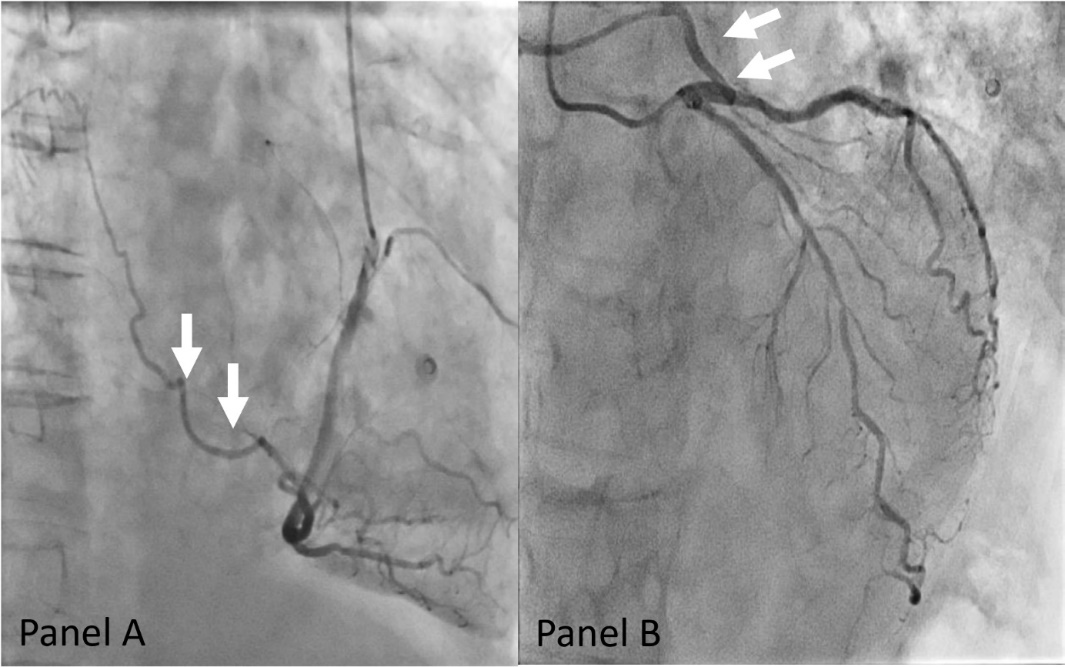
Panel A: Right anterior oblique view of the CAF originating from the right coronary artery.
Panel B: Right anterior oblique view of the CAF originating from the left anterior descending artery.
Figure 1: CCA images from both CAF
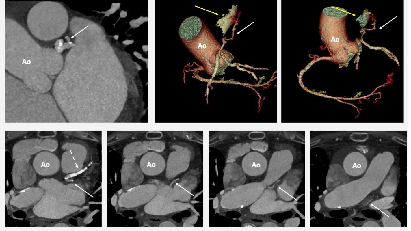
Upper left image: Coronal oblique view derived from CTA – The coronary-pulmonary fistula originates from the proximal left anterior descending artery (white arrowhead).
Upper right and mid images: 3D reconstruction of the CAF. The path of the fistula is illustrated with white arrowheads, while yellow arrowheads indicate the presence of a pulmonary sequestration.
Lower images: Set of axial views derived from CTA The path of the fistula is illustrated with white arrowheads, while dashed white arrowheads indicate the stent in the proximal LAD
Ao indicates aorta, x pulmonary sequester
Figure 2. CTA images from the left sided CAF.
Movie 1.
CCA movie illustrating CAF originating from the right coronary artery (right anterior oblique projection).
Movie 2.
CCA movie demonstrating CAF originating from the left anterior descending artery (right anterior oblique projection).
A retrospective analysis of the coronary angiogram performed during the PCI confirmed the presence of these CAFs, ruling out any connection to iatrogenic dissection or complications related to the PCI. Furthermore, the morphology of these CAFs appeared to be unchanged.
Because the patient's syncope could not be attributed to the CAFs, and an electrophysiological study revealed no abnormalities except for the pre-existing permanent atrial fibrillation and normal HV conduction, an observational approach was undertaken with the implantation of a loop recorder. After one month, a decision was made to implant a VVI pacemaker due to the documentation of a ventricular pause lasting over 3 seconds, despite the absence of symptoms, and the patient's history of unexplained and recurrent syncopal episodes. Initially, the patient's condition showed improvement, and there were no further instances of syncope following pacemaker implantation. However, two years later, he passed away because of respiratory failure caused by SARS-CoV-19 infection.
In this case report we describe a rare case of congenital asymptomatic bilateral coronary artery fistulas feeding a pulmonary sequester in a patient with idiopathic pulmonary fibrosis. Although coronary artery fistulas arising from both the right and left coronary artery are well described and certain types of CAF have been associated with abnormalities in the lung parenchyma or pulmonary vasculature, the co-existence of 2 CAFs originating from both left and right coronary artery and feeding a pulmonary sequester is extremely rare and has to the best of our knowledge never been described. [5,9]
Various hypotheses explain the embryogenesis of CAF. The most widely accepted one is the Hackensellner involution-persistence hypothesis. This hypothesis states that only two of the six branches of the truncus persist and form the coronary arteries, while the other four involute. CPAF develop when there is an abnormal persistence of the branch from the pulmonary sinus that connects to the normal branches forming the coronary arteries. [13]
Another hypothesis suggests the presence of congenital, small, non-functional anastomoses between the bronchial arteries and the coronary arteries that grow and enlarge due to several factors. This may occur when there is a shunting of blood from the coronary to the bronchial circulation, for example, in the presence of a supravalvular aortic stenosis causing increased coronary artery pressure or when the bronchial artery pressure drops, for example, in pulmonary atresia. Similarly, in obstructive coronary artery disease, the fistula may fill the coronary artery distal to the obstruction. [7]
CAF can be acquired and have been documented after a myocardial infarction or penetrating trauma. Recurrent inflammation, observed in bronchiectasis, may stimulate angiogenesis and the formation of an acquired CAF. [14] In this case, CAFs were already present five years before the diagnosis of idiopathic pulmonary fibrosis (IPF) at the time of a percutaneous coronary intervention (PCI). This timeframe makes it less plausible to establish a causal relationship between the two conditions.
There are no prospective randomized trials comparing the various management options, and all recommendations are based on small retrospective series. The management strategy is influenced by multiple factors, such as the size and anatomy of the CAFs, presence of symptoms, associated cardiovascular abnormalities, patient's age and comorbidities. [10,13]
Small CAFs tend to close on their own over time, so a monitored approach is usually sufficient. The choice between transcatheter closure (TC) and surgical intervention is still debated. However, with advancements in transcatheter equipment and techniques, TC has become the preferred method for closure of CAFs. Specifically, for patients with favorable anatomy (single narrow drainage site, proximal fistula origin, absence of multiple fistulas or large branch vessels) and no concomitant cardiac disorders, TC is now considered the first choice. [15]
Despite being less invasive, TC is not without complications such as residual leakage or recanalization of the fistulas, coil migration, thromboembolism and distal embolization. Therefore, it is recommended to thoroughly evaluate both the CAFs and the patient before starting therapy. [13]
In this case, the discovery of a pulmonary sequestration was incidental. Thanks to the normal FFR value, which ruled out a connection between coronary steal and ventricular rhythm issues, a conservative approach was chosen. [4,12]
Coronary artery fistulas remain a remarkable entity with many unanswered questions about its pathophysiology, natural history and treatment. It is important to acknowledge their existence and detect possible complications to facilitate the most optimal, patient-tailored management. In a patient with sequestration, suspicion of coronary origin is important and requires preoperative angiography when surgical ligation of the feeding artery is considered. Injury or proximal ligation may produce myocardial ischemia, infarction or death. Although this entity is extremely rare, knowledge of it can prevent inappropriate management and fatal results.
The main limitation of this study is its inability to generalize the findings beyond the specific case being reported. Therefore, the use and interpretation of these results should be approached with caution and considered within a broader context.
AM, MV contributed to the conception and writing of the manuscript. All authors contributed to manuscript revision, read and approved the submitted version.
Conflicts of Interest: none declared.
Funding Statement: none declared.
Patient Consent Statement
We declare that an informed consent, in compliance with COPE guidelines, has been obtained from the involved patient and that the patient has given approval for the information to be published in this case report.
Author Data
The data underlying this article are available in the article and in its online supplementary material. We have no conflicts of interest to disclose.
Dear Editorial Team, Clinical Medical Reviews and Reports. My experience with the journal was highly positive. The peer-review process was rigorous, constructive, and completed in a timely manner. The reviewers provided valuable comments that helped improve the quality and clarity of our manuscript. The editorial office was professional, responsive, and supportive throughout all stages of the publication process. Communication was clear and efficient, and any questions were addressed promptly. Overall, I found the journal to maintain high scientific standards and an excellent publication workflow. I would be pleased to consider submitting future work to this journal. Best wishes from, Elena Popa.

It was my pleasure to submit my testimonial concerning the Reviewer Board of our Scientific Journal “Brain and Neurological Disorders”. The Reviewers focused on some modifications and their contribution was helpful. The ladies of our Editorial Office were also supported my efforts. It was my honor to have such a co-operation and I am looking forward for more collaboration.

Dear Grace Pierce, Editorial Coordinator of Journal of Clinical Research and Reports, Thank you for the speedy and efficient peer review process. I appreciate the fact that your peer reviewers do not take months to respond like with some other journals. I would also like to thank the editorial office for responding quickly to my questions. It is an excellent journal. I plan to submit more manuscripts in the future. Best wishes from, Robert W. McGee

Dear Grace Pierce, Editorial Coordinator of Journal of Clinical Research and Reports, Working with you and your team on our recent publication in JCRR has been a truly wonderful and enjoyable experience. The responses were prompt, and the reviewers were patient, constructive, and highly professional. One reviewer in particular gave me the feeling that a professor was carefully reading and commenting on my coursework, which was deeply touching. The entire process was straightforward and hassle‑free, with no tedious online forms to complete. I highly recommend this journal. Best wishes from, DR Aibing Rao, Head of R&D

I Appreciate the Opportunity to Share my Experience with the Journal of Clinical Research and Reports. The peer review process was timely and constructive, and the feedback provided helped improve the quality of our manuscript. The editorial office was professional, responsive, and supportive throughout the process, ensuring smooth communication and efficient handling of the submission. Overall, it was a positive experience collaborating with your team.

Dear Mercy Grace, Editorial Coordinator of Obstetrics Gynecology and Reproductive Sciences, We would like to express our gratitude for your help at all stages of publishing and editing the article. The editors of the magazine answer all the necessary questions and help at every stage. We will definitely continue to cooperate and publish other works in the Obstetrics Gynecology and Reproductive Sciences! Best wishes from, Alla Konstantinovna Politova,
