
Case Report | DOI: https://doi.org/10.31579/2578-8965/128
1 Obstetrics department, Children's hospital El Harrouch, University Hospital of Casablanca
*Corresponding Author: Haddout S., Obstetrics department, Children's hospital El Harrouch, University Hospital of Casablanca.
Citation: Haddout S, Jalal M, Ikouch K, Lamrissi A, Bouhya. (2022). Bilateral Adnexal Torsion During the Third Trimester of Pregnancy, J. Obstetrics Gynecology and Reproductive Sciences, 6(5) DOI: 10.31579/2578-8965/128
Copyright: © 2022 Haddout S. This is an open-access article distributed under the terms of The Creative Commons Attribution License, which permits unrestricted use, distribution, and reproduction in any medium, provided the original author and source are credited.
Received: 01 July 2022 | Accepted: 20 July 2022 | Published: 26 July 2022
Keywords: adnexal torsion during pregnancy; adnexal torsion and 3rd trimester; multicystic ovaries and adnexal torsion.
Acute pelvic pain during pregnancy can pose a problem for differential diagnosis. We report a case of bilateral adnexal torsion in the third trimester of pregnancy. Indeed, this diagnosis may go unnoticed in front of pelvic pain associated with a pregnancy and only an early management can avoid irreversible lesions due to ischemia, which may jeopardize the subsequent prognosis of fertility. Our patient is 28 years old, primiparous, with polycystic ovary syndrome, followed for a pregnancy stimulated by clomiphene citrate. She developed preeclampsia with intrauterine growth retardation during the follow-up of her pregnancy. She presented to the maternity emergency room with sustained uterine contractions at 36 weeks' gestation. The patient had a code red cesarean section for suspected retroplacental hematoma. Intraoperatively, no retroplacental hematoma was found but bilateral torsion of the adnexa with huge polycystic ovaries. Conservative treatment was recommended and the postoperative follow-up of the cesarean section was simple. Adnexal torsion is an emergency that should not be ignored in the presence of any acute pelvic pain in a pregnant woman.
Adnexal torsion during pregnancy is a rare emergency with an incidence of 4 per 10000 pregnancies. It usually occurs in the first trimester on a pathological ovary. However, the diagnosis remains difficult, because of the ascension of the ovary in advanced pregnancies, which can mimic other surgical emergencies such as acute appendicitis, cholecystitis or acute pyelonephritis, and in cases of chronic pelvic pain. We report here a case of bilateral adnexal torsion on polycystic ovaries during the 3rd trimester of pregnancy. The interest of this situation lies in its diagnostic difficulty, and in the choice of the therapeutic attitude to adopt.
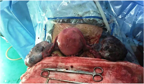
Our patient is 28 years old, with a history of polycystic ovaries and 5 years of infertility. She is followed in our structure for her pregnancy obtained by stimulation with clomiphene citrate. During her pregnancy, she developed severe pre-eclampsia, stabilized under treatment, complicated by intrauterine growth retardation. She consulted for sustained uterine contractions at 36 weeks of amenorrhea. In addition, she reported chronic pelvic pain that had been evolving since her second trimester. On examination, the patient was apyretic, in good general condition with discrete oedemas of the lower limbs and a blood pressure of 130/93 on monotherapy. She had sustained uterine contractions without cervical changes. There was no external bleeding and fetal heart sounds were heard. Abdominal ultrasound found a progressive monofetal pregnancy with a biometry lower than the 3rd percentile, a homogeneous placenta normally inserted, amniotic fluid in normal quantity, a normal umbilical doppler. The cardiac rhythm recording showed a fetal bradycardia, hence the indication of a code red cesarean section for suspicion of retroplacental hematoma in view of the uterine hypertonia and the fetal bradycardia. On exploration, macroscopic examination of the placenta did not reveal any cupules or haematomas. On the other hand, the adnexa were twisted (one turn of spiral) with a purplish-blue color and in the early stages of necrosis, with ovaries 14 cm in length (Figure 1).
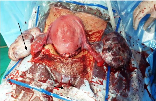
Detorsion of the adnexa allowed revascularization and recolouring of the adnexa (Figure 2).
Ovariopexy was performed with fixation of the ovaries at the level of the broad ligament. Post-operative follow-up is simple. At three months postpartum, a control ultrasound was performed in the department with a decrease in the volume of the ovaries, without any othernotable anomaly.
During pregnancy, adnexal torsion is a rare emergency. Its incidence varies from 3 to 5 per 10,000 pregnancies [1, 2]. Between 8 and 28% of twists occur during pregnancy [3, 4], mostly in the first trimester but can be diagnosed at any stage of pregnancy [4]. Torsion usually occurs on a pathological ovary: malignant or benign tumour, corpus luteum cyst, or on the occasion of an ovarian hyperstimulation syndrome in early pregnancy [3]. Symptoms are characterized by sharp lateral pelvic pain associated with nausea and vomiting. Its diagnosis during pregnancy is made complex because it requires the elimination of classic differential diagnoses but also those that may be related to pregnancy (miscarriage, retro-placental hematoma, uterine rupture). Moreover, the clinical examination like the imaging examinations becomes more difficult because of the uterine volume and the concomitant rise of the ovary in the abdominal cavity. Ultrasound is the reference examination. It eliminates differential diagnoses, and looks for factors that may favor torsion and indirect signs of ischemia. The first interruption of the venous flow leads to a reactive edema which is identifiable by the increase in ovarian volume compared to the contralateral side [5, 6]. Moreover, the increase in the number of cortical follicles is a non-specific aspect but which has been found many times in the case of torsion on a healthy ovary. It would be due to fluid transudation secondary to ovarian congestion [6, 7]. This aspect of homogeneous and peripheral follicular structure was found in our case.
Finally, images of hemorrhagic infarction can be visualized late, and a blade of nonspecific ascites is often associated [8]. Ultrasound has the advantage of locating the ovary in the abdominal cavity and looking for elective pain when passing the probe. The usefulness of ovarian vessel Doppler is controversial. Although the absence of Doppler signal confirms the absence of arterial or venous flow and therefore torsion, the reverse is not true [9]. MRI is a satisfactory complementary exploration technique in pregnant women, which has the same benefit as ultrasound with greater precision [10]. The combination of Doppler and MRI is useful but should not delay surgical management. Classically, the prevention of a recurrence initially involves etiological treatment: cystectomy, follicular puncture or adnexectomy. In the case of a healthy ovary, the situation is different. The recent retrospective study by Pansky et al. shows in women in the post-pubertal period a recurrence rate of 67% when the torsion concerns a healthy ovary (including 50% on the same side) against 8% when the ovary is pathological [6]. Bilateral oovariopexy therefore seems to be necessary [6, 11]. Many techniques have been described: fixation of the ovary to the broad ligament or on the lateral wall, shortening of the utero-ovarian ligament or fixation of the ovary to the uterus [12–14]. The latter does not seem to be reasonably applicable during pregnancy. Only two cases of ovarian fixation during pregnancy have been reported in the literature [11, 13]. Djavadjan et al performed a plication of the utero-ovarian ligament at 15 weeks by suturing the most proximal portion of this ligament to its most distal portion. In a case of recurrent torsion in a 10 WA pregnancy obtained by IVF, Weitzman et al describe the shortening of the utero-ovarian ligament using an endoloop due to the risk of hemorrhage due to hyper-vascularization. The realization of these techniques is especially problematic in the 2nd and 3rd trimester due to the increase in size of the uterus. On the other hand, the efficacy and harmlessness of oovariopexy have not been demonstrated. The shortening of the utero-ovarian ligament could be the cause of a decrease in ovarian vascularization and therefore an alteration of ovarian function [6]. We can also imagine that the lateral parietal fixation could be the cause of an anatomical disturbance of the tubo-ovarian relationships, and of flange occlusion [6]. Thus, the choice of technique is left to the operator according to the specific anatomical constraints of each patient. Whichever one is chosen; contralateral preventive treatment could prevent contralateral recurrences during pregnancy. The laparoscopic management of adnexal pathologies in the first and second trimester of pregnancy is no longer discussed. Oelsner et al compared the outcomes of 197 laparotomies and 192 laparoscopies in 17 centers [14]. Laparoscopy does not increase the risk of spontaneous abortion, premature delivery, intrauterine growth retardation.
Classically described in pathological ovaries or tubes, adnexal torsion can however rarely occur in healthy ovaries [18]. Chronic pelvic pain should be treated with caution, as it may conceal gynaecological emergencies and delay management.
Dear Editorial Team, Clinical Medical Reviews and Reports. My experience with the journal was highly positive. The peer-review process was rigorous, constructive, and completed in a timely manner. The reviewers provided valuable comments that helped improve the quality and clarity of our manuscript. The editorial office was professional, responsive, and supportive throughout all stages of the publication process. Communication was clear and efficient, and any questions were addressed promptly. Overall, I found the journal to maintain high scientific standards and an excellent publication workflow. I would be pleased to consider submitting future work to this journal. Best wishes from, Elena Popa.

It was my pleasure to submit my testimonial concerning the Reviewer Board of our Scientific Journal “Brain and Neurological Disorders”. The Reviewers focused on some modifications and their contribution was helpful. The ladies of our Editorial Office were also supported my efforts. It was my honor to have such a co-operation and I am looking forward for more collaboration.

Dear Grace Pierce, Editorial Coordinator of Journal of Clinical Research and Reports, Thank you for the speedy and efficient peer review process. I appreciate the fact that your peer reviewers do not take months to respond like with some other journals. I would also like to thank the editorial office for responding quickly to my questions. It is an excellent journal. I plan to submit more manuscripts in the future. Best wishes from, Robert W. McGee

Dear Grace Pierce, Editorial Coordinator of Journal of Clinical Research and Reports, Working with you and your team on our recent publication in JCRR has been a truly wonderful and enjoyable experience. The responses were prompt, and the reviewers were patient, constructive, and highly professional. One reviewer in particular gave me the feeling that a professor was carefully reading and commenting on my coursework, which was deeply touching. The entire process was straightforward and hassle‑free, with no tedious online forms to complete. I highly recommend this journal. Best wishes from, DR Aibing Rao, Head of R&D

I Appreciate the Opportunity to Share my Experience with the Journal of Clinical Research and Reports. The peer review process was timely and constructive, and the feedback provided helped improve the quality of our manuscript. The editorial office was professional, responsive, and supportive throughout the process, ensuring smooth communication and efficient handling of the submission. Overall, it was a positive experience collaborating with your team.

Dear Mercy Grace, Editorial Coordinator of Obstetrics Gynecology and Reproductive Sciences, We would like to express our gratitude for your help at all stages of publishing and editing the article. The editors of the magazine answer all the necessary questions and help at every stage. We will definitely continue to cooperate and publish other works in the Obstetrics Gynecology and Reproductive Sciences! Best wishes from, Alla Konstantinovna Politova,
