
Editorial | DOI: https://doi.org/10.31579/2578-8868/358
Neurosurgeon Consultant, Neurosurgery Department, Policlínica Juaneda Miramar, Palma de Mallorca, Balearic Islands, Spain.
*Corresponding Author: Gonçalo Januário, Neurosurgeon Consultant, Neurosurgery Department, Policlínica Juaneda Miramar, Palma de Mallorca, Balearic Islands, Spain.
Citation: Gonçalo Januário, (2025), Basic Concepts and Materials used in Unilateral Biportal Endoscopy, J. Neuroscience and Neurological Surgery, 17(2); DOI:10.31579/2578-8868/358
Copyright: ©, 2025, Gonçalo Januário. This is an open-access article distributed under the terms of The Creative Commons Attribution License, which permits unrestricted use, distribution, and reproduction in any medium, provided the original author and source are credited
Received: 09 January 2025 | Accepted: 24 January 2025 | Published: 05 February 2025
Keywords: minimal invasive; materials; biportal; endoscopy; spine surgery
Unilateral Biportal Endoscopy (UBE) is a minimally invasive spinal surgical technique that use two small incisions; one for an endoscope and another for surgical instruments. This approach allows surgeons to perform spinal procedures with enhanced precision, improved visualization, and reduced tissue disruption compared to open surgeries. The basic concept behind UBE is to access the spinal structures through a single side of the body, reducing muscle dissection with faster recovery times. Key materials used in UBE include specialized endoscopes, which provide high-definition, real-time visualization of the surgical site, and working instruments designed to perform decompression, disc removal, or nerve root release with minimal invasiveness. Endoscopes are typically rigid or flexible, with a small diameter, allowing for easy maneuverability through narrow spaces in the spinal canal. The surgical instruments used in UBE, such as forceps, shavers, and laser probes, are specifically designed for precision and to fit through small portals. Frequently we use common instruments, this is a important advantage of this technique. UBE permit access the spinal structures through the interlaminar space, with minimal trauma to surrounding tissues. The procedure’s minimally invasive nature offers benefits including reduced blood loss, decreased postoperative pain, and faster recovery times. However, the technique requires advanced training and skill, as well as familiarity with the equipment and anatomy. This abstract explores the basic principles and materials integral to UBE, highlighting its clinical applications and its growing role in modern spinal surgery.
The French physician Antoine Jean Desormeaux, lived in the 19th-century, considered the "father of endoscopy” developed an instrument to examine the urinary tract and bladder named “endoscope”. It was the first time in history that this term was used.1
Throughout the last century, many technological advances were achieved with increasingly sophisticated devices and surgical approaches. Particularly during the last decade the endoscopic surgery began to increase the applicability for the treatment of spine pathologies.
Actually is not yet considered gold standard but is progressing quickly to become the reference technique maybe during the next years. Like all surgical techniques present advantages and disadvantages, ideal for decompression and discectomy in cases of stenosis or disc herniation respectively. At this time it is increasingly used in the 3 segments of the spine (cervical, dorsal, lumbar) and in different pathologies. However, we must not forget that it has a limited indication in cases of high-grade listhesis as an isolated technique.
Two endoscopic surgical techniques are currently available, uniportal and biportal. Uniportal, full- endoscopic spine surgery is a minimally invasive procedure that uses a keyhole approach for the decompression or the discectomy. For other part the Unilateral biportal endoscopy (UBE), can be performed more widely, use two portals that permit high range of movements and amplified vision in the surgical field. When compared both techniques the muscle destruction is similar.2
All the endoscopic procedures require a working space, but the spine from an anatomical point of view present a limited work space especially by the bone and osteoarticular structures.
A important meaning in the endoscopic spine surgeries is the Kambin’s triangle, an anatomical reference, a natural corridor that allows the ideal approach to the lumbar canal.3
As in all spine surgeries, also in both endoscopic techniques, the manipulation of the duramater and particularly the nerve roots should be avoided to reduce complications.
The UBE requires two or more channels, in cases with a multilevel approach, to create sufficient work spaces around the levels we want to treat. The approaches in UBE are interlaminar, between the two laminae, or extraforaminal, depending the pathology location.
The anatomical corridor for the interlaminar approach, present two potential ways. The interfascicular related with the two small muscles of the multifidus, and the space containing fat and connective tissue located between the multifidus and the lamina. When these two potential spaces can be converted to an atraumatic working path, UBE guarantees the same indications as conventional spinal surgical treatments but with concepts of minimally invasive surgery.
In our center we don’t use dilators to do the approach and find the interlaminar space, we respect the anatomical corridors. For the extraforaminal approach, the surrounding area of the foramen with fat and connective tissue acts as the working space.4
As a big difference for open surgery the endoscopic spinal surgery is a fluid-medium procedure, similar to orthopedic arthroscopy. The operational concept is similar in both procedures.
In UBE we need to create two surgical portals on the same side of the spine, one for the endoscope and the other for the instruments used during the procedure.
The irrigating with saline solution enters in the endoscopic portal and leaves through the working portal (Figure 1).

Figure 1: illustration of fluid input and output in UBE.
A continuous and controlled circulation of the saline solution fluid is a critical factor for performing a successful UBE. The ideal pressure in the working space is 30–50 mmHg. The correct circulation of the saline solution between the channels allows a clearer view of the surgical field and in addition reduce the infection rate.
Therefore, to prevent complications related to circulation and working in a liquid medium, which are one of the main problems for beginners, it´s important facilitate the free circulation of the flow between the portals. In UBE several structures can influence and limit the free flow of saline solution, such as the fascia and the posterior paravertebral muscles. Furthermore, the spinal canal is surrounded by bony and ligamentous structures that also reduce the free circulation of the flow. Once the yellow ligament is removed, in the interlaminar approaches, the epidural space is directly entered without a separating structure in addition to the thin layer of epidural fat. Attention in this point for evitate dural tears or leaks As in all surgical techniques, knowledge of anatomy and anatomical landmarks is essential. It allows a better understanding and guidance during the procedure, avoiding complications and facilitating the workflow. It certainly reduces the need for intraoperative x-rays or the use of the neuronavigation system. Mastery of anatomical references can allow the surgeon to realize that the navigation system may be presenting a shift on relation to the real location. All the technology available in the operating room is a great advantage but the degree of accuracy must always be verified at all times. This is an important point for all those who begin to perform this technique.
Another key point that we must take into account is the distance from the skin to the spine. This aspect, especially in obese or with highly developed muscles patients, makes it difficult for the flow of saline solution to
circulate. And it can be a confusion factor or complications during surgery, especially in the first steps of the surgical procedure. If this happens, do not forget the basic anatomical points, identifying them at all times and of course confirming them with the intraoperative imaging techniques that we have available in our operating rooms. Do not forget that it is very important confirm the correct opening of the fascia.
If the circulation is not correct or the pressure increases a high hydrostatic pressure may cause headache in the postoperative period. To prevent complications the use of a controlled pressure pump or using the effect of gravity with the principle of communicating vessels is recommended. Thus avoiding high intracanal pressure in our work space. The optimal hydrostatic pressure, depend the authors, is located between 30–50 mmHg, which varies according to the distance between the irrigating saline bag and the working space. The working space in the patient is fixed less then 1 cm and the height of the saline bag decides the pressure.
For calculate the pressure (mmHg) we dividing the distance (cm) between the patient and the saline bag by 1.36. [height(cm) ÷ 1.36 = pressure (mmHg)].[5]
In our center we don’t use pump-controlled system, we use the effect of the gravity for a natural input and output of the saline solution. Sometimes, if necessary, we temporarily place a drainage to help the output flow. One important point for facilitate the flow between the portals, in our opinion, is a correct open of the fascia.
The principal factors related to fluid complications are a excessive saline pressure in the epidural space impacts the neural system and can cause headache, muscle pain, transient dysesthesia or paresthesias, nuchal pain, long-level epidural bleeding, delayed awakening from general anesthesia, seizure, intracranial hematoma, blindness due to retinal bleeding.
Are also described as a rare complication like a massive abdominal fluid collection could occur while performing the paraspinal approach. Is caused by massive damage to the inter-transverse membrane and ligaments, which function as a barrier between the back and abdominal cavity.
Another complication related with the fluid could be hypothermia caused by the prolonged use of cold saline, for prevention we recommend a tepid saline and a heating pad on the surgical table.
1. Endoscope:
2. Surgical Instruments:
3. Trocars and Cannulas: in our center we don’t use, but we make reference.
4. Surgical Navigation and Imaging:
5. Coagulation devices:
6. Materials for decompression, discectomy:
7. Saline Solution or Irrigation System:
8. Suture Materials:
The UBE approach can be done in a operation room with a team composed for surgeon and a assistant, anesthesiologist, scrub nurse and circulating nurse. Commonly we use the endoscope of 0º with the benefits that this lens reduces the vision distortion that could be caused by an angled lens. Sometimes we need to use also the 30º degrees angulation view especially in case of bilateral decompression and to observe the dead angles. The other characteristics of the endoscope are 176 mm length and 4 mm diameter. During the approach the drilling system is very helpful, we use the drill burr with a sheath as in arthroscopy. However, occasionally, a high-speed drill without a sheath is applied for areas near sensitive cord levels. In our center we use a standard endoscopic drill burr with a sheath, could be effective for controlling the water pressure in the working space and for bone dust drainage.
For the procedure we use as equipment: endoscopic system (endoscope, camera, cable, and monitor) and drilling system (Figure 2).

Figure 2: endoscopic (A,B,C) and drilling system (D,E).
For localize the level to operate we use a C-arm fluoroscopy system or neuronavigation, we use the fusion of both system as a guide to select the correct entry points and also to accurate the position/location during surgery (Figure 3).

Figure 3: A: c-arm device; B: neuronavigation system; C: intraoperative image with fusion or radiography and neuronavigation.
In UBE surgery we use two different types of coagulation systems monopolar and bipolar. The monopolar coagulation is used in all the procedures except in patient with a pacemaker.
The irrigation type is also very important, the saline solution is a very effective source and cooperate in the coagulation process with greater conductivity.
For control the hemostasis and maintain a clear vision we use monopolar device outside the medular canal, inside the canal and near the duramater or nerve roots we use bipolar coagulation.
Outside the canal, during the first steps of the procedure, with a intensity between 15-25, the monopolar system (for muscles, bone and soft tissues) permit a good bleeding control. Inside the canal we use bipolar coagulation between 20-25 or less near the nerve root 10-15. High temperature can be caused for high intensity power and is risky for tissue damage. Respecting the recommended power limits avoids potential
complications. In both systems a short-term and brief use is recommended rather than one prolonged application.
As a possibility, available in other centers, the radiofrequency system is also frequently used for control the hemorrhage during the surgery. The use of bone wax its also a possibility for specific situations. The coagulation devices used during UBE (Figure 4).
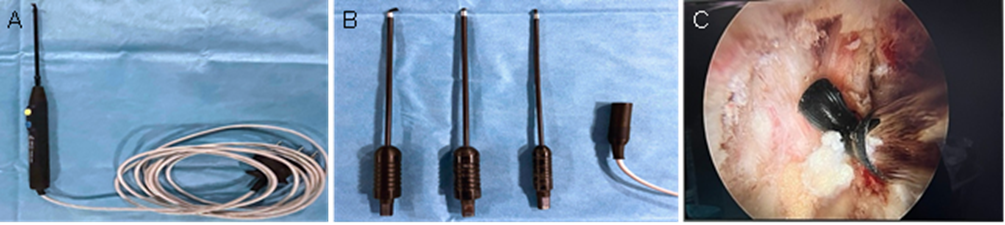
Figure 4: A: monopolar device; B: bipolar devices; C: intraoperative image with bipolar cautery.
During the surgery the fluid quality and type, temperature, infusion pressure and also the input and out put of the fluid are very important.
As described above the UBE its a surgery performed “underwater” and the correct circulation of the fluid in the surgical field permit a better vision and consequently a correct identification of anatomical structures and can avoid many complications. In our center we use a saline solution infused by gravity, with a infusion rate less than 30 mmHg.
All the procedures are under general anesthesia, we consider the benefit much more high then the risk. In other centers they use different methods such as local, epidural, and also general anesthesia.
The surgical procedures are performed in a specific operating table and support device for spinal surgery. The distribution in our operating room, the surgeon are located in the side of the mainly symptom presented by the patient and the endoscopic system in front of the surgeon. The irrigating saline lines in the distal portion of the surgical table.
In the case of a right-handed surgeon, the left hand the non-dominant holds the endoscope. In this case the endoscope-related lines, camera cable, and irrigating saline line, should be tucked to the left side of the operator. The drilling system should be fixed at the right side of the surgeon in left sides approaches.
After after anesthetic induction a very important point is the patient position, the first step of the surgical procedure. A correct prone position avoid complications and is mandatory check all the points and the correct distribution of the patient body, to obtain no pressure in eyes, and reduce in all articulations. The abdominal hight pressure will cause more bleeding, particularly in epidural space. Reducing the pressure to this level can make the entire procedure easier.
We recommend also flex the legs to indirectly open the vertebral space, very important particularly for interlaminal approaches.
The placement of surgical drapes, with all care to ensure asepsis, is equally important taking into account that the UBE surgery use many bags of saline fluid to irrigation. In this context ideally a kit with a collection bag included should be available. If not we created a simple aseptic working field of 10 cm around the skin incisions with the surgical drapes and place a antimicrobial film. After that we make a bulge around the surgical site with compresses and place sterilized bags on the side to collect the used saline solution. Finally we also recommend the application of a second antimicrobial film to isolate the surgical field.
Related with saline solution irrigation, the hydraulic pressure is a important point for a safe and smooth operation, in our case we can controlled this point using the gravity as described above. We placed the saline solution bag 50–70 cm above the patient’s back, for obtain a goal of water pressure inside the body around 30 mmHg.
The UBE procedure use many instruments common to open spine surgery, is a real advantage for the hospitals because represent a reduction in the costs if we compare with uniportal endoscopy.
About the surgical instruments as commented previously in our center we use a common instruments but we also have exclusive instruments for UBE.
5 Actually we have also available many specially designed UBE toolset, which is comprised common tools and also devices for establishing the standard UBE surgical pathway and performing all surgical steps. Particular instruments as rotating Kerrison punch, curved Kerrison punch, curved Curets, Indian knife and pituitary rongeurs all of them are essential for do UBE surgery (Figure 5).
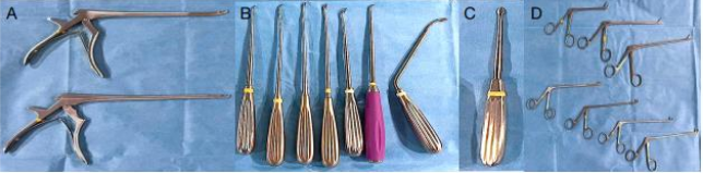
Figure 5 : A: Kerrison punch ; B: curets; C: Indian knife, D: Pituitary rongeurs.
Many of the surgical advances that have emerged over the time are dependent and related to anesthetic advances. Like in all the surgical procedures the blood pressure is a important parameter, in UBE surgery is a crucial point and a optimal blood pressure (BP) is essential for a bloodless endoscopic view. For this endoscopic procedures we recommend a optimal systolic BP between 90–100 mmHg in general anesthesia, based in our experience. Further randomized and systemized study is needed.
For perform all surgical techniques and particularly for UBE is mandatory a complete understanding of the Basic Concepts. An adequate knowledge of the anatomy of the area to be operated is the main concept, and after all the learning curve like for all the surgical techniques.
At the beginning its mandatory understand and utilize the new basic concepts, take care of each of the steps in order to reduce the rate of complications, which is usually higher in this phase of the training process. These basic concepts should naturally include knowledge of the function of the different instruments used during the procedure. Every surgeon has their own learning curve, it is up to each one to do the best possible to provide greater benefit to the patient with each particular technique. By doing so, a consistently desirable surgical outcome is guaranteed while minimizing the possibility of complications.
The quality of the learning curve are related with many factors for examples the number of cases in a period of time, a professor for clarify any type of doubts, previous knowledges and personal experience.
The pathology to be treated, with indication for surgical treatment, will determine the use of one approach or another one, adjusting the technique to the patient's needs.
It is equally important to know the objectives to be achieved with the technique as well as its limitations, complications or controversies.
The majority of spine surgeons have experience with many different methods for treat the different types of spine pathologies, and they use the most appropriate for each specific cases. Frequently they often use the one they know better and have high control and fewer complications.
All surgical technique present advantages and disadvantages and has its own distinct basic concepts.
1. Two-Port Approach:
2. Unilateral Access:
3. Endoscopic Visualization:
4. Minimally Invasive:
5. Indications:
Advantages of UBE:
Challenges:
UBE is an evolving field, and with advancements in technology, its applications are becoming more widespread in spinal and other types of surgeries.
In resume we divide the UBE technique in 5 basic concepts as follows:
1. Unilateral biportal endoscopy
Previously called biportal endoscopic spine surgery (BESS) name related with the use of two portals for do the surgery. Actually the nomenclature in use is Unilateral biportal endoscopy (UBE).
This technique applies the knee arthroscopy surgical concept to the spine. In this approach we made two portals in the same side, unilateral, of the spine pathology. One of them is the endoscopic portal and the other is the working portal for introduce all the materials that we need during the procedure.
The greats benefits of the UBE are that uses the same surgical route as in open surgery, and also very important the movements of the endoscopic and the different tools have independent ways in separate portals. This point reduce visual limitations and permit a high range of movement for all the tools and devices.
2. Surgery in liquid medium
The UBE uses irrigating saline fluid. This solution in comparison with other increase the conduction and finally the high precision on the homeostasis devices.
During the surgical procedure, the work space is filled with saline solution that enters through the endoscope portal. Continuous irrigation is very important, is essential control the input and output of the solution to balance the hydrostatic pressure in the work space. Its important control the pressor inside the canal for reduce the complications rate.
This balance is critical for maintain a clear vision of the surgical field and facilitating the surgery and turn on more safety. It has also been proven that continuous fluid is associated with a lower rate of infections.
3. Working movements in Triangulation
In the endoscope the lens are located in the front position of the device. In the beginning stage of the learning curve it’s hard to place the different instruments under endoscopic view.
During the procedure, the endoscope and the different instruments must be moved in a triangular or convergent manner. In this way the distal end of each instrument is visible allowing safe work. Triangulation means placing the ends of the instruments just in front of the endoscope lens. If the skin incision are made in the correct place the endoscope and the different tools don’t cross or shock. When this situation occur and the endoscope and instruments are crossing each other in the shaft, each interferes with the other’s movement complicating the performance of the procedure.
This situation is named ‘early triangulation’ and in this situation is mandatory or recommended a repositioning, using for them a c-arm or O-arm fluoroscopy or with neuronavigation.
The most extreme situation would be when the endoscope cannot face the instruments at the tip, it will not be possible to find the instrument in the endoscopic view. This situation is called “open triangulation”. The opening of the fascia should be reviewed and, if necessary, repositioning should also be carried out, using the intraoperative imaging devices available in each operating room.
The UBE is a minimal invasive approach the muscle destruction is reduced, during the approach a important point is a correct open the muscular fascia. In our case we open on cruce, in both portals, and after with a muscle detacher create a cavity through the soft tissues and multifidus muscle. This initial step is very important and guarantees better output flow and a clear vision.
With this approach we obtain a free movement of the different devices and tools with less destruction, and safety, with small skin incisions, less then 1 cm.
4. Two hands procedure, the coordination.
In UBE the non-dominant hand supports the main acting hand for obtain safe performances. In this technique the non-dominant hand holds the endoscope, and the dominant hand operates the surgical instruments. The dominant hand should function without any support from the non-dominant hand. The most important point is do the surgery safely and respect the ergonomic recommendations.
To access certain points, blunt angled instruments or changing the angulation of the endoscope lens are sometimes necessary, as 30 º.
Surgical skills must be developed to obtain the division and elevation technique, while removing the yellow ligament and ensuring sufficient surgical space, performing all movements smoothly to achieve a safe surgery particularly when we are inside the spinal canal, close to the spinal canal, near the duramater or the nerve roots.
In UBE the instrument and endoscope should be handled with pivot movements. This type of movements should be made, using the two portals as the central pivot point. As an example, if you plan to move the tip of the instruments forward, then you should move the tip of the other instruments, which are outside of the patient’s body, backward. This theory applies to every movement performed during the procedure.
This is a specific knowledge that you need to introduce and become accustomed to this during the learning curve period.
5. Image quality
After introducing the endoscope inside the patient's body, the distance to the area to be treated is reduced, this is one of the great differences and advantages in relation to surgery performed with a microscope. The advantage is even more evident in cases of bilateral approaches in which by entering from one side, in cases of interlaminar approaches, it is possible to reach the contralateral side, achieving adequate central canal and lateral recess decompression with release of the nerve roots in the contralateral foramen.
For obtain a good vision its also important that the irrigation saline solution flows continuously through the tip of the endoscope sheath, and the flow pressure controls bleeding to some extent and removes hematoma to leave a clear surgical field.
For obtain a estable vision without trembling caused by the surgeon harm, did placing the arm close to the body.
Working in triangulation formation, as described in the point 3, the endoscope and the instruments work independently but very adjacent to each other this point permit a wide vision but exit the risk of causing damage to the endoscope lens particularly by speeding drill burrs. To avoid this situation its important maintain a distance between the endoscope and the instruments.
As recommendation remember that a small movement (rotation, introduction and extraction of the endoscope) of the tip of the endoscope results in very different surgical vision. The UBE technique is the present and the future for the treatment of vertebral pathologies, in the different spine levels. It’s available in more and more places around the world and maybe soon will be the gold standard technique for the treatment of disc herniation and other degenerative diseases. Mainly in stand alone decompression surgeries and in other cases associate with minimal invasive, percutaneous fusion techniques.
All the patients, all my colleagues during my career especially to my mentor in endoscopic spine surgery Dr. Pedro Llinás. To my family for all the support.
Dear Editorial Team, Clinical Medical Reviews and Reports. My experience with the journal was highly positive. The peer-review process was rigorous, constructive, and completed in a timely manner. The reviewers provided valuable comments that helped improve the quality and clarity of our manuscript. The editorial office was professional, responsive, and supportive throughout all stages of the publication process. Communication was clear and efficient, and any questions were addressed promptly. Overall, I found the journal to maintain high scientific standards and an excellent publication workflow. I would be pleased to consider submitting future work to this journal. Best wishes from, Elena Popa.

It was my pleasure to submit my testimonial concerning the Reviewer Board of our Scientific Journal “Brain and Neurological Disorders”. The Reviewers focused on some modifications and their contribution was helpful. The ladies of our Editorial Office were also supported my efforts. It was my honor to have such a co-operation and I am looking forward for more collaboration.

Dear Grace Pierce, Editorial Coordinator of Journal of Clinical Research and Reports, Thank you for the speedy and efficient peer review process. I appreciate the fact that your peer reviewers do not take months to respond like with some other journals. I would also like to thank the editorial office for responding quickly to my questions. It is an excellent journal. I plan to submit more manuscripts in the future. Best wishes from, Robert W. McGee

Dear Grace Pierce, Editorial Coordinator of Journal of Clinical Research and Reports, Working with you and your team on our recent publication in JCRR has been a truly wonderful and enjoyable experience. The responses were prompt, and the reviewers were patient, constructive, and highly professional. One reviewer in particular gave me the feeling that a professor was carefully reading and commenting on my coursework, which was deeply touching. The entire process was straightforward and hassle‑free, with no tedious online forms to complete. I highly recommend this journal. Best wishes from, DR Aibing Rao, Head of R&D

I Appreciate the Opportunity to Share my Experience with the Journal of Clinical Research and Reports. The peer review process was timely and constructive, and the feedback provided helped improve the quality of our manuscript. The editorial office was professional, responsive, and supportive throughout the process, ensuring smooth communication and efficient handling of the submission. Overall, it was a positive experience collaborating with your team.

Dear Mercy Grace, Editorial Coordinator of Obstetrics Gynecology and Reproductive Sciences, We would like to express our gratitude for your help at all stages of publishing and editing the article. The editors of the magazine answer all the necessary questions and help at every stage. We will definitely continue to cooperate and publish other works in the Obstetrics Gynecology and Reproductive Sciences! Best wishes from, Alla Konstantinovna Politova,
