
Case Report | DOI: https://doi.org/10.31579/2640-1053/238
1Guelmim Faculty of Medicine and Pharmacy - Ibn Zohr Agadir University Guelmim- Morocco 81000.
2Pediatric surgery Department, Guelmin Regional Hospital, Morocco 81000.
3Guelmim Military Hospital Moulay El Hassan General Surgery Department. Guelmim-Morocco 81000.
4Pathological Anatomy Laboratory, Guelmim- Morocco 81000.
5Mohammed VI Polytechnic University, Benguerir-Morocco 43150.
6Faculty of Medicine and Pharmacy of Marrakech, Cadi Ayyad University, Marrakech 40000, Morocco.
7Private practice physician, Marrakesh- Morocco 40000.
8Faculty of Medicine and Pharmacy of Rabat, University Mohammed V - Souissi, Rabat-Morocco 10120. N. CHIBOUB.
9Guelmim Faculty of Medicine and Pharmacy - Ibn Zohr Agadir University Guelmim- Morocco 81000.
*Corresponding Author: Imane Boujguenna. Guelmim Faculty of Medicine and Pharmacy - Ibn Zohr Agadir University Guelmim- Morocco 81000.
Citation: Imane B., M. E. Lamghari, M. E. Ramraoui, F. Boukis, S. Malki, (2025), Atypical Superficial Presentations and Locations of a Rare Entity: Fibrolipoma- A Report of Eight Cases, J. Cancer Research and Cellular Therapeutics. 9(3); DOI: 10.31579/2640-1053/238
Copyright: © 2025, Imane Boujguenna. This is an open-access article distributed under the terms of the Creative Commons Attribution License, which permits unrestricted use, distribution, and reproduction in any medium, provided the original author and source are credited.
Received: 05 May 2025 | Accepted: 14 May 2025 | Published: 21 May 2025
Keywords: fibrolipoma; histopathology; superficial presentations
Fibrolipoma is a rare histological variant of lipoma, characterized by mature adipose tissue interspersed with dense fibrous connective tissue. We report eight cases of fibrolipomas in Moroccan patients with atypical locations. Clinically, fibrolipomas can mimic various lesions, posing a diagnostic challenge. Histopathological examination is essential to confirm the diagnosis. Given the rarity of this lesion, further case documentation is necessary to enhance awareness among healthcare professionals regarding its clinical and histopathological characteristics
Lipomas are benign mesenchymal neoplasms composed of mature adipocytes, usually surrounded by a thin fibrous capsule [1]. Fibrolipoma is a rare variant of lipoma, characterized by a mixture of adipose and fibrous connective tissue [2]. We report eight cases of fibrolipomas in Moroccan patients with atypical superficial locations.
Case 1:
An 11-year-old Moroccan male, with no significant medical history, presented with a superficial subcutaneous swelling on the forearm, evolving over several months. Clinical examination revealed a roughly rounded mass, initially suggestive of an epidermoid cyst. Surgical excision was performed, and macroscopic examination revealed fragmented beige-yellowish tissue with a soft-to-firm consistency. Histological analysis showed a biphasic tumor proliferation (Figure 1), with adipose lobules separated by regular septa (Figure 2). The fibrous component consisted of regular fibroblasts interspersed with adipose lobules, without signs of malignancy (Figure 3). The postoperative course was uneventful, with no recurrence.
Figures:

Figure 1: Biphasic tumor proliferation (H&E, x25).
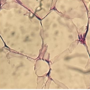
Figure 2: Adipose lobules (H&E, x25).
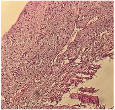
Figure 3: Fibrous component (H&E, x25).
Case 2:
A 42-year-old Moroccan male presented with a superficial subcutaneous swelling on the trunk, evolving for one year. Clinical examination revealed a well-defined, rounded mass. Macroscopic analysis after excision showed a 4 cm beige-yellowish fragment with a soft-to-firm consistency. Histopathological examination confirmed a biphasic tumor with adipose and fibrous components, without malignancy. The postoperative course was uncomplicated, and no recurrence was noted.
Case 3:
A 33-year-old Moroccan male presented with a superficial subcutaneous swelling on the back for four months. A well-circumscribed, rounded mass was noted on clinical examination. Surgical excision yielded a 4.5 cm beige-yellowish tissue fragment. Microscopic examination confirmed a fibrolipoma with adipose and fibrous components, without malignant features. Postoperative evolution was favorable, with no recurrence.
Case 4:
An 18-year-old Moroccan female presented with a three-month history of a superficial subcutaneous mass on the cheek. The mass was excised, and macroscopic examination showed a 0.6 cm beige-yellowish soft-to-firm fragment. Histopathological analysis revealed a fibrolipoma without signs of malignancy. The postoperative period was uneventful, with no recurrence.
Case 5:
A 27-year-old Moroccan male presented with a superficial subcutaneous mass on the arm, evolving for two years. Excision revealed a 6 cm beige-yellowish mass with a soft-to-firm consistency. Histological examination confirmed a fibrolipoma with adipose and fibrous proliferation. The postoperative course was uneventful, with no recurrence.
Case 6:
A 26-year-old Moroccan male presented with a superficial subcutaneous swelling on the forehead, evolving for one year. Surgical excision was performed, yielding a 1 cm beige-yellowish fragment. Histological analysis confirmed the diagnosis of fibrolipoma. The postoperative course was favorable, with no recurrence.
Case 7:
A 30-year-old Moroccan female presented with a superficial subcutaneous mass on the trunk for five months. The excised specimen measured 2 cm, with histological confirmation of a fibrolipoma. The postoperative course was uncomplicated, with no recurrence.
Case 8:
A 5-year-old Moroccan male presented with a one-year history of a superficial subcutaneous mass in the lumbar region. The excised specimen measured 4 cm, and histopathology confirmed a fibrolipoma. The postoperative period was uneventful, with no recurrence.
According to the 2020 WHO classification [3], fibrolipoma is a rare microscopic variant of lipoma, characterized by mature adipose tissue interspersed with dense fibrous connective tissue [4]. Its exact etiology remains controversial, with endocrine, dysmetabolic, genetic, and traumatic factors being considered [5]. Clinically, fibrolipomas lack specific symptoms and may be asymptomatic, painful, functionally impairing, or aesthetically concerning. In rare cases, they can cause respiratory or esophageal obstruction [5,6,7,8]. Magnetic resonance imaging (MRI) is a valuable tool for diagnosing all lipoma variants. Despite their benign nature, fibrolipomas can pose surgical challenges depending on their location and size. Microscopically, fibrolipomas consist of benign adipocyte lobules resembling a meshwork, with broad bands of dense collagen. They are generally well-circumscribed and sometimes finely encapsulated, like classical lipomas [5]. The clinical differential diagnosis includes all benign or cystic neoplastic masses. Histologically, fibrolipomas must be distinguished from conventional lipomas and atypical lipomatous tumors, sometimes requiring fluorescence in situ hybridization (FISH) analysis for MDM2 amplification [2]. Multiple fibrolipomas should prompt consideration of genetic syndromes, as seen with classical lipomas in conditions such as neurofibromatosis, Gardner syndrome, encephalo-cranio-cutaneous lipomatosis, multiple familial lipomatosis, Proteus syndrome, Cowden syndrome, multiple hamartoma syndrome, and Dercum disease [6]. In such cases, oncogenetic consultation is recommended. Treatment is based on surgical excision, with low recurrence rates [9,10,11,12]. Malignant transformation is rare [13]. Fibrolipoma is a rare histological variant of lipoma that can present in atypical superficial locations. Given its non-specific clinical presentation, it may mimic other soft tissue masses, necessitating histopathological confirmation. While surgical excision remains the treatment of choice, recognizing this entity is crucial to avoiding misdiagnosis, especially in cases of multiple or large lesions.
To anyone who has participated in the care of this patient directly or indirectly
None declared.
No funding sources
ethics approval was not required for this study
"Written informed consent was obtained from the patients for publication of this case series and any accompanying images.
Dear Editorial Team, Clinical Medical Reviews and Reports. My experience with the journal was highly positive. The peer-review process was rigorous, constructive, and completed in a timely manner. The reviewers provided valuable comments that helped improve the quality and clarity of our manuscript. The editorial office was professional, responsive, and supportive throughout all stages of the publication process. Communication was clear and efficient, and any questions were addressed promptly. Overall, I found the journal to maintain high scientific standards and an excellent publication workflow. I would be pleased to consider submitting future work to this journal. Best wishes from, Elena Popa.

It was my pleasure to submit my testimonial concerning the Reviewer Board of our Scientific Journal “Brain and Neurological Disorders”. The Reviewers focused on some modifications and their contribution was helpful. The ladies of our Editorial Office were also supported my efforts. It was my honor to have such a co-operation and I am looking forward for more collaboration.

Dear Grace Pierce, Editorial Coordinator of Journal of Clinical Research and Reports, Thank you for the speedy and efficient peer review process. I appreciate the fact that your peer reviewers do not take months to respond like with some other journals. I would also like to thank the editorial office for responding quickly to my questions. It is an excellent journal. I plan to submit more manuscripts in the future. Best wishes from, Robert W. McGee

Dear Grace Pierce, Editorial Coordinator of Journal of Clinical Research and Reports, Working with you and your team on our recent publication in JCRR has been a truly wonderful and enjoyable experience. The responses were prompt, and the reviewers were patient, constructive, and highly professional. One reviewer in particular gave me the feeling that a professor was carefully reading and commenting on my coursework, which was deeply touching. The entire process was straightforward and hassle‑free, with no tedious online forms to complete. I highly recommend this journal. Best wishes from, DR Aibing Rao, Head of R&D

I Appreciate the Opportunity to Share my Experience with the Journal of Clinical Research and Reports. The peer review process was timely and constructive, and the feedback provided helped improve the quality of our manuscript. The editorial office was professional, responsive, and supportive throughout the process, ensuring smooth communication and efficient handling of the submission. Overall, it was a positive experience collaborating with your team.

Dear Mercy Grace, Editorial Coordinator of Obstetrics Gynecology and Reproductive Sciences, We would like to express our gratitude for your help at all stages of publishing and editing the article. The editors of the magazine answer all the necessary questions and help at every stage. We will definitely continue to cooperate and publish other works in the Obstetrics Gynecology and Reproductive Sciences! Best wishes from, Alla Konstantinovna Politova,
