
Case Report | DOI: https://doi.org/10.31579/2692-9759/068
1Consultant Paediatric Endocrinologist, Yenepoya Medical College and Hospital, Yenepoya University, Mangalore, Karnataka, India.
2Consultant Obstetrician and Gynaecologist, Yenepoya Medical College and Hospital, Yenepoya University, Mangalore, Karnataka, India.
*Corresponding Author: Diksha Shirodkar, Assistant professor, MD Paediatrics, Fellow in Clinical genetics (SIAMG), Advanced training in Pediatric Endocrinology (Manipal University), Consultant Paediatrician and Paediatric Endocrinologist, Yenepoya Medical College and Hospital,
Citation: Shirodkar D, Sadiq Unnisa A. (2021) Atypical Genotype in a typical phenotype. Journal of Endocrinology and Disorders. 5(3): Doi:10.31579/2640-1045/068
Copyright: © 2021 Diksha Shirodkar. This is an open-access article distributed under the terms of the Creative Commons Attribution License, which permits unrestricted use, distribution, and reproduction in any medium, provided the original author and source are credited.
Received: 30 March 2021 | Accepted: 26 April 2021 | Published: 07 May 2021
Keywords: turner syndrome; trisomy x; mosaic turner syndrome; three cell lines; 47,XXX/46,XX/45,X
Introduction: Usually, onset of thelarche heralds puberty. Delayed puberty is worrisome and needs medical attention. Our patient in her late adolescence presented with primary amenorrhea, whose evaluation left us surprised.
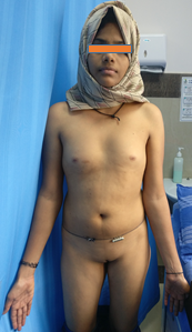
Case report: An eighteen-year-old scholastically backward girl, presented with complaints of not attaining menarche. Physical examination included a height of 156 cm(10th-25thcentile),weight 51 kg(50th centile), wide carrying angle, multiple nevi and a broad chest, however no other Turner stigmata was noted. Her sexual maturity rating (SMR) was A2P2B1 Laboratory investigations revealed increased gonadotropins (FSH:77mIU/ml; LH:25.4mIU/ml), low estradiol (14 pg/ml) and vitamin-D deficiency (21ng/ml). Ultrasonography of abdomen-pelvis showed small infantile uterus with streak ovaries. Karyotype (50 metaphases) demonstrated mosaicism [47,XXX (29)/45,X(19)/46,XX(2)]. Hormone replacement therapy and vitamin D replacement was initiated.
Conclusion: 30-40% of the Turner syndrome are mosaics, the most common being 45,X/46,XX. Mosaicism is the presence of 2 or more cell lines with different chromosomal constitutions. The cell lines are derived due mostly to postzygotic mitotic nondisjunction. X/XX/XXX can present with or without classical turner stigmata. Trisomy X has a spectrum of presentation from normal menses and fertility to recurrent abortions and primary/secondary amenorrhea (primary ovarian insufficiency). Varied clinical phenotype due to three cell lines in a Turner mosaic makes this case unique.
Puberty in females is determined by the onset of thelarche. Whenever an adolescent girl presents with short stature, primary amenorrhea and absence of secondary sexual characteristics, the first diagnosis which comes to our mind is Turner syndrome (TS). It affects approximately 1 in 2,500 live female births [1]. This syndrome occurs due to either monosomy X (the most common form of TS with the classical phenotype) resulting from meiotic nondisjunction in the parental gamete or in the early embryonic divisions (the latter causing mosaicism). The severity of clinical manifestations is related to the type of chromosomal abnormalities, the time at which chromosome disjunction occurred and the proportion of compromised cells in each tissue [2]. Here we present to you a case of an adolescent female who presented with primary amenorrhea, with a not-so classical genotype.
Case report:
18 year old adolescent girl, presented with primary amenorrhea with poor development of secondary sexual characteristics. She was born to a non-consanguineous wedlock and was the 7th child of 8 children. No history of delayed puberty in any family member or decreased sense of smell. Academically, she was a school dropout and preferred helping her mother at home. On examination her vitals were stable with no hyper/hypotension. She weighed 51 kg (50th centile), was 156 cm (10-25th centile) tall and with normal body proportions. She had specific Turner stigmata which included a broad shield-like chest, wide spaced nipples, a wide carrying angle, multiple nevi (10 in number) and bilateral fifth finger clinodactyly. Her sexual maturity rating as per Tanner classification was A2B1P2M0. She had no cardiac murmur, no goiter, normal neck length, no evidence of brachydactyly. In view of some of the clinical features of Turner syndrome and the primary amenorrhea laboratory investigations (Table 1) and a Karyotype evaluation (Figure 2) was done. The laboratory investigations proved that the child was having primary ovarian insufficiency (FSH levels were menopausal with low estradiol) and vitamin D level insufficiency. The sonogram of the pelvis showed an infantile uterus with streak gonads. The karyotyping left us amazed with the presence of 3 cell lines(47,XXX/46,XX/45,X), majority of which was 47,XXX. The treatment was hormone replacement therapy with estradiol valerate and Vitamin D supplementation.


Trisomy X (47,XXX) is a sex chromosome disorder with a variable phenotype, incidence being 1 in 1,000 female births [3].This de-arrangement also occurs due to meiotic non-disjunction, the risk increasing with maternal age. The most common physical features include normal to tall stature, epicanthal folds, occasional hypotonia and clinodactyly. Seizures, genitourinary abnormalities, learning disabilities (verbal processing defects, decrement in IQ and global delays) and motor delays are also seen. The pubertal onset can be normal or delayed and fertility can range anywhere from being normal to premature ovarian failure [4, 7]. Birthweight and length are usually normal for gestational age, however, stature increases in early childhood and by adolescence most girls with 47,XXX are at or above the 75th percentile for height [8]. On the other hand, Turner Syndrome is characterized cytogenetically by X chromosome monosomy, the presence of an abnormal X chromosome (isochromosome/ring chromosome/Xp or Xq deletion) or mosaicism of a 45,X cell line with another cell line. Mosaicism(45,X/46,XX or 45,X/47,XXX cell lineages) has been detected in 30% of the Turner syndrome cases [4,6,7].This condition has classical phenotypical features with short stature, delayed puberty, ovarian dysgenesis (hypergonadotropic hypogonadism), infertility, congenital malformations of the heart, hearing loss, ocular problems, endocrine disorders such as type 1 and type 2 diabetes mellitus, osteoporosis and autoimmune disorders [5]. The clinical presentation in our case was typical of a Turner phenotype which is consistent with gonadal dysgenesis causing primary amenorrhea, however the absence of short stature could be attributed to the huge proportion of the trisomy X(47,XXX) cell line. Although hypergonadotropic hypogonadism is a known entity in Turner syndrome, there are case reports of primary ovarian insufficiency in Trisomy X also. Since the height of the patient was normal compared to the mid-parental height, medical attention was never sought for short stature and the girl presented to us only because she had not attained menarche. Cases with X/XX/XXX have been described to report in adult life, during evaluation of recurrent pregnancy loss and short stature accounts for only 14% of diagnoses. Similarly, there have been case reports, albeit few, of triple mosaic TS with spontaneous pubarche, thelarche, and menarche [6].
This harps the fact that any adolescent presenting with delayed puberty has to be evaluated with a karyotype, even if she has classical Turner stigmata. Mosaicism is the one entity which will decide the age and the mode of presentation. More so, it is all the more essential for the karyotype in any patient with primary amenorrhea to look for Y cell line so as to prevent a gonadoblastoma in future. Appropriate genetic counselling has to be provided to these patients as aneuploidy in off-springs and its consequences is a possibility. Trisomy X has a heterogenous presentation from normal menses, tall stature and normal fertility to recurrent abortions and primary/secondary amenorrhea (primary ovarian insufficiency). A mixed phenotype in our case due to three cell lines (Triple mosaic TS) in varying proportions makes this case unique.
Acknowledgement: None
Conflict of interest: None
Dear Editorial Team, Clinical Medical Reviews and Reports. My experience with the journal was highly positive. The peer-review process was rigorous, constructive, and completed in a timely manner. The reviewers provided valuable comments that helped improve the quality and clarity of our manuscript. The editorial office was professional, responsive, and supportive throughout all stages of the publication process. Communication was clear and efficient, and any questions were addressed promptly. Overall, I found the journal to maintain high scientific standards and an excellent publication workflow. I would be pleased to consider submitting future work to this journal. Best wishes from, Elena Popa.

It was my pleasure to submit my testimonial concerning the Reviewer Board of our Scientific Journal “Brain and Neurological Disorders”. The Reviewers focused on some modifications and their contribution was helpful. The ladies of our Editorial Office were also supported my efforts. It was my honor to have such a co-operation and I am looking forward for more collaboration.

Dear Grace Pierce, Editorial Coordinator of Journal of Clinical Research and Reports, Thank you for the speedy and efficient peer review process. I appreciate the fact that your peer reviewers do not take months to respond like with some other journals. I would also like to thank the editorial office for responding quickly to my questions. It is an excellent journal. I plan to submit more manuscripts in the future. Best wishes from, Robert W. McGee

Dear Grace Pierce, Editorial Coordinator of Journal of Clinical Research and Reports, Working with you and your team on our recent publication in JCRR has been a truly wonderful and enjoyable experience. The responses were prompt, and the reviewers were patient, constructive, and highly professional. One reviewer in particular gave me the feeling that a professor was carefully reading and commenting on my coursework, which was deeply touching. The entire process was straightforward and hassle‑free, with no tedious online forms to complete. I highly recommend this journal. Best wishes from, DR Aibing Rao, Head of R&D

I Appreciate the Opportunity to Share my Experience with the Journal of Clinical Research and Reports. The peer review process was timely and constructive, and the feedback provided helped improve the quality of our manuscript. The editorial office was professional, responsive, and supportive throughout the process, ensuring smooth communication and efficient handling of the submission. Overall, it was a positive experience collaborating with your team.

Dear Mercy Grace, Editorial Coordinator of Obstetrics Gynecology and Reproductive Sciences, We would like to express our gratitude for your help at all stages of publishing and editing the article. The editors of the magazine answer all the necessary questions and help at every stage. We will definitely continue to cooperate and publish other works in the Obstetrics Gynecology and Reproductive Sciences! Best wishes from, Alla Konstantinovna Politova,
