
Case Report | DOI: https://doi.org/10.31579/2578-8868/139
Department of Orthopaedic Surgery, Changzheng Hospital, Second Military Medical University, 415 Fengyang Road, Shanghai 200003, P. R. China
*Corresponding Author: Ye Tian M.D. and Wen Yuan M.D. Department of Orthopaedic Surgery, Changzheng Hospital, Second Military Medical University, 415 Fengyang Road, Shanghai 200003, P. R. China
Citation: Gang Liu., Peng Cao., Xiaolong Shen., Ye Tian., Wen Yuan., (2020) Atlantoaxial subluxation due to os odontoideum combined with cervical spondylotic myelopathy: case report and literature review. J. Neuroscience and Neurological Surgery. 7(1); DOI:10.31579/2578-8868/139
Copyright: © 2020 Ye Tian and Wen Yuan, This is an open-access article distributed under the terms of The Creative Commons Attribution License, which permits unrestricted use, distribution, and reproduction in any medium, provided the original author and source are credited
Received: 06 October 2020 | Accepted: 20 October 2020 | Published: 28 October 2020
Keywords: Os odontoideum; cervical spondylotic myelopathy; spinal cord compression
Study Design: This was a case report and literature review
Objective: We describe a case of os odontoideum combined with cervical spondylotic myelopathy (CSM), both of which require surgical treatment.
Summary of Background Data: Cervical spondylotic myelopathy is often a disease of the older population, while os odontoideum is a well known disease mainly diagnosed in children and young adults but rarely in the middle-aged population. Os odontoideum combined with cervical spondylotic myelopathy, both of which require surgical treatment is even more rare, there was only one such case in the literature.
Methods: We describe a 68-year-old male who underwent C1–C2 posterior screw-rod fixation for os odontoideum and cervical posterior single open-door laminoplasty for cervical spondylotic myelopathy.
Results: Twelve months after surgery, the patient showed improvement and the plain radiographs showed no loss of correction or instrumentation failure.
Conclusions: To our best knowledge, this is the second case of surgical stabilization for both cervical spondylotic myelopathy and myelopathy atlantoaxial subluxation due to os odontoideum.
Cervical spondylotic myelopathy (CSM)
rheumatoid arthritis (RA)
Magnetic resonance imaging (MRI)
Os odontoideum is defined as an ossicle with smooth circumferential cortical margins representing the odontoid process that has no osseous continuity with the body of C2. Here, we describe a case of os odontoideum combined with cervical spondylotic myelopathy (CSM), which require surgical treatment. We performed C1–C2 posterior screw-rod fixation and cervical posterior single open-door laminoplasty for this patient. To our best knowledge, this is the second case of surgical stabilization for both cervical spondylotic myelopathy and myelopathy atlantoaxial subluxation due to os odontoideum.
History and Presentation: A 68-year-old male was admitted to our hospital with complaints of numbness and weakness in right upper extremity for 14 months. He mentioned an accident about one month ago when he had a mild head trauma from behind. Since then, This patient experienced numbness in all limbs, more in the right extremity. In December 2017, he visited our hospital and was diagnosed with cervical spondylotic myelopathy combined with os odontoideum. He had no past history of rheumatoid arthritis (RA), Turner’s syndrome, Down’s syndrome or gonadal dysgenesis.
Physical examination revealed a thin male with normal vital signs and limited neck movement. Neurological examination disclosed decreased sensation in the bilateral upper extremities, below the level of the C5-7 dermatome on the right and C7 on the left. Manual muscle testing revealed generalized muscle weakness throughout his left limbs (grade 4 of 5) and right limbs (grade 2-3 of 5). bilateral hyperactive deep tendon reflexes a positive Hoffmann sign in right were also disclosed.
Serial plain radiographs (Figure.1.) of the cervical spine showed os odontoideum and anterior atlantoaxial subluxation plus increased interspinous C1-C2 distance. The subluxation could be reduced with the neck in extension.
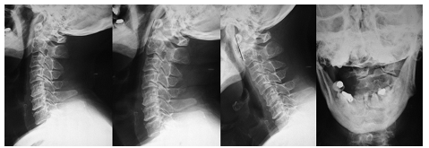
Figure.1:
a Preoperative plain radiograph in neutral position demonstrating os odontoideum and anterior atlantoaxial subluxation.
b The subluxation is reduced with the neck in extension.
c The subluxation is more severe with the neck in flexion.
CT (Figure.2.) and MRI (Figure.3.) of the cervical spine showed os odontoideum and anterior atlantoaxial subluxation, pinal cord compression due to the subluxation and cervical spondylosis of the lower cervical spine at multiple levels. Hyperintense signal changes were also revealed.

Figure 2: Reformatted sagittal CT shows round os odontoideum.
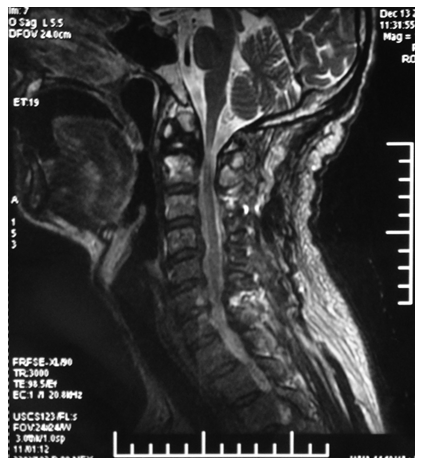
Figure. 3: MR T2 weighted images showing marked Compression at cervicomedullary region, with cervical spondylosis at C3-C6.
Operation and Postoperative Course: After anesthesia, the patient was turned over together with the protected tailor-made plaster bed to the operation table. C1–2 reduction was routinely checked under C-arm fluoroscopy just before operation. The surgery was carried out with a posterior approach and exposure of occiput to axis (C2). Initially, C1–C2 posterior screw-rod fixation with an iliac crest autograft were performed. Next, cervical posterior single open-door laminoplasty was performed for this patient (Figure.4.).
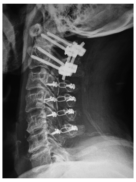
Figure. 4: Postoperative plain radiograph shows C1/C2 fusion and cervical posterior single open-door laminoplasty at C3-6 levels. The subluxation was also reduced.
He was discharged 8 days after surgery. At this time he was able to walk and free of neck pain. A soft neck collar was applied for 2 months. Twelve months after surgery, although slight muscle weakness was observed, numbness of the extremities improved. No related complications were detected within 2 years of follow-up.
Os odontoideum, is an ossicle with smooth circumferential cortical margins representing the odontoid process that has no osseous continuity with the body of C2 [16,27].
There is a considerable debate in the literature regarding the etiology of os odontoideum The main etiology of os odontoideum is the congenital anomalies of the odontoid process or trauma [20,28,30]. However, the etiology of the pathology does not play an important role in its diagnosis or management.
Most patients of Os odontoideum are diagnosed in the first three decades of life but rarely in the middle-aged population [9,10]. A review of the literature revealed 10 cases (including our case)of this disorder older than 66 years of age (table 1).

Table 1: Os odontoideum patients older than 66 years
Os odontoideum can be classified into two types, orthotopic and dystopic. Orthotopic defines an ossicle that staying in the normal position of the odontoid; whereas the so called dystopic defines an ossicle settle more cranially.
Os odontoideum can be symptomatic or asymptomatic. Symptomatic cases can present with a wide range of signs and symptoms. These symptoms can be divided into three groups: occipitocervical pain, myelopathy and intracranial symptoms or signs from vertebrobasilar ischemia. Of the various symptoms, occipitocervical pain is the most frequent and myelopathy is the most serious. These symptoms are thought to be caused by the static compression of the spinal cord or repeated minor trauma to the spinal cord [26].
Standard radiological techniques are usually adequate to obtain diagnose. The addition of pain dynamic radiographs in the form of flexion and extension are recommended to provide valuable information regarding the degree of instability [29].
However, the degree of C1–C2 instability does not seem to correlate with neurological status in patients with os odontoideum; in the mean while, sagittal spinal canal diameter on plain x-rays of 13 mm or less may be associated with myelopathy [6,26,27].
Beyond plain spine x-rays and flexion-extension x-rays, computed axial tomog-raphy in conjunction with 3-dimensional reconstruction can provide information about the bony anatomy at the craniocervical junction, including the position of the transverse foramina at C1 and C2 and the completeness of the atlas ring [23], which can greatly enhance the precision of the preoperative assessment. MRI is the modality of choice to asses the compression of spinal cord. It is particularly important to measure signal changes within the cord.
Surgical treatment is not required for every patient in whom os odontoideum is identified. There is a definite role for conservative treatment in patients with a stable noncompressive and neurologically stable os odontoideum, which has already been proved by numerous reports [6,25,27].
However, a lack of C1–C2 instability at initial diagnosis does not guarantee that instability will not develop in these patients. There are reports in the literature of patients with stable os odontoideum in whom instability developed later [2] or of a conservatively treated patient presenting with paraplegia after minor trauma [8]. So it is strongly recommended that these individuals should be followed closely.
Although there is still a debate in the literature regarding surgical stabilization of an unstable os odontoideum in neurologically free subjects and some authors do believe that observation rather than surgery was should be recommended for unstable os odontoideum in asymptomatic patients[4,11,18].
Considering the increased likelihood of future spinal cord injury, more and more investigators of this disorder favor operative stabilization and fusion of C1–C2 instability associated with os odontoideum [14,16,22,31].
For those os odontoideum patients with signs and symptoms of spinal cord compression and/or progressive myelopathy, there is no doubt that they should be treated surgically.
The universal theme of the various surgical approach and the techniques has two main goals: to remove the compression at the craniocervical junction area and to confirm or to secure cervical spinal stability in relation to subaxial spine and in some cases to the fixed skull.
Os odontoideum should be considered carefully for surgery. In planning the surgery, except for the preoperative clinical symptoms and image findings of the patient, the surgeons should also take into account evidence of atlantoaxial instability,anatomy favorable for surgical instrumentation and the surgeon’s preference, experience and comfort level with the particular approach.
Most cases of atlantoaxial instability associated with os odontoideum are anterior subluxation [6,26] and posterior C1–C2 arthrodesis in the treatment of os odontoideum provides effective stabilization of the atlantoaxial joint in the majority of cases [9,10,13,15,32].
In the previous reports of posterior C1–C2 arthrodesis due to os odontoideum, sublaminar wiring fusion, atlantoaxial transarticular screw Fusion and C1-C2 screw rod fusion had been performed.
According to previous literature, we and other authors had reported successful fusion rate for posterior wiring and fusion techniques such as the Gallie and Brooks techniques to be in 40 to 100% of cases [1,3,4,7,27,33].
Placement of transarticular C1–2 screws with strut graft have shown superior fusion rates over sublaminar wiring [5,19]. However, the use of transarticular screws has become less popular. Because it has been reported that about one fifth of patients with craniovertebral junction abnormalities have vertebral artery anomalies, which indicating this technique involved a risk of injuring the VA [9,23]
Beside transarticular screws, Other types of screws, such as C-1 lateral mass screws, C-2 pedicle or pars screw are available. Harms and Melcher [13] had reported 6 of the 37 patients had os odontoideum, after surgery, all of them showed radiological evidence of fusion. Similar results were obtained by many other authors [12,17,24]
These reports had further confirm the superiority of fusion in this technique compared with the wiring methods such as the Gallie and Brooks techniques. Furthermore, this technique has less chance of vertebral artery injury compared to transarticular screws. We believe that Harms and Melcher technique of posterior atlantoaxial screw fusion is the current option of choice for the patients of os odontoideum.
In our case, MRI demonstrated severe compression and hyperintense signal changes at C1/2 level, indicating that decompression at this level was essential. X-ray showed reduction could be achieved with the neck in extesion. Moreover, since MRI also demonstrated compression at levels C3-6 and decreased sensation was observed in the dermatome of the lower cervical levels, after we performed C1–C2 posterior screw-rod fixation with an iliac crest autograft, considering the stenosis of C3-C6 level, aervical posterior single open-door laminoplasty to decompress the spinal cord were performed. To the best of our knowledge, this is the second case of surgical stabilization for both cervical spondylotic and myelopathy atlantoaxial subluxation due to os odontoideum (table2).
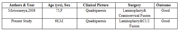
Table 2: Surgical stabilization for both os odontoideum and cervical spondylotic myelopathy
Neither author has a financial or personal relationship with another person or organization that could inappropriately influence (bias) her or his work. This work was prepared without sponsorship from any funding agency.
Dear Editorial Team, Clinical Medical Reviews and Reports. My experience with the journal was highly positive. The peer-review process was rigorous, constructive, and completed in a timely manner. The reviewers provided valuable comments that helped improve the quality and clarity of our manuscript. The editorial office was professional, responsive, and supportive throughout all stages of the publication process. Communication was clear and efficient, and any questions were addressed promptly. Overall, I found the journal to maintain high scientific standards and an excellent publication workflow. I would be pleased to consider submitting future work to this journal. Best wishes from, Elena Popa.

It was my pleasure to submit my testimonial concerning the Reviewer Board of our Scientific Journal “Brain and Neurological Disorders”. The Reviewers focused on some modifications and their contribution was helpful. The ladies of our Editorial Office were also supported my efforts. It was my honor to have such a co-operation and I am looking forward for more collaboration.

Dear Grace Pierce, Editorial Coordinator of Journal of Clinical Research and Reports, Thank you for the speedy and efficient peer review process. I appreciate the fact that your peer reviewers do not take months to respond like with some other journals. I would also like to thank the editorial office for responding quickly to my questions. It is an excellent journal. I plan to submit more manuscripts in the future. Best wishes from, Robert W. McGee

Dear Grace Pierce, Editorial Coordinator of Journal of Clinical Research and Reports, Working with you and your team on our recent publication in JCRR has been a truly wonderful and enjoyable experience. The responses were prompt, and the reviewers were patient, constructive, and highly professional. One reviewer in particular gave me the feeling that a professor was carefully reading and commenting on my coursework, which was deeply touching. The entire process was straightforward and hassle‑free, with no tedious online forms to complete. I highly recommend this journal. Best wishes from, DR Aibing Rao, Head of R&D

I Appreciate the Opportunity to Share my Experience with the Journal of Clinical Research and Reports. The peer review process was timely and constructive, and the feedback provided helped improve the quality of our manuscript. The editorial office was professional, responsive, and supportive throughout the process, ensuring smooth communication and efficient handling of the submission. Overall, it was a positive experience collaborating with your team.

Dear Mercy Grace, Editorial Coordinator of Obstetrics Gynecology and Reproductive Sciences, We would like to express our gratitude for your help at all stages of publishing and editing the article. The editors of the magazine answer all the necessary questions and help at every stage. We will definitely continue to cooperate and publish other works in the Obstetrics Gynecology and Reproductive Sciences! Best wishes from, Alla Konstantinovna Politova,
