
Research Article | DOI: https://doi.org/10.31579/2690-4861/476
1Doctor of Medical Sciences, Professor, Head of the Department of Pediatrics, IPO, Krasnoyarsk State Medical University named after Professor V.F. Voino-Yasenetsky; Krasnoyarsk, Russian Federation
2Doctor of Physics and Mathematics. Sciences, Professor of the Department of General Physics, Moscow Institute of Physics and Technology (National Research University) MIPT, Moscow, Russian Federation.
*Corresponding Author: Taranushenko Tatyana Evgenievna, Doctor of Medical Sciences, Professor, Head of the Department of Pediatrics, IPO, Krasnoyarsk State Medical University named after Professor V.F. Voino-Yasenetsky; Krasnoyarsk, Russian Federation
Citation: Taranushenko T. Evgenievna , Salmin V. Valerievich , Salmin V. Valerievich, Kiseleva N. Gennadievna, (2024), Assessment of Skin Autofluorescence in Children with Diabetes Mellitus Type 1, International Journal of Clinical Case Reports and Reviews, 18(4); DOI:10.31579/2690-4861/476
Copyright: © 2024, Taranushenko Tatyana Evgenievna. This is an open-access article distributed under the terms of the Creative Commons Attribution License, which permits unrestricted use, distribution, and reproduction in any medium, provided the original author and source are credited.
Received: 15 May 2024 | Accepted: 24 May 2024 | Published: 01 August 2024
Keywords: autofluorescence; type 1 diabetes mellitus; age; gender; experience; complications
Objective: to measure skin autofluorescence in children and adolescents suffering from type 1 diabetes mellitus and evaluate its relationship with gender, age, experience and chronic complications of the disease.
Materials and methods: the study group included 47 children and adolescents with type 1 diabetes. Autofluorescence of the skin from the inner surface of the shoulder and nail of patients was measured using an original compact spectrofluorimeter based on the STS-VIS OCEAN OPTICS © USA microspectrometer with UVA excitation. Statistical analysis was carried out using StatsoftStatistica 12.0 software. The data is presented as a two-dimensional array. The UV LED signal was averaged and smoothed using the moving average method with a 10 nm window. Then the spectra were renormalized taking into account the found coefficients. The result of applying additional normalization is a decrease in the standard deviation.
Results and discussion: significant differences were revealed in the skin fluorescence spectra of children of different ages. between age groups (5-7) and (8-12) is most significant in the region of the alpha band of oxyhemoglobin (540 nm) (p <0.005). When using I-normalization, the NADH peak region (p < 0.02) is significant with increasing disease duration. When studying the influence of gender factors on the level of skin autofluorescence, the most significant differences are found in the area of only the isosbestic points of deoxy and oxyhemoglobin 442 nm (p<10-7) and 491 nm (p<10-8). Significant differences in skin autofluorescence at the reference length were also obtained waves in the autofluorescence spectrum of 500 nm correspond to p<10-14, depending on the presence of complications.
Conclusion: in Russia, as well as throughout the world, there is an increase in the incidence of type 1 diabetes mellitus. For early diagnosis of changes in carbohydrate metabolism and complications of the disease, a simple, accessible, non-invasive research method is needed. Taking into account the results of our study, when creating non-invasive methods for monitoring the state of carbohydrate metabolism, it is necessary to take into account gender and age characteristics, experience and the presence of complications of type 1 diabetes mellitus.
Diabetes mellitus type 1 (DM 1t)
Diabetes Complications and Control Trial (DCCT)
Epidemiologyof Diabetes Interventions and Complications (EDIC)
Advanced glycation end products (AGEs)
Gender (SEX)
Duration of disease (DD)
Continuous subcutaneous insulin infusion (CSII)
Continuous glucose monitoring (CGM)
Autofluorescence spectra (SAF)
Maintaining
Type 1 diabetes mellitus (T1DM) in children and adolescents is a complex medical and social problem.
In 2021, there were 108,300 children and adolescents under 15 years of age with newly diagnosed type 1 diabetes and 651,700 children and adolescents with T1DM worldwide [1]. The average increase in the incidence of T1DM is 3-4% per year. Studying the epidemiology of the disease in Russia shows a steady increase in the incidence of type 1 diabetes mellitus. According to the results of the federal register, as of January 1, 2023, the total number of patients with diabetes mellitus in Russia registered at the dispensary according to the federal register of diabetes mellitus was 4,962,762 people (3.31% of the population of the Russian Federation), of which: T1DM - 5.58% (277.1 thousand), children and adolescents accounted for 48,031 people [2]. The results of the T1DM Diabetes Complications and Control Trial (DCCT) and the subsequent Epidemiology of Diabetes Interventions and Complications (EDIC) study confirmed the association of chronic hyperglycemia with the risk of developing microvascular complications.
To date, it has been proven that the development of endothelial dysfunction underlies the development of vascular complications in diabetes mellitus. In addition to hyperglycemia and oxidative stress, the accumulation of advanced glycation end products (AGEs) plays an important role in its progression. The reaction of protein glycosylation was first described by L. Maillard in 1913. Glycosylation is a non-enzymatic process in which glucose combines with residues of almost all proteins, which leads to a change in their structure and, as a consequence, function. To date, the process of hemoglobin glycation has been well studied. During short-term incubation of the protein with glucose, unstable intermediate compounds, the so-called Schiff bases, are formed. As the process continues for up to several weeks, they become more stable, but still reversible, Amadori products. Subsequently, long-term hyperglycemia will lead to the conversion of ketamines into advanced glycation end products (AGEs). Advanced glycation end products are a unique skin marker in diabetes; they accumulate in proteins with a longer half-life. The accumulation of AGEs leads to disruption of the barrier function of the vascular wall, the accumulation of reactive oxygen species, the production of pro-inflammatory cytokines and a number of other processes that contribute to the development of endothelial dysfunction. Accumulation of AGEs in the skin causes an increase in skin autofluorescence, which correlates with microcirculatory disorders. The level of skin autofluorescence is considered as an integral indicator of dysmetabolic changes in the development of diabetes mellitus, pathology of the kidneys, brain, endocrine, vascular and respiratory systems [3,4].
According to clinical recommendations, one of the main components of the treatment of T1DM is teaching patients self-monitoring of glycemia. The only method for preventing the development of microvascular complications is to achieve and maintain optimal target glycemic levels [5,6].
Patients' adherence to self-control depends primarily on the level of pain, accessibility and simplicity of the method for studying glycemia. Therefore, it is still relevant to create a non-invasive, accessible, accurate method for monitoring the state of carbohydrate metabolism.
Purpose of the study: to identify diagnostically significant indicators of skin autofluorescence in children suffering from T1DM depending on indicators such as age (AGE), gender (SEX), disease duration (DD), and the presence of complications. To evaluate correlations between skin autofluorescence parameters depending on age, duration of the disease, and complications.
The study was conducted on the basis of Federal State Budgetary Institution Clinical Hospital No. 51 of the FMBA of Russia, a branch of the Federal State Budgetary Institution FSNKTs FMBA of Russia Clinical Hospital No. 42.
All patients signed informed consent to participate in the study. The study was approved by the ethics committee of the Federal State Budgetary Educational Institution of Higher Education Krasnoyarsk State Medical University named after. prof. V.F. Voino-Yasenetsky Ministry of Health of Russia (protocol No. 114 of 10/05/2022)
The study group included 47 patients suffering from type 1 diabetes mellitus. Of these, the group of children included 29 (61.7%), and the group of adolescents included 18 people (38.3%). At the same time, slightly more than half of the respondents were boys (57.4% and 42.5% girls). The average length of illness of patients at the time of examination was 4.47 min. HbA1c 6.0, max 18.7%. All children were on constant insulin replacement therapy from the moment the disease was diagnosed: 10 patients (21.2%) were on continuous subcutaneous insulin infusion (CSII) and 37 people (78.7%) were on a pen syringe. Growth disturbances were noted in one child (2.1%), underweight was detected in 24 cases (51%), excess weight in 3 cases (6.4%). All those observed during the study period were on continuous glucose monitoring (CGM), with a predominance of Libra flash monitoring. When observing patients, chronic complications were identified in 8 cases (17%) in the form of diabetic neuropathy in 6 cases, diabetic nephropathy at the stage of microalbuminuria in two cases. Background diseases in the form of thyroid pathology were found in 9 patients.
Statistical analysis was carried out using StatsoftStatistica 12.0 software.
Autofluorescence (SAF) spectra were recorded from the inner surface of the patient's upper arm for 30 seconds using an original compact spectrofluorimeter based on the STS-VIS OCEAN OPTICS © USA microspectrometer with UVA excitation created by a LED (375 nm) [7].
Fluorescence spectra of skin from the inner surface of the forearm, obtained using the device, consist of two wide contours. The first, in the range of 400-700 nm, represents, in fact, the autofluorescence of the skin, and the second 700-820 nm, the spectrum of the UV LED excitation 375 nm, in the second order of diffraction of the diffraction grating (Figure. 1).
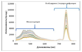
Figure 1. Skin fluorescence spectra.
For further analysis, the fluorescence spectra were normalized to the average value of the UV LED signal and smoothed using the moving average method with a 10 nm window. The resulting normalized spectra are presented in Figure. 2
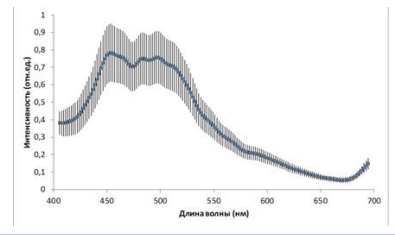
Figure 2: Normalized spectra
This method of normalizing spectra will be further called D-normalization.
To more accurately compare the shapes of the spectra, we applied additional normalization. To do this, the average spectrum for the entire group of patients F (λ) was calculated and for each spectrum Fi(λ) the linear regression coefficients ai, bi were calculated using the least squares method so that after subsequent renormalization the specified spectra were as close as possible to the average: 
Then the spectra were renormalized taking into account the found coefficients:
The result of applying additional normalization is a decrease in the standard deviation of Figure. 3. This normalization will be called I-normalization.
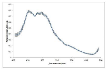
Figure 3: Result of I-normalization
To assess the capabilities of UV-induced fluorescence spectroscopy in diagnosing the course of diabetes mellitus in children of different ages, we analyzed the significance of differences in fluorescence spectra between different age groups, gender differences, differences associated
with the duration of the disease, as well as differences in metabolic indicators of the course of diabetes mellitus: the level of glycated hemoglobin, glycemic variability, average daily glycemia. For analysis, we plotted the dependence of the Z-score of differences between selected
pairwise groups on the wavelength obtained using the Mann-Whitney test. Exceeding the specified indicator modulo the critical value of 1.96 corresponds to significant differences at the p <0>
In the age groups that are traditionally accepted for pediatric practice, we have identified three age groups: 1.5-7 years; 2.8-12 years; 3.13-18 years.
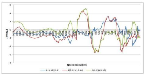
Figure 4: Pairwise Z-scores in age groups
As can be seen from the presented diagram in Fig. 4, the most pronounced age-related differences in the skin fluorescence spectra are recorded when using I-normalization, and these differences are maximum in the region of the peak of the Soret band of hemoglobin (410-445 nm) (p <10>
noteworthy is the presence of a significant difference between the older age group (13-18) years and the two younger ones (5-7) and (8-12) in the region of the fluorescence peak of the main tissue fluorophore NADH (460-510 nm) (p <10>.

Differences are statistically significant at p <0>
Table 1: Significance of differences in fluorescence intensity at the indicated wavelengths according to the Mann-Whitney test for different age groups
Considering the influence of the duration of type 1 diabetes mellitus in the study group, two subgroups were identified:
1. up to 5 years 2. over 5 years.
In our study, as can be seen in the diagram in Figure. 5, significant differences in the spectra are also found when using I-normalization, and just as for comparing age groups, the NADH peak area (p<0>.
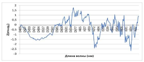
Figure 5: Autofluorescence spectra depending on the duration of T1DM
| Wavelengths | Comparison groups Median [1Q; 3Q] | р | ||
| Experience<5> | Experience >5 years | |||
I483 | 0,749 [0,739; 0,761] | 0,755 [0,748; 0,763] | 0,02 | |
| 0,064 [0,058; 0,070] | 0,068 [0,061; 0,075] | 0,007 | |
Differences are statistically significant at p <0>
Table 2: Significance of differences in fluorescence intensity at the indicated wavelengths according to the Mann-Whitney test for different groups according to the length of the disease.
In addition, the work studied the influence of gender as one of the factors influencing the level of skin autofluorescence in diabetes mellitus. Not many studies have been devoted to studying the influence of gender factors on the level of autofluorescence in diabetes mellitus; their contribution is about 0.4–1.9%.
Based on the results of our work, a diagram demonstrating gender differences in skin fluorescence spectra is shown in Fig. 6. As follows from the presented diagram, gender differences are significant with both types of spectra normalization, but also, as before, they become most significant with I-normalization.
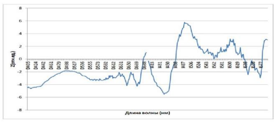
Figure 6: Sex differences in fluorescence spectra
| Wavelengths | Comparison groups Median [1Q; 3Q] | Р по Манна-Уитни | ||
| М | Ж | |||
I442 | 0,649 [0,626; 0,675] | 0,676 [0,653; 0,694] | <10> | |
| 0,750 [0,741; 0,758] | 0,733 [0,721; 0,746] | <10> | |
Differences are statistically significant at p <0>
Table 3: Significance of differences in fluorescence intensity at the indicated wavelengths according to the Mann-Whitney test for comparison by gender
We also analyzed changes in skin autofluorescence in children and adolescents suffering from T1DM depending on the presence of microvascular complications.
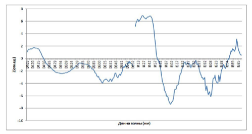
Figure. 7 Autofluorescence spectra of the skin of children with T1DM depending on the presence of microvascular complications.
As follows from the diagram above, significant areas of the spectrum that distinguish different groups of glycemic variability are present in both normalization methods.
As can be seen from the presented analyses, the most significant differences detected by spectrofluorimetry methods correspond to groups with complications of diabetes mellitus in comparison with their absence. The reference wavelength in the autofluorescence spectrum of 500 nm corresponds to p <10>.
| Wavelengths | Comparison groups Median [1Q; 3Q] | Mann-Whitney significance | ||
| There are complications | No complications | |||
| 0,406 [0,380; 0,416] | 0,433 [0,420; 0,454] | <10> | |
| 0,770 [0,762; 0,787] | 0,747 [0,738; 0,757] | <10> | |
| I610 | 0,166 [0,158; 0,176] | 0, 156 [0,150; 0,160] | <10> | |
Differences are statistically significant at p <0>
Table 4: Significance of differences in fluorescence intensity at the indicated wavelengths according to the Mann-Whitney test between the presence or absence of complications
Thus, in children of different ages with type 1 diabetes mellitus there are significant age-related differences in the skin fluorescence spectra. According to most authors, age is the most important factor influencing SAF, ranging from 23.8–28.5% [8,9]. These differences may be due to both age-related anatomical changes in the skin, changes in total hemoglobin, and the development of irreversible microcirculatory changes, accompanied by an increase in hypoxic changes in the skin. Taking into account the most significant parts of the spectrum, we determined the intensities at the indicated wavelengths and compared the fluorescence intensities.
When studying the effect of diabetes experience on the level of autofluorescence, attention is drawn to the significant spectral region of 656 nm (p <0>
The bulk of the work has proven a direct connection between the length of T1DM disease and the level of skin autofluorescence [11,12,13].
When considering the results of the identified publications, a gender-specific feature of SAF was revealed: the level was higher in girls compared to boys with diabetes mellitus [14,15].
In our study, the most significant gender differences are found in the region of only the isosbestic points of deoxy and oxyhemoglobin 442 nm (p <10>
When analyzing the level of skin autofluorescence depending on the presence of microvascular complications, a more pronounced structure of the spectrum is realized when using I-normalization. The most pronounced deviations in the spectrum of Z-scores are observed in the region of the Soret band of hemoglobin 424 nm (isosbestic point of oxy- and deoxyhemoglobin), 500 nm (also isosbestic point of oxy- and deoxyhemoglobin at the peak of NADH luminescence (the most significant changes), 610 nm - not interpreted peak. According to publications, the accumulation of glycation products in the skin of patients suffering from T1DM is described and their correlation with the progression of the disease is proven [16,17].
In our work, we found a significant increase in the level of autofluorescence in the skin of patients suffering from T1DM with age, duration of diabetes, female gender, and the presence of microvascular complications.
The detected connection between these same chromophores and fluorophores with gender and age differences and the duration of the disease should be taken into account when developing these methods.
It should be noted that the discovered relationship between fluorescence intensity at reference wavelengths corresponding to the main chromophores and fluorophores of the skin can be used in the development of non-invasive methods for analyzing laboratory parameters during diabetes mellitus.
Dear Editorial Team, Clinical Medical Reviews and Reports. My experience with the journal was highly positive. The peer-review process was rigorous, constructive, and completed in a timely manner. The reviewers provided valuable comments that helped improve the quality and clarity of our manuscript. The editorial office was professional, responsive, and supportive throughout all stages of the publication process. Communication was clear and efficient, and any questions were addressed promptly. Overall, I found the journal to maintain high scientific standards and an excellent publication workflow. I would be pleased to consider submitting future work to this journal. Best wishes from, Elena Popa.

It was my pleasure to submit my testimonial concerning the Reviewer Board of our Scientific Journal “Brain and Neurological Disorders”. The Reviewers focused on some modifications and their contribution was helpful. The ladies of our Editorial Office were also supported my efforts. It was my honor to have such a co-operation and I am looking forward for more collaboration.

Dear Grace Pierce, Editorial Coordinator of Journal of Clinical Research and Reports, Thank you for the speedy and efficient peer review process. I appreciate the fact that your peer reviewers do not take months to respond like with some other journals. I would also like to thank the editorial office for responding quickly to my questions. It is an excellent journal. I plan to submit more manuscripts in the future. Best wishes from, Robert W. McGee

Dear Grace Pierce, Editorial Coordinator of Journal of Clinical Research and Reports, Working with you and your team on our recent publication in JCRR has been a truly wonderful and enjoyable experience. The responses were prompt, and the reviewers were patient, constructive, and highly professional. One reviewer in particular gave me the feeling that a professor was carefully reading and commenting on my coursework, which was deeply touching. The entire process was straightforward and hassle‑free, with no tedious online forms to complete. I highly recommend this journal. Best wishes from, DR Aibing Rao, Head of R&D

I Appreciate the Opportunity to Share my Experience with the Journal of Clinical Research and Reports. The peer review process was timely and constructive, and the feedback provided helped improve the quality of our manuscript. The editorial office was professional, responsive, and supportive throughout the process, ensuring smooth communication and efficient handling of the submission. Overall, it was a positive experience collaborating with your team.

Dear Mercy Grace, Editorial Coordinator of Obstetrics Gynecology and Reproductive Sciences, We would like to express our gratitude for your help at all stages of publishing and editing the article. The editors of the magazine answer all the necessary questions and help at every stage. We will definitely continue to cooperate and publish other works in the Obstetrics Gynecology and Reproductive Sciences! Best wishes from, Alla Konstantinovna Politova,
