AUCTORES
Globalize your Research

Research Article | DOI: https://doi.org/10.31579/2641-0419/115
1Departments of Cardiothoracic and Vascular Surgery,
2Cardiology and Cardiac Anaesthesia
3All India Institute of Medical Sciences, New Delhi
*Corresponding Author: Ujjwal Kumar Chowdhury, MCh, Diplomate NB ProfessorDepartment of Cardiothoracic and Vascular Surgery AIIMS, New Delhi-110029, INDIA
Citation: Lakshmi K. Sankhyan, Vikrant Yadav, Srikant Sharma, Mrigank Choubey, Niwin George, Sushamagayatri, Vishwas Malik, Ujjwal K. Chowdhury (2021) Assessment of Myocardial Mechanics in Patients Undergoing Pericardiectomy for Chronic Constrictive Pericarditis by Tissue Doppler Imaging and 2D Speckled Tracking Echocardiography: A Prospective Observational (Cohort) Study. J. Clinical Cardiology and Cardiovascular Interventions, 4(2); Doi:10.31579/2641-0419/115
Copyright: © 2021 Ujjwal Kumar Chowdhury, This is an open-access article distributed under the terms of the Creative Commons Attribution License, which permits unrestricted use, distribution, and reproduction in any medium, provided the original author and source are credited.
Received: 23 November 2020 | Accepted: 11 January 2021 | Published: 18 January 2021
Keywords: tissue doppler imaging; two-dimensional speckle echocardiography; chronic constrictive pericarditis; Pericardiectomy; echocardiography
Background: This study was designed to prospectively evaluate the changes in tissue Doppler imaging (TDI) at mitral and tricuspid annuli and two dimensional speckle tracking echocardiography in patients undergoing pericardiectomy for chronic constrictive pericarditis and identify the relationship if any of the tissue Doppler imaging and speckle echocardiographic derived variables with patient’s symptomatic status following surgery.
Patients and Methods: Twelve patients undergoing pericardiectomy for constrictive pericarditis aged 7 years to 70 years (median 21; IQR: 19.75-26.5 years) were studied for 2-36 months (median 19 months). They underwent Doppler flow velocity, TDI, and 2D-speckle echocardiographic studies. Friedman’ test was used to test the changes in TDI-derived mitral and tricuspid annular velocities and speckle derived parameters in postoperative period from baseline.
Results: Despite congestive heart failure, all patients had normal left ventricular ejection fraction and increased medial mitral and tricuspid early diastolic septal velocity (e¢) with “annulus reversus”. This pattern of annular velocity improved maximally in the immediate postoperative period. At closing interval, 2 (16.7%) patients continued to be in New York Heart Association class II and both of them continued to remain in atrial fibrillation. There was statistical significant improvement in the Global cirumferential strain than in global longitudinal and global radial strain after pericardiectomy.
Conclusions: We conclude that tissue Doppler imaging and speckle tracking echocardiography are useful investigative modalities for serial evaluation of patients undergoing pericardiectomy. It can be performed serially with a high degree of reproducibility.
Pericardiectomy is usually the only accepted curative treatment for constrictive pericarditis and several studies including ourselves have shown its efficacy in improving symptoms with normalization of hemodynamics in the majority of cases. [1-7]
However, the outcome after pericardiectomy is variable for multifactorial reasons. [6,7] This could be because constrictive pericarditis is a heterogeneous disease and some patients have concomitant myocarditis or myocardial fibrosis. Another reason could be due to incomplete pericardiectomy due to its severity and calcification. [6.31]
Non-invasive assessment of regional myocardial function by magnetic resonance imaging and computed tomography imaging are useful diagnostic alternatives. Echocardiography remains advantageous for widespread clinical use because of its portability, low risk, and comparatively high temporal resolution.[14,22,32]
Doppler myocardial imaging is an echocardiographic technique that has the potential to enhance diagnostic information available from Doppler blood-flow indices. [7-11] Specifically, tissue Doppler imaging (TDI) has allowed the determination of discrete amplitude cut- off points at the lateral mitral annulus to distinguish constrictive pericarditis from restrictive cardiomyopathy without overlap.[33-39,40-47]
Because the mechanoelastic properties of the myocardium are preserved in constrictive pericarditis, the longitudinal mitral annular velocities are normal. Tissue Doppler imaging can measure mitral or tricuspid annular motion which reflects ventricular systolic and diastolic motion in the long axis.[10,33-39] In constrictive pericarditis, early diastolic septal velocity (medial e) is preserved or even increased, [10,33-39] due to limitation of lateral expansion by the constricting pericardium, and early diastolic lateral mitral annular velocity (mitral lateral e) tends to be lower than medial e¢ which is a reversal of their normal relationship. [10,33-39] This mitral annular velocity pattern is relatively specific for constrictive pericarditis in patients with heart failure since e¢ velocity is usually reduced in patients with myocardial disease whether left ventricular ejection fraction (LVEF) is preserved or reduced.[48-60]
In our previous investigation, we had prospectively evaluated the changes in tissue Doppler imaging (TDI) at mitral and tricuspid annuli in patients undergoing pericardiectomy for chronic constrictive pericarditis (CCP) and identified the relationship of all the TDI-derived variables with the patients symptomatic status following pericardiectomy.10 Our previous study evaluated the relationship of TDI-derived mitral and tricuspid annular velocities with the postoperative functional status and concluded that TDI is a useful investigative modality for diagnosis of constrictive pericarditis and are non-predictors of postoperative outcome following pericardiectomy.[10]
Heart performs complex rotational and translational movement inside the chest, thus distorting the measurements of myocardial velocities. In our previous study, we only recorded tissue Doppler imaging of longitudinal axis motion in the 4-chamber view. [10] Due to the local tethering effect, analysis of multiple annular regions could have provided additional helpful data.[40-47]
Myocardial regional mechanics assessed by echocardiographic approaches have been described by 4 principal types of strain or deformation: longitudinal, radial, circumferential, and rotational. Although myocardial fibre orientation results in these strain vectors occurring three dimensionally in an integrated manner, most investigative works have been done using individual strain assessments. The term strain applied to echocardiography in a simplistic sense is to describe lengthening, shortening, or thickening, also known as regional deformation.[40-47]
Speckle-tracking-derived deformation analysis can provide not only strain (and strain rate) but displacement and rotation of the myocardium.[40-47] In addition to the short-axis rotation, more recently, speckle-tracing echocardiography could assess longitudinal septal-to-lateral rotation displacement (SLRD), which can quantify the rocking or swinging motion of the whole heart. [40-47]
Although several studies have evaluated left ventricular mechanics of patients with constrictive pericarditis quantitatively, there are limited data on the assessment of change before and after pericardiectomy. There are no studies either on the comparison of myocardial mechanics following pericardiectomy performed via median sternotomy or modified anterolateral thoracotomy. There are no data either on the degree and timing of reduction of the speckle-tracking derived myocardial mechanics and their relationship following surgery. After total and radical pericardiectomy, the heart loses its support from the pericardium which limits undue cardiac displacement and starts to swing vigorously. [40-47]
Since the degree of restriction in myocardial motion in CCP is never uniform, and there may be underlying myocardial fibrosis, restricting the analysis only to the annular myocardial segment will miss the complete picture of the disease. Therefore, the inclusion of multilevel myocardial segment will provide a global picture rather than regional.
In the immediate postoperative period, the myocardial function is impaired by numerous factors like myocardial oedema, use of inotropic agents and arrhythmia. Therefore, it's prudent to study the parameters at multiple stages i.e. preoperative (baseline), immediate postoperative, day 4 postoperative, at discharge, at 3 and 6 months.
This prospective non-randomised study aims to: i) serially evaluate the immediate and late effects of total and radical pericardiectomy on the clinical outcome and left ventricular size and function, ii) serially assess the effect of total and radical pericardiectomy on the speckle tracking derived myocardial mechanics, namely, longitudinal displacement (LD), longitudinal strain (LS) and septal-to-lateral rotational displacement (SLRD), iii) analyze the relationship of the speckle tracking derived parameters with the patients symptomatic status in the pre- and postoperative period, and iv) compare the speckle tracking derived parameters after total and radical pericardiectomy via median sternotomy and modified left lateral thoracotomy and objectively assess the adequacy of pericardiectomy.
Criterions of decision-making and selection of patients
This study included diagnosed patients with CCP with raised central venous pressure (CVP)/ right atrial pressure (RAP) more than 12mmHg, with or without hepatic dysfunction, renal dysfunction, pleural effusion and massive ascites. This also included patients with constrictive pericarditis with hemodynamic decompensation requiring inotropes, and ventilator support in the preoperative period, and patients with focal/patchy calcific pericarditis. Echocardiographically, pericardial thickness of ≥ 3 mm was considered significant.
Patients with i) annular constrictive pericarditis, ii) calcific pericardial patch compressing predominantly the right atrium and right ventricular outflow tract, iii) circumferential “cocoon” calcification encompassing all cardiac chambers, iv) calcific spurs penetrating the ventricular chambers, v) recurrent pericarditis following previous partial pericardiectomy, vi) constrictive pericarditis following mediastinal irradiation, vii) extracardiac intrapericardial mass, vii) previous open heart surgery and those with a gradient between superior and inferior venacava and right atrium >2mmHg were preferably considered for median sternotomy approach. Median sternotomy was preferred in this subset of patients for improved surgical exposure and easy institution of cardiopulmonary bypass if required, for inadvertent cardiac injury and bleeding.
A modified left anterolateral thoracotomy was the preferred approach in the remaining patients of CCP. In general, it is our institutional protocol to select the patients for a left anterolateral thoracotomy in cases of purulent pericarditis and CCP. Thoracotomy was the preferred option in these patients because of the presence of concomitant pyothorax and the concerns of sternal infection. We could achieve total pericardiectomy in these patients because of loculations and poorly formed adhesions which could be easily peeled off. [3,4,8,10,11]
Exclusion criteria: Patients with concomitant congenital or acquired heart disease were excluded.
Study design: Prospective observational (cohort) study
This study conforms to the principles outlined in the declaration of Helsinki and was approved by the Institutional Ethics Committee. Patients were enrolled in the study protocol after obtaining informed written consent from patients/parents/guardians.
Between January 2018 and August 2020, 12 consecutive patients (9 males) undergoing total pericardiectomy via median sternotomy (n=7), and modified left anterolateral thoracotomy (n=5) without cardiopulmonary bypass for CCP operated by a single surgeon were included in this study. Patients’ age at operation ranged from 7 to 70 years (median: 21; IQR:19.75-26.5 years).
Preoperatively, 9 (75%) patients were males. Preoperatively 9 (75%) and 2 (16.7%) patients were in New York Heart Association Class III and IV respectively with congestive heart failure (CHF) as the presenting symptom.
Six (50%) patients had ascites and atrial fibrillation was found in 5 (41.7%) patients. Two (16.7%) patients exhibited evidence of grade II mitral and tricuspid regurgitation. Demographic details are summarized in table 1.
Eight out of 12 (66.7%) patients had history of pulmonary tuberculosis and all patients received multidrug therapy (isoniazid, rifampicin, ethambutol, pyrazinamide) for 3 months followed by triple drug therapy for 9 months after operation. Preoperatively, all patients were administered digitalis and diuretics.
The etiology was considered tubercular if the histopathology of the excised pericardium showed granulomas, caseation, giant cells (n=8, 66.7%), or if the debris removed at surgery was positive for acid fast bacilli (n=8, 66.7%). A history of pulmonary and lymph node tuberculosis was present in all (n=12, 100%) patients.
Chest roentgenogram revealed pleural effusion (n=7, 58.3%), and pulmonary infiltrates (n=9, 75%). A lateral chest roentgenogram and computed tomogram demonstrated islands of focal/oblique patchy calcification over the anterior and diaphragmatic surfaces of the heart in 5 (41.7%) patients.
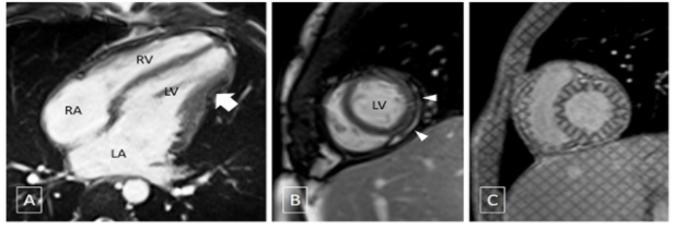
None had mitral annular calcification. Five of 12 (41.7%) patients with atrial fibrillation were in New York Heart Association class IV (Table 1). The clinical profile, Doppler echocardiography, tissue Doppler imaging and computed-tomography conclusively established the diagnosis of CCP in all 12 (100%) patients (Figures 1 and 2). Five (41.7%) patients underwent cardiac magnetic resonance (CMR) to define both morphological and functional changes (Figures 3A-3C).
Preoperative cardiac catheterization was performed on 3 (25%) patients to quantify right and left heart pressures, assess coronary anatomy, and obtain endomyocardial biopsy. The rest did not have catheterization because of their class III and IV symptoms with hepatic dysfunction, renal dysfunction or the echocardiographic findings were unequivocal. All demonstrated the findings considered diagnostic of constrictive pericarditis: an elevated right atrial pressure usually with a M-or-W shaped contour, and abnormally high right ventricular end-diastolic pressure with a characteristic dip-plateau diastolic configuration, and a ratio of right ventricular end-diastolic pressure to right ventricular systolic pressure of ≥ 0.30 (Table 1).

Echocardiographic Studies and Measurements
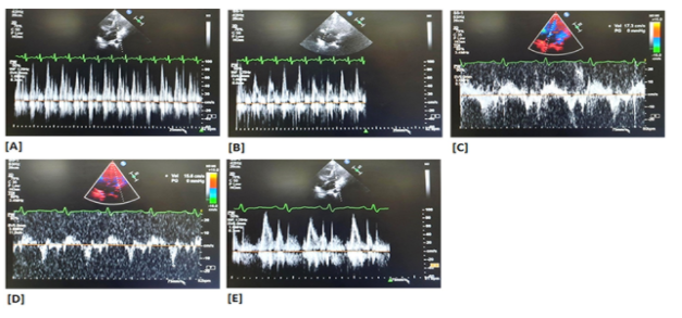
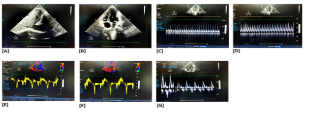
All patients had comprehensive evaluation with M-mode, two-dimensional (2-D) and pulsed-wave Doppler echocardiography with a respirometer recording and tissue Doppler imaging (TDI) before and after pericardiectomy using a Phillips iE 33 with 2.0 to 5.0 MHz transducer. Left ventricular ejection fraction (LVEF) was calculated by 2-D echocardiography with a modification of the method of Quinones and colleagues. [18] Left atrial volume was measured by the modified biplane area-length method. [19] Right ventricular systolic function was visually assessed. By using pulsed wave Doppler echocardiography, the following variables were measured: trans-mitral and trans-tricuspid peak velocities of early (E) and late filling (A) and E wave deceleration time (DT). On TDI, peak annular velocities were measured from the apical four chamber view at systole (s'), early (e') and late (a') diastole with a 2- 5 mm tissue Doppler sample volume placed at the septal corner and at the mitral and tricuspid lateral annuli. In patients with atrial fibrillation, five consecutive signals were measured and averaged. Inferior vena caval (IVC) diameter was assessed in subcostal sagittal view.
On Doppler, two flow velocity envelopes can be seen during diastole in persons with sinus rhythm: the E-wave, representing the early, passive filling of the ventricle, and the A-wave, that happens late in diastole, representing the active filling, the atrial contraction. For both mitral and tricuspid valve E and A wave measured. Mitral or tricuspid regurgitation was assessed semi-quantitatively as grade 1+ to 4+. A constrictive pattern was defined as 25% or greater increase in mitral E-velocity with expiration as compared with inspiration and an augmented (25% or more) diastolic flow reversal in the hepatic vein after the onset of expiration compared with inspiration. On tissue Doppler imaging, lateral mitral e¢, represents early diastolic myocardial relaxation velocity below the baseline as the annulus ascends away from the apex with cursor at lateral annulus; medial mitral e¢ and lateral tricuspid e¢ are same velocities measured at mitral medial annulus and tricuspid lateral annulus respectively. The mitral lateral s¢ velocity represents the systolic myocardial velocity at lateral mitral annulus. The medial mitral s¢ and lateral tricuspid s¢ are same velocities measured at mitral medial annulus and tricuspid lateral annulus respectively
Constrictive pericarditis was considered to be hemodynamically significant when there were clinical features of constriction with supportive echocardiographic and hemodynamic criteria as outlined earlier.
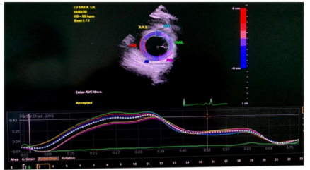
Strain by Speckle Tracking
A more recent echocardiographic approach to strain analysis is speckle tracking. Speckle tracking is a post-processing computer algorithm that uses the routine greyscale digital images. Although several manufacturers have devised speckle-tracking echocardiographic approaches, the fundamental approach is similar. [40-47]
Briefly, routine greyscale digital images of the myocardium contain unique speckle patterns. A user-defined region of interest is placed on the myocardial wall. Within this region of interest, the image-processing algorithm automatically subdivides regions into blocks of pixels tracking stable patterns of speckles. Subsequent frames are then automatically analyzed by searching for the new location of the speckle patterns within each of the blocks using correlation criteria and the sum of absolute differences. The location shift of these acoustic markers from frame to frame representing tissue movement provides the spatial and temporal data used to calculate velocity vectors. Temporal alterations in these stable speckle patterns are identified as moving farther apart or closer together and create a series of regional strain vectors.
Because strain information is not dependent on the Doppler angle of incidence like tissue Doppler imaging strain, several more strain analyses are possible, including longitudinal, circumferential, radial, and rotational. Currently, most echocardiography laboratories continue to use the subjective visual assessment of wall motion for resting and stress imaging for everyday clinical use, and strain imaging has been more often regarded as a research tool. Adoption of strain imaging in clinical practice appears to have been gaining momentum more recently so do forconstrictive pericarditis. [40-47]
We had assessed improvement in myocardial mechanics in CCP patients undergoing pericardiectomy using speckle tracking.This recently developed technique for characterization and quantification of myocardial deformation provided data noninvasively to better evaluation of the effectiveness of pericardiectomy. (i.e. in CCP, the epicardial dysfunction leads to depressed Global Circumferential Strain (GCS) and Left Ventricular Torsion (LVT), whereas Global Longitudinal Strain (GLS) and Global Radial Strain (GRS) are preserved and its strengths and weakness, and the potential present and future clinical applications.
Postoperative Studies
These included 3-monthly clinical examinations, electrocardiogram and chest radiographs. A minimum of 2 months follow-up was mandatory for this study. Preoperative studies were performed within 7 days before surgery. Postoperatively, all survivors were followed echocardiographically at the time of discharge and at 3 months. All late echoes have been grouped into one time period (3 months) with a range of no greater than 3 months. Echocardiographic data were measured according to American Society of echocardiographic criteria. [36]
Definitions and Acceptable Normal Values (Electronics)
For uniformity with other studies, total pericardiectomy was defined as wide excision of the pericardium with the phrenic nerves defining the posterior extent, the great vessels including the intrapericardial portion of superior vena cava and superior vena cava‑right atrial junction defining the superior extent, and the diaphragmatic surface, including the inferior vena cava‑right atrial junction defining the inferior extent of the pericardial resection.3,4,8 Radical pericardiectomy was defined as excision of the pericardium as defined under total pericardiectomy including the removal of the pericardium posterior to the phrenic nerve and the diaphragmatic pericardium. Constricting layers of the epicardium were removed whenever possible. The atria and venae cavae were decorticated as a routine in all cases in this study group. Pericardiectomy was considered partial if both ventricles could not be decorticated completely because of dense myopericardial adhesions or calcification. [3,4,8]
The importance of unrecognized constricting epicardial (visceral pericardial) peel was described by Harrington in 1944 and successful pericardiectomy requires decortication of the ventricular epicardium and relief of all constricting layers. [3,4,7]
Transthoracic two‑dimensional, color‑flow Doppler echocardiographic studies, speckle tracking echocardiography were performed on all patients before and after the operation. Mitral, tricuspid, superior vena cava, hepatic vein, and pulmonary flow velocities were measured. Mitral or tricuspid regurgitation was assessed semi‑quantitatively as Grade 1 + to 4+. Ejection fraction was calculated using modified Quinones method. A constrictive pattern was defined as 25% or greater increase in mitral E‑velocity with expiration as compared with inspiration and an augmented (25% or more) diastolic flow reversal in the hepatic vein after the onset of expiration compared with inspiration.
Low output syndrome was diagnosed if the patient required inotropic support dopamine (4–10 μg/kg/min), dobutamine (5–10 μg/kg/min), epinephrine (0.01–0.1 μg/kg/min), milrinone (50 μg/kg intravenous bolus followed by 0.375–0.75 μg/kg/min), either isolated or in combination, in the operating room or intensive care unit to maintain stable hemodynamics in the absence of mechanical external compression after correction of all electrolytes or blood gas abnormalities and after adjusting the preload to its optimal value. Low output syndrome was also diagnosed if there was an increasing requirement of the above‑mentioned inotropes along with afterload reduction with sodium nitroprusside. Patients who received < 4 μg/kg/min of dopamine to increase renal perfusion were not considered to have low‑output syndrome. [3,4,8]
Accordingly, under the definition of low output syndrome after pericardiectomy, an integration of relevant clinical, laboratory and bedside echocardiographic criteria were used. The criteria for diagnosis were as follows: cold extremities, absent pedal pulses, decreased toe temperature, reduced systolic pressure, impaired renal function and oliguria (<1.0 mL.kg-1.h-1), metabolic acidosis, increased serum lactate levels >2.0 mmol/L, >2 hours), low mixed venous oxygen saturation (<50%), and blunt sensorium. [3,4,8]
Perioperative mortality was defined as that occurring within 30 days after surgery. Cardiac-related death was defined as death due to cardiac causes, such as progressive congestive cardiac failure.6-10 Hypoproteinemia was defined as serum albumin level < 3.5 gm/dl. Renal dysfunction was defined as serum creatinine >2.0 gm/dl. [3,4,8,48]
Statistical Analysis
Statistical analysis was carried out using Stata 11.0 (College Station, Texas, USA). Continuous data were presented as mean±standard deviation, whereas categorical variables were presented as frequency distribution and percentage. Qualitative data were analysed by using 2 test or student’s t test. Normality assumptions for continuous variables were assessed using Shapiro-Wilks test. Comparisons between two groups were done with the t-test. Echocardiographic parameters over a period of time between various clinical parameters were tested using Friedman’s test. The correlation between mitral annular systolic velocities and left ventricular ejection fraction was assessed using Spearman’s rank correlation. The p value of <0.05 was considered as statistically significant.
There was no early death. Eleven (91.6%) patients had low-cardiac-output in the immediate postoperative period. All patients were routinely started on dopamine (4µg.kg-1.min-1) to increase renal perfusion on operation table after completing excision of the thickened pericardium. Patients with normal renal function were administered oral angiotensin-converting enzyme (ACE) inhibitors before weaning from inotropic agents. Postoperatively, digoxin, diuretics and ACE-inhibitors were weaned at varying time intervals.
Patients considered to have low output syndrome (n=11) required dopamine (4-10 µg.kg-1.min-1), epinephrine (0.01-0.1 µg.kg-1.min-1) and milrinone (50 µg/kg intravenous bolus followed by 0.375-0.75 µg.kg-1.min-1) either isolated or in combination. Median duration of inotrope requirement was 4 days (range 2-7 days) in these patients. Patients with normal renal function were administered oral angiotensin-converting enzyme inhibitors before weaning from inotropic agents. Two (16.66%) patients required intraoartic balloon counter pulsation as an additional support. There was marked reduction of filling pressure within 24 hours in the majority of patients (n=10) after total pericardiectomy [mean= right atrial pressure (RAP) 19.82±4.6 (18-29) to 6.11±0.85 (6-9); p<0.001]. Echocardiographically, diastolic filling characteristics remained abnormal in 3 (25%) patients of the study group in the immediate postoperative period. There was no late death. Reoperation was not required for any patients.
Follow-up
Follow-up was 100% complete (range 2-36 months, median 19) and yielded 19 patients-years of data.
At closing interval, 2 (16.6%) patients continued to remain in NYHA class II, and had persistent abnormalities of the diastolic filling pattern (p<0.05) on Doppler echocardiography. Pairwise comparison between symptomatic (n=2, 16.7%) and asymptomatic (n=10, 83.3%) patients revealed significant abnormality of the indexed IVC diameter (p<0.05) and increased left ventricular end-diastolic internal diameter (LVID) (p<0.05) in all patients of the symptomatic group. Nine of these symptomatic patients continued to remain in atrial fibrillation. Preoperatively, these symptomatic patients (n=2) were in NYHA class IV and were in atrial fibrillation. Thus, 2 (16.7%) of 5 patients who had preoperative atrial fibrillation continued to remain in atrial fibrillation. This could be the causative factor for alteration of left atrial mechanics and the left ventricular filling pressure which could lead to ongoing symptoms. Surgical techniques did not affect the outcome of atrial fibrillation.
These symptomatic patients (n=2, 16.7%) had significantly higher right atrial pressure in the immediate preoperative period compared to the asymptomatic group (n=10, 83.3%) [mean RAP=21.8±3.8 (symptomatic) vs 19.82±4.6 mmHg (asymptomatic), p<0.05]. Postoperatively, despite total pericardiectomy, the right atrial pressure of the symptomatic group continued to remain higher than the asymptomatic group [mean RAP=9.1±0.7 (symptomatic) vs 6.1±0.85 mmHg (asymptomatic), p<0.001)]. There were no differences of TDI-derived systolic and diastolic annular velocities of the mitral and tricuspid valves between symptomatic and asymptomatic patients in the preoperative period. Tissue Doppler imaging-derived mitral and tricuspid annular velocities failed to predict the postoperative outcome of patients undergoing pericardiectomy.
Data analyses and study interpretation of echocardiographic data (Tables 2 & 3)

To assess the characterization of the mitral and tricuspid annular velocity changes and speckle echocardiographic derived myocardial mechanics in patients undergoing pericardiectomy for constructive pericarditis, Friedman’s test analysis revealed the following results:
Global circumferential strain (GCS)
Non-parametric tests (Friedman test) were used to make statistical inference as data were not normally distributed. Friedman test was used to explore whether the GCS changed significantly over time.
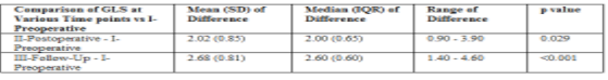
The mean GCS increased from a minimum of 24.43 at the I-preoperative timepoint to a maximum of 30.08 at the III-follow-up timepoint. This change was statistically significant (Friedman Test: χ2 = 24.0, p = <0.001).
As a significant change was observed in GCS over time using the Friedman Test, post-hoc pairwise analysis was performed to explore at which timepoints the GCS differed significantly from the I-preoperative timepoint (Table 4)
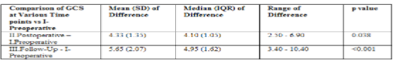
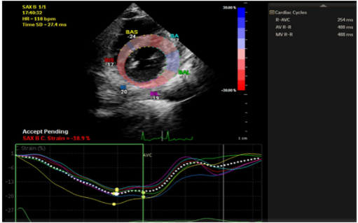
Table 4: Global circumferential strains (GCS)
The GCS differed significantly from the I-preoperative timepoint at the following timepoints: II-Postoperative, III-follow-up. The maximum change from the I-preoperative timepoint was observed at the 3 follow-up time point (Figure 4).
Global longitudinal strain (GLS)
Non-parametric tests (Friedman test) were used to make statistical inference as data were not normally distributed. Friedman test was used to explore whether the GLS changed significantly over time.
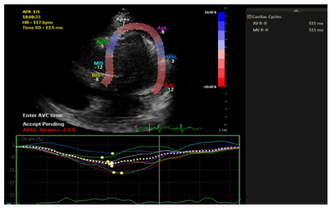
The mean GLS increased from a minimum of 19.63 at the I-preoperative timepoint to a maximum of 22.31 at the III-follow-up timepoint. This change was statistically significant (Friedman Test: χ2 = 23.5, p = <0.001).
As a significant change was observed in GLS over time using the Friedman Test, post-hoc pairwise analysis was performed to explore at which timepoints the GLS differed significantly from the I-preoperative timepoint (Table 5).
The GLS differed significantly from the I-preoperative timepoint at the following timepoints: II-postoperative, III-follow-up. The maximum change from the I-preoperative timepoint was observed at the III-Follow-Up timepoint (Figures 5 and 6).
Global radial strain (GRS)
Non-parametric tests (Friedman test) were used to make statistical inference as data were not normally distributed. Friedman test was used to explore whether the GRS changed significantly over time.
The mean GRS increased from a minimum of 46.28 at the I-preoperative timepoint to a maximum of 54.26 at the III-follow-up timepoint. This change was statistically significant (Friedman Test: χ2 = 23.4, p = <0.001).
As a significant change was observed in GRS over time using the Friedman Test, post-hoc pairwise analysis was performed to explore at which timepoints the GRS differed significantly from the I-preoperative timepoint (Table 6)
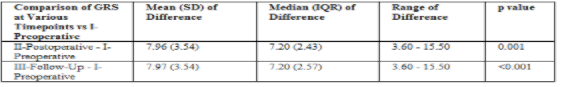
The GRS differed significantly from the I-preoperative timepoint at the following timepoints: II-Postoperative, III-follow-up. The maximum change from the I-preoperative timepoint was observed at the III-Follow-Up timepoint (Figure 7).
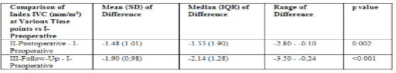
Indexed IVC diameter (mm/m2)
Non-parametric tests (Friedman test) were used to make statistical inference as data were not normally distributed. Friedman test was used to explore whether the Index IVC (mm/m2) changed significantly over time.
The mean Index IVC (mm/m2) decreased from a maximum of 15.58 at the I-preoperative timepoint to a minimum of 13.68 at the III-Follow-Up timepoint. This change was statistically significant (Friedman Test: χ2 = 20.8, p = <0.001).
As a significant change was observed in Index IVC (mm/m2) over time using the Friedman Test, post-hoc pairwise analysis was performed to explore at which timepoints the Index IVC (mm/m2) differed significantly from the I-preoperative timepoint (Table 7).
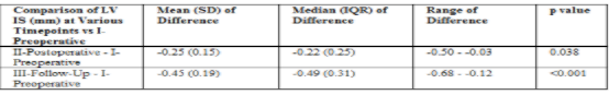
Left ventricular end-systolic internal diameter (LVIS)
Non-parametric tests (Friedman test) were used to make statistical inference as data were not normally distributed. Friedman test was used to explore whether the LV IS (mm) changed significantly over time.
The mean LVIS (mm) decreased from a maximum of 22.99 at the I-preoperative timepoint to a minimum of 22.54 at the III-Follow-Up timepoint. This change was statistically significant (Friedman Test: χ2 = 24.0, p = <0.001).
As a significant change was observed in LV IS (mm) over time using the Friedman Test, post-hoc pairwise analysis was performed to explore at which timepoints the LV IS (mm) differed significantly from the I-preoperative timepoint (Table 8)
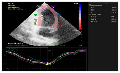
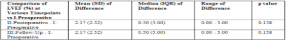
Left ventricular ejection fraction (LVEF)
Non-parametric tests (Friedman test) were used to make statistical inference as data were not normally distributed. Friedman test was used to explore whether the LVEF (%) changed significantly over time.
The mean LVEF (%) increased from a minimum of 47.83 at the I-preoperative timepoint to a maximum of 50.00 at the III-Follow-Up timepoint. This change was statistically significant (Friedman Test: χ2 = 12.0, p = 0.002).
As a significant change was observed in LVEF (%) over time using the Friedman Test, post-hoc pairwise analysis was performed to explore at which timepoints the LVEF (%) differed significantly from the I-preoperative timepoint (Table 9) There was no significant difference between any of the timepoints as compared to the I-preoperative timepoint in terms of LVEF (%).
Left ventricular end-diastolic internal diameter (LVID)
The mean LVID (mm) decreased from a maximum of 38.99 at the I-preoperative timepoint to a minimum of 38.40 at the III-Follow-Up timepoint. This change was statistically significant (Friedman Test: χ2 = 24.0, p = <0.001).
Mitral medial E'
The mean Medial E' (cm/sec) increased from a minimum of 14.07 at the I-preoperative timepoint to a maximum of 14.95 at the III-Follow up timepoint. This change was statistically significant (Friedman Test: χ2 = 12.9, p = 0.002).
Mitral lateral E'
The mean Mitral Lateral E' (cm/sec) increased from a minimum of 11.86 at the I-preoperative timepoint to a maximum of 14.09 at the III-Follow up timepoint. This change was statistically significant (Friedman Test: χ2 = 23.5, p = <0.001).
Transmitral late diastolic filling velocity (MVA)
The mean MVA (cm/sec) increased from a minimum of 42.33 at the I-preoperative timepoint to a maximum of 61.35 at the III-Follow-Up timepoint. This change was statistically significant (Friedman Test: χ2 = 24.0, p = <0.001)
Transmitral early diastolic filling velocity (MVE)
The mean MVE (cm/sec) increased from a minimum of 79.57 at the I-preoperative timepoint to a maximum of 115.97 at the III-Follow-Up timepoint. This change was statistically significant (Friedman Test: χ2 = 24.0, p = <0.001).
MVE (Respiratory Variation) (cm/sec) over time
The mean MVE (Respiratory Variation) (cm/sec) decreased from a maximum of 43.10 at the I-preoperative timepoint to a minimum of 22.39 at the III-Follow-Up timepoint. This change was statistically significant (Friedman Test: χ2 = 24.0, p = <0.001).
Assessment of change in MVE/A over time
The mean MVE/A increased from 1.88 at the I-preoperative timepoint to a maximum of 2.01 at the II-Postoperative timepoint, and then decreased to 1.87 at the III-Follow-Up timepoint. This change was not statistically significant (Friedman Test: χ2 = 5.1, p = 0.076).
Change in Tricuspid Lateral E' (cm/sec)
The mean Tricuspid Lateral E' (cm/sec) increased from a minimum of 12.75 at the I-preoperative timepoint to a maximum of 14.39 at the III-Follow-Up timepoint. This change was statistically significant (Friedman Test: χ2 = 22.0, p = <0.001).
Assessment of change in transtricuspid late diastolic filling velocity
The mean TVA (cm/sec) increased from a minimum of 28.86 at the I-preoperative timepoint to a maximum of 43.12 at the III-Follow-Up timepoint, and then decreased to 42.68 at the II-Postoperative timepoint. This change was statistically significant (Friedman Test: χ2 = 21.6, p = <0.001).
Transtricuspid early diastolic filling velocity (TVE)
The mean TVE (cm/sec) decreased from 52.94 at the I-preoperative timepoint to a minimum of 51.04 at the II-Postoperative timepoint, and then increased to 53.71 at the III-Follow-Up timepoint. This change was statistically significant (Friedman Test: χ2 = 8.7, p = 0.013).
Assessment of change in TVE (Respiratory Variation) (%) over time
The mean TVE (Respiratory Variation) (%) decreased from a maximum of 26.33 at the I-preoperative timepoint to a minimum of 21.02 at the II-Postoperative timepoint, and then increased to 26.33 at the III-Follow-Up timepoint. This change was statistically significant (Friedman Test: χ2 = 24.0, p = <0.001).
TVE/A
The mean TVE/A decreased from a maximum of 1.84 at the I-preoperative timepoint to a minimum of 1.21 at the II-Post Operative timepoint, and then increased to 1.26 at the III-Follow-Up timepoint. This change was statistically significant (Friedman Test: χ2 = 17.9, p = <0.001).
So far as we are aware, there have been few published studies in the literature investigating the role of tissue Doppler imaging-derived parameters of mitral and tricuspid annular motion on global and regional ventricular function and speckle echocardiographic derived variables of myocardial mechanics and their role in differentiating CP from RCM.[7-17]
The principal findings of this investigation include:
Currently, tissue Doppler imaging is an integral part of an echocardiography examination in various areas of cardiology. Tissue Doppler imaging offers a quantitative measurement of regional and global myocardial tissue function. In particular, the assessment of longitudinal mitral annular motion provides an accurate estimate of global left ventricular function [12-16] and it has further facilitated the detection of constrictive pericarditis. Since the mechanoelastic properties of the myocardium are preserved in constrictive pericarditis, the longitudinal mitral annular velocities remain normal or can be exaggerated as lateral expansion in constrictive pericarditis is limited. Garcia and colleagues were the first to report that measurement of longitudinal axis expansion by tissue Doppler imaging provided a clinically useful distinction between constrictive pericarditis and restrictive cardiomyopathy. [16] Rajagopalan and colleagues showed that a peak e' velocity ≥ 8 cm/s could discriminate between constrictive pericarditis and restrictive cardiomyopathy with high sensitivity (89%) and specificity (100%). [51] Studies by Ha and colleagues and by Sohn and colleagues recommended that e' velocity can provide a helpful diagnostic indicator and should be measured routinely in the evaluation of heart failure or suspected constrictive pericarditis. [30,31] Ha and colleagues recommended the same 8 cm/s cut off value for diagnosis of constrictive pericarditis, where e' velocity is equal or greater than 8 cm/s, with 95% sensitivity and 96% specificity.[49] Ha and colleagues also evaluated the role of tissue Doppler imaging in the diagnosis of constrictive pericarditis in patients without diagnostic respiratory variation of transmitral early diastolic filling velocity. It was confirmed that e' velocity was well-preserved independent of any respiratory variation in mitral inflow velocities. Other studies suggested that e' should be used with caution if constrictive pericarditis is combined with myocardial diseases, extensive annular calcification or segmental non-uniform myocardial velocities. [35] Several investigators have shown that E/e' ratio correlates well with left ventricular filling pressure. [36] E/e' >15 identifies increased left ventricular filling pressure while E/e' <8 describes normal filling pressure. Ha introduced the concept of annulus paradoxus, which describes the paradoxical behavior of the mitral annulus in constrictive pericarditis. [49] Ha found that an inverse relationship exists between E/e' and left ventricular filling pressure, which can be explained by the fact that in constrictive pericarditis the mitral annulus has an exaggerated longitudinal motion leading to an increase in e', despite high filling pressures.[49]
In normal subjects, the mitral lateral annulus e' velocity is higher than the medial annulus e' velocity. Reuss and colleagues identified the reversal of the normal relationship of mitral lateral e' and medial e' velocities in constrictive pericarditis, where mitral lateral e' velocity is lower than medial e' velocity, therefore lateral/medial e' ratio is inverted and called, “annulus reversus". [33] This finding is based on the tethering of the adjacent fibrotic and scarred pericardium, which influences sthe lateral mitral annulus in patients with constrictive pericarditis. In a patient with preserved mitral e' velocities (> 8 cm/sec) and a low E/e' ratio (< 8) with high left ventricular filling pressure, the recognition of, “annulus reversus” should alert to the diagnosis of constrictive pericarditis.[11-18]
In general, tissue Doppler imaging offers incremental diagnostic information to M-mode, 2D echo and transmitral flow Doppler for detecting constrictive physiology, with a reported sensitivity and specificity of 88.8% and 94.8%, respectively. [11-18]
Kim JS and colleagues examined the medial annular velocities in patients with constrictive pericarditis after pericardiectomy in 16 patients and found that e' decreased significantly after pericardiectomy. [38] However, there is no substantive data on mitral annulus systolic velocity and tricuspid annulus velocity in constrictive pericarditis and no data on the effect of pericardiectomy on these annular velocities. The effect on pericardiectomy on mitral and tricuspid annular velocities, which may provide further insight into the mechanism of annulus motion, is unknown.
Building on these observations from a small number of patients, our aim was to provide a comprehensive evaluation of tissue Doppler imaging at both mitral and tricuspid annuli in a larger number of patients and follow their evolution after pericardial resection.
Early diastolic mitral annulus velocity
We confirmed the presence of “annulus reverses” in patients with constrictive pericarditis. Based on the hypothesis that the lateral annulus motion is restricted by the constricting pericardium and that medial annulus diastolic motion increases in compensation, it may be anticipated that medial mitral annulus velocity decreases and lateral annulus velocity increases after pericardiectomy, and that mitral lateral/medial e' ratio normalizes. In this study, both was confirmed.
Early diastolic tricuspid annulus velocity
In this study, all patients exhibited a reduction in tricuspid lateral e' velocity after pericardiectomy. The phenomenon of “annulus reversus” was observed in all patients in this study. There was reduced lateral tricuspid annular velocity (e') in all patients and normalization of the tricuspid lateral/medial e' ratio following pericardiectomy during the follow up period. Therefore, the above mentioned mechanisms operative at the mitral annulus may as well be responsible for findings at the tricuspid annulus.
Garcia and colleagues were the first to report that the measurement of longitudinal axis expansion by tissue Doppler imaging provided a clinically useful distinction between constrictive pericarditis and restrictive cardiomyopathy.20 Our studies have confirmed that medial e' velocity was relatively normal or even accentuated in all patients with constrictive pericarditis irrespective of characteristic respiratory variation in mitral E velocity. Characteristic respiratory variation across the mitral valve and tricuspid valve was present in all patients in this study group.
In this study, there was proportionately greater postoperative reduction in tricuspid lateral e' velocity compared to mitral annulus values. As the pericardial disease process is often asymmetric, being more pronounced over the right ventricle, annular motion here may be expected to be most exaggerated before pericardiectomy and following decortication approximate normality.
Similar observations were noted by other investigators. [39] Sengupta and colleagues found higher net twist but no significant increase in torsion post-pericardiectomy, a conclusion limited by small number of patients and early timing of the postoperative studies when restoration of function may have been incomplete.[34,35]
To explain this paradoxical relationship between s' and stroke volume in constrictive pericarditis, Veress and colleagues postulated that systolic and diastolic motion of the mitral annulus are closely coupled in part via elastic recoil mechanisms. [39]
Systolic annulus velocity
s' by tissue Doppler imaging reflects the peak velocity of myocardial fiber shortening in the longitudinal direction and provides a more sensitive assessment of global left ventricle and right ventricle systolic function than 2-D or M-mode imaging. s' has been correlated with peak positive dP/dt and left ventricular ejection fraction in patients with dilated cardiomyopathy, hypertensive heart disease and myocardial infarction. There is little information on mitral and tricuspid s' velocities in patients with constrictive pericarditis. Studies in very small patient population have either compared s' velocity between constrictive pericarditis and restrictive cardiomyopathy or measured changes in s' velocity pre- and post-pericardiectomy. [39]
The mean s' velocity in all patients in this study was lower both before and after pericardiectomy than published normative values [42] and also lower, especially prepericardiectomy. These observations are consistent with previous smaller studies. This finding seems counterintuitive since s' velocity is expected to increase with augmented stroke volume after pericardiectomy.
We postulated that stroke volume in constrictive pericarditis is closely coupled, in part via elastic recoil mechanisms. Thus, in the pre-pericardiectomy setting, both longitudinal systolic and diastolic motion of the annuli are exaggerated while following release of pericardial constraint, both decrease in tandem. This hypothesis is supported by the moderate to high correlation between annular s' and e' as well as s' and a', especially before pericardiectomy when restorative forces may be most operative.
There appeared to be proportionately greater postoperative reduction in tricuspid lateral or right ventricle s' and e' compared to mitral annulus values. As the pericardial disease process is often asymmetric, being most pronounced over the right ventricle, annular motion here may be expected to be most exaggerated before pericardiectomy, and following decortication, approximate normality. However, the disproportionate reduction in tricuspid lateral s' and e' probably seems also from postoperative right ventricular dysfunction, which was moderate in 7 (21.2%) patients.
Left ventricular ejection fraction did not change despite the expected increase in stroke volume after pericardiectomy. It is postulated that after pericardial resection, left ventricular filling increases and other elements of left ventricular shortening including torsion are recruitable, contributing to better cardiac output and compensating for abnormal longitudinal function. [28] Sengupta and colleagues found higher net twist but no significant increase in torsion post pericardiectomy, a conclusion limited by small patient numbers and early timing of the postoperative studies when restoration of function may have been incomplete. [28] To confirm this hypothesis, detailed analysis of myocardial mechanics in a larger number of patients pre- and post-pericardiectomy will be required. [34,35]
Monitoring of intracardiac pressures during pericardiectomy has been proposed to evaluate the result of decortications but Viola [40] argued against the value of this assessment because further recovery of myocardial failure may occur late after pericardiectomy. In this study, we showed that there is a relationship between the degree of decrease in atrial pressure after pericardiectomy and postoperative diastolic function. Further, early abnormalities in diastolic filling pattern may improve in the late follow-up; however, the long-term hemodynamic result may not be predicted by the immediate postoperative Doppler echocardiographic findings.
Symptoms may persist after successful pericardiectomy in patients with mixed constrictive-restrictive disease because of abnormalities in intrinsic myocardial compliance. In our study group, 2 (16.6%) patients continued to have NYHA Class II symptoms late postoperatively. These patients exhibited higher right atrial pressure (mean±SD= 10.25±1.48 mmHg), raised indexed inferior vena caval diameter, (mean±SD= 14.09±3.01 mm), higher LVID (mean±SD= 40.27.4.7 mm) and persistently abnormal transmitral early diastolic filling velocity (MVE, mean±SD=115.97±11.09 cm/sec, MVE/A 1.87±0.22) in the 6 month follow up.
There are limited studies to assess the extent of myocardial damage with two-dimensional speckle tracking echocardiography (2DSTE). Longitudinal, radial, and circumferential mechanics of the LV were quantified by 2DSTE on 26 patients with constrictive pericarditis and 19 patients with restrictive cardiomyopathy by Sengupta and associates. In comparison with controls, constrictive pericarditis patients had impaired left ventricle circumferential strain (ε) (base; −16±6 vs −9±6%; P.01) significantly reduced in constrictive pericarditis patients when compared to a control group.55 Amaki and associates validated 2DSTE and cardiac magnetic resonance imaging (CMR) on 28 patients with constrictive pericarditis and 30 patients with restrictive cardiomyopathy.
The global longitudinal scale was higher in patients with constrictive pericarditis than in those with restrictive cardiomyopathy [−18.5% (−20.1 to −15.2) vs −11.6% (−14.6 to −9.3); P<.001], and both techniques were found to have similar diagnostic value (area under the curve, 0.84 vs 0.88 for cardiac magnetic resonance imaging and echocardiography, respectively). [56,57] The ratio between lateral and septal longitudinal ε was not significantly different among the 3 groups. Patients with RCM had marginally lower circumferential ε when compared with CP [−23.9 (−28.3 to −20.2) vs −19.3 (−23.3 to −16.0)%, P=.07].[57]
Negishi and colleagues investigated 83 patients with CCP whether pericardiectomy improves myocardial mechanics using 2DSTE. Besides left ventricle ε, the authors studied left ventricle longitudinal and rotational displacement. Longitudinal displacement of left ventricle opposing walls was similar, but decreased in constrictive pericarditis compared to controls. After pericardiectomy, septal displacement decreased, but lateral displacement increased. Septal longitudinal ε was similar between two groups, but lateral longitudinal ε was lower in the constrictive pericarditis group. Septal longitudinal ε decreased significantly (−20.3±5.0% vs −17.7±4.6%, P=.032) after surgery, but lateral longitudinal ε did not (−14.7±5.8% vs −15.2±3.4%, P=.51). Patients with constrictive pericarditis had lower absolute values of Global longitudinal strain (−20.1±1.9 vs −16.2±3.3%, P<.01) and Global circumferential strain (−20.7±5.1 vs −14.7±5.0%, P<.01), with no significant difference in Global radial strain (50.4±16.2 vs40.8±18.8%, P=.07) compared with controls. No significant difference in SLRD between controls and constrictive pericarditis (−2.3±3.3 vs −0.6±3.0°, P=.07). After pericardiectomy, SLRD increased significantly (−0.8±3.3% vs 2.1±3.0, P<.01). No changes in GLS (−15.6±3.9% vs −15.8±3.2, P=.88) and GRS (37.4±18.9% vs 39.1±16.5%, P=.73) after pericardiectomy. GCS increased (−13.5±5.7 vs −17.6±5.5, P<.01) after pericardiectomy.[59]
In this study, speckle echocardiographic derived parameters revealed: i) significant increase of the Global Circumferential Strain (GCS) from preoperative value of 24.43±3.17 to 28.77±2.55 and further improvement to 30.08±2.61 on 6 month follow-up, ii) slight increase of the Global Longitudinal Strain (GLS) from preoperative value of 19.63±2.98 to 21.66±2.61and on 6 month follow-up to 22.31±2.62, and iii) increase of the Global Radial Strain (GRS) from preoperative value of 46.28±7.39 to postoperative of 54.24±5.53 with no significant improvement on 6 month follow- up.
This is in accordance with the study conducted by Negishi and associates who demonstrated increase of GCS among 83 patients with constrictive pericarditis undergoing pericardiectomy 13.5±5.7 to 17.6±5.5, p<.01); with no significant difference in GLS and GRS
In this study, 5 (41.7%) patients had extensive pericardial calcification over the anterior and inferior surfaces of the right and left ventricle. However, total pericardiectomy including removal of the calcified pericardium overlying the anterolateral and diaphragmatic surface of the right ventricle was achieved in all patients of the study group. These patients in the immediate postoperative period required higher inotropic support because of low cardiac output. This phenomenon may reflect underlying myocardial damage or atrophy secondary to long standing encasement and penetration of the myocardium by calcium spurs, and persistent inflammation. We believe that subjecting the newly liberated right, and perhaps left ventricle to even moderately elevated filling pressure led to increased wall stress and deteriorating cardiac function possibly due to fibrous invasion of the myocardium and varying grades of myocardial atrophy. We concur with the observations of other investigators regarding the possibility of residual constriction, fibrous invasion of the myocardium and abnormal ventricular compliance secondary to myocardial alterations.[4,5,40,41] The utility of speckle tracking echocardiography and tissue Doppler imaging in identifying residual constrictive pericarditis requires further investigation on a large cohort of patients correlating the clinical outcomes.
Clinical Implications
With a GCS, GLS and GRS data and mitral inflow profile of increased filling pressure and expiratory hepatic vein diastolic reversals as well as abnormal ventricular septal motion in constrictive pericarditis, the disease can be diagnosed by echocardiography with assistance of cardiac catheterization. The characteristic pattern of annulus velocities revert to normal after pericardiectomy in asymptomatic patients.
It will be of clinical interest to investigate if the extent of postoperative changes in annular velocities can predict clinical outcome after pericardiectomy. Further evaluation of these indices as a marker of successful treatment of constrictive pericarditis is warranted. Further studies are underway to compare these speckled tracking and tissue Doppler derived parameters achieved by median sternotomy and anterolateral thoracotomy approaches on a large number of patients.
Study Limitations
This study included only a small number of patients and 7 (53.8%) patients underwent pericardiectomy via median sternotomy. Hence, the tissue Doppler imaging and speckle derived variables could not be compared with anterolateral thoracotomy approach.
Secondly, we only recorded tissue Doppler imaging of longitudinal axis motion in the 4-chamber view. Due to the local tethering effect, analysis of multiple annular regions could have provided additional helpful data. It would also be helpful to characterize radial and circumferential function for a better understanding of the mechanics of the unique annular motion in constrictive pericarditis and effects of pericardiectomy.
Thirdly, due to its limited resolution, 2D speckle tracking echocardiography might be suboptimal in patients with calcific CCP. Other limiting factors are data procured from postsurgical patients with postoperative adhesions, and patients with atrial fibrillation.
Conclusions
This study demonstrates that patients with congestive heart failure and normal left ventricular ejection fraction, preserved or increased medial e' velocity strongly suggests constrictive pericarditis. The diagnosis is further supported if medial e' is higher than lateral e'. The annulus velocity study demonstrates that in patients with constrictive pericarditis:
We conclude that tissue Doppler imaging and speckle tracking echocardiography are useful investigative modalities for serial evaluation of patients undergoing pericardiectomy. It can be performed serially with a high degree of reproducibility. It can be used for late postoperative assessment, obviating the need for frequent cardiac catheterization. In addition, we propose routine and serial utilization of this modality may be the investigation of choice for assessment of adequacy of pericardial resection and its subsequent effect on ventricular remodeling.
Clearly Auctoresonline and particularly Psychology and Mental Health Care Journal is dedicated to improving health care services for individuals and populations. The editorial boards' ability to efficiently recognize and share the global importance of health literacy with a variety of stakeholders. Auctoresonline publishing platform can be used to facilitate of optimal client-based services and should be added to health care professionals' repertoire of evidence-based health care resources.

Journal of Clinical Cardiology and Cardiovascular Intervention The submission and review process was adequate. However I think that the publication total value should have been enlightened in early fases. Thank you for all.

Journal of Women Health Care and Issues By the present mail, I want to say thank to you and tour colleagues for facilitating my published article. Specially thank you for the peer review process, support from the editorial office. I appreciate positively the quality of your journal.

Journal of Clinical Research and Reports I would be very delighted to submit my testimonial regarding the reviewer board and the editorial office. The reviewer board were accurate and helpful regarding any modifications for my manuscript. And the editorial office were very helpful and supportive in contacting and monitoring with any update and offering help. It was my pleasure to contribute with your promising Journal and I am looking forward for more collaboration.

We would like to thank the Journal of Thoracic Disease and Cardiothoracic Surgery because of the services they provided us for our articles. The peer-review process was done in a very excellent time manner, and the opinions of the reviewers helped us to improve our manuscript further. The editorial office had an outstanding correspondence with us and guided us in many ways. During a hard time of the pandemic that is affecting every one of us tremendously, the editorial office helped us make everything easier for publishing scientific work. Hope for a more scientific relationship with your Journal.

The peer-review process which consisted high quality queries on the paper. I did answer six reviewers’ questions and comments before the paper was accepted. The support from the editorial office is excellent.

Journal of Neuroscience and Neurological Surgery. I had the experience of publishing a research article recently. The whole process was simple from submission to publication. The reviewers made specific and valuable recommendations and corrections that improved the quality of my publication. I strongly recommend this Journal.

Dr. Katarzyna Byczkowska My testimonial covering: "The peer review process is quick and effective. The support from the editorial office is very professional and friendly. Quality of the Clinical Cardiology and Cardiovascular Interventions is scientific and publishes ground-breaking research on cardiology that is useful for other professionals in the field.

Thank you most sincerely, with regard to the support you have given in relation to the reviewing process and the processing of my article entitled "Large Cell Neuroendocrine Carcinoma of The Prostate Gland: A Review and Update" for publication in your esteemed Journal, Journal of Cancer Research and Cellular Therapeutics". The editorial team has been very supportive.

Testimony of Journal of Clinical Otorhinolaryngology: work with your Reviews has been a educational and constructive experience. The editorial office were very helpful and supportive. It was a pleasure to contribute to your Journal.

Dr. Bernard Terkimbi Utoo, I am happy to publish my scientific work in Journal of Women Health Care and Issues (JWHCI). The manuscript submission was seamless and peer review process was top notch. I was amazed that 4 reviewers worked on the manuscript which made it a highly technical, standard and excellent quality paper. I appreciate the format and consideration for the APC as well as the speed of publication. It is my pleasure to continue with this scientific relationship with the esteem JWHCI.

This is an acknowledgment for peer reviewers, editorial board of Journal of Clinical Research and Reports. They show a lot of consideration for us as publishers for our research article “Evaluation of the different factors associated with side effects of COVID-19 vaccination on medical students, Mutah university, Al-Karak, Jordan”, in a very professional and easy way. This journal is one of outstanding medical journal.

Dear Hao Jiang, to Journal of Nutrition and Food Processing We greatly appreciate the efficient, professional and rapid processing of our paper by your team. If there is anything else we should do, please do not hesitate to let us know. On behalf of my co-authors, we would like to express our great appreciation to editor and reviewers.

As an author who has recently published in the journal "Brain and Neurological Disorders". I am delighted to provide a testimonial on the peer review process, editorial office support, and the overall quality of the journal. The peer review process at Brain and Neurological Disorders is rigorous and meticulous, ensuring that only high-quality, evidence-based research is published. The reviewers are experts in their fields, and their comments and suggestions were constructive and helped improve the quality of my manuscript. The review process was timely and efficient, with clear communication from the editorial office at each stage. The support from the editorial office was exceptional throughout the entire process. The editorial staff was responsive, professional, and always willing to help. They provided valuable guidance on formatting, structure, and ethical considerations, making the submission process seamless. Moreover, they kept me informed about the status of my manuscript and provided timely updates, which made the process less stressful. The journal Brain and Neurological Disorders is of the highest quality, with a strong focus on publishing cutting-edge research in the field of neurology. The articles published in this journal are well-researched, rigorously peer-reviewed, and written by experts in the field. The journal maintains high standards, ensuring that readers are provided with the most up-to-date and reliable information on brain and neurological disorders. In conclusion, I had a wonderful experience publishing in Brain and Neurological Disorders. The peer review process was thorough, the editorial office provided exceptional support, and the journal's quality is second to none. I would highly recommend this journal to any researcher working in the field of neurology and brain disorders.
Dear Agrippa Hilda, Journal of Neuroscience and Neurological Surgery, Editorial Coordinator, I trust this message finds you well. I want to extend my appreciation for considering my article for publication in your esteemed journal. I am pleased to provide a testimonial regarding the peer review process and the support received from your editorial office. The peer review process for my paper was carried out in a highly professional and thorough manner. The feedback and comments provided by the authors were constructive and very useful in improving the quality of the manuscript. This rigorous assessment process undoubtedly contributes to the high standards maintained by your journal.

International Journal of Clinical Case Reports and Reviews. I strongly recommend to consider submitting your work to this high-quality journal. The support and availability of the Editorial staff is outstanding and the review process was both efficient and rigorous.

Thank you very much for publishing my Research Article titled “Comparing Treatment Outcome Of Allergic Rhinitis Patients After Using Fluticasone Nasal Spray And Nasal Douching" in the Journal of Clinical Otorhinolaryngology. As Medical Professionals we are immensely benefited from study of various informative Articles and Papers published in this high quality Journal. I look forward to enriching my knowledge by regular study of the Journal and contribute my future work in the field of ENT through the Journal for use by the medical fraternity. The support from the Editorial office was excellent and very prompt. I also welcome the comments received from the readers of my Research Article.

Dear Erica Kelsey, Editorial Coordinator of Cancer Research and Cellular Therapeutics Our team is very satisfied with the processing of our paper by your journal. That was fast, efficient, rigorous, but without unnecessary complications. We appreciated the very short time between the submission of the paper and its publication on line on your site.

I am very glad to say that the peer review process is very successful and fast and support from the Editorial Office. Therefore, I would like to continue our scientific relationship for a long time. And I especially thank you for your kindly attention towards my article. Have a good day!

"We recently published an article entitled “Influence of beta-Cyclodextrins upon the Degradation of Carbofuran Derivatives under Alkaline Conditions" in the Journal of “Pesticides and Biofertilizers” to show that the cyclodextrins protect the carbamates increasing their half-life time in the presence of basic conditions This will be very helpful to understand carbofuran behaviour in the analytical, agro-environmental and food areas. We greatly appreciated the interaction with the editor and the editorial team; we were particularly well accompanied during the course of the revision process, since all various steps towards publication were short and without delay".

I would like to express my gratitude towards you process of article review and submission. I found this to be very fair and expedient. Your follow up has been excellent. I have many publications in national and international journal and your process has been one of the best so far. Keep up the great work.

We are grateful for this opportunity to provide a glowing recommendation to the Journal of Psychiatry and Psychotherapy. We found that the editorial team were very supportive, helpful, kept us abreast of timelines and over all very professional in nature. The peer review process was rigorous, efficient and constructive that really enhanced our article submission. The experience with this journal remains one of our best ever and we look forward to providing future submissions in the near future.

I am very pleased to serve as EBM of the journal, I hope many years of my experience in stem cells can help the journal from one way or another. As we know, stem cells hold great potential for regenerative medicine, which are mostly used to promote the repair response of diseased, dysfunctional or injured tissue using stem cells or their derivatives. I think Stem Cell Research and Therapeutics International is a great platform to publish and share the understanding towards the biology and translational or clinical application of stem cells.

I would like to give my testimony in the support I have got by the peer review process and to support the editorial office where they were of asset to support young author like me to be encouraged to publish their work in your respected journal and globalize and share knowledge across the globe. I really give my great gratitude to your journal and the peer review including the editorial office.

I am delighted to publish our manuscript entitled "A Perspective on Cocaine Induced Stroke - Its Mechanisms and Management" in the Journal of Neuroscience and Neurological Surgery. The peer review process, support from the editorial office, and quality of the journal are excellent. The manuscripts published are of high quality and of excellent scientific value. I recommend this journal very much to colleagues.

Dr.Tania Muñoz, My experience as researcher and author of a review article in The Journal Clinical Cardiology and Interventions has been very enriching and stimulating. The editorial team is excellent, performs its work with absolute responsibility and delivery. They are proactive, dynamic and receptive to all proposals. Supporting at all times the vast universe of authors who choose them as an option for publication. The team of review specialists, members of the editorial board, are brilliant professionals, with remarkable performance in medical research and scientific methodology. Together they form a frontline team that consolidates the JCCI as a magnificent option for the publication and review of high-level medical articles and broad collective interest. I am honored to be able to share my review article and open to receive all your comments.

“The peer review process of JPMHC is quick and effective. Authors are benefited by good and professional reviewers with huge experience in the field of psychology and mental health. The support from the editorial office is very professional. People to contact to are friendly and happy to help and assist any query authors might have. Quality of the Journal is scientific and publishes ground-breaking research on mental health that is useful for other professionals in the field”.

Dear editorial department: On behalf of our team, I hereby certify the reliability and superiority of the International Journal of Clinical Case Reports and Reviews in the peer review process, editorial support, and journal quality. Firstly, the peer review process of the International Journal of Clinical Case Reports and Reviews is rigorous, fair, transparent, fast, and of high quality. The editorial department invites experts from relevant fields as anonymous reviewers to review all submitted manuscripts. These experts have rich academic backgrounds and experience, and can accurately evaluate the academic quality, originality, and suitability of manuscripts. The editorial department is committed to ensuring the rigor of the peer review process, while also making every effort to ensure a fast review cycle to meet the needs of authors and the academic community. Secondly, the editorial team of the International Journal of Clinical Case Reports and Reviews is composed of a group of senior scholars and professionals with rich experience and professional knowledge in related fields. The editorial department is committed to assisting authors in improving their manuscripts, ensuring their academic accuracy, clarity, and completeness. Editors actively collaborate with authors, providing useful suggestions and feedback to promote the improvement and development of the manuscript. We believe that the support of the editorial department is one of the key factors in ensuring the quality of the journal. Finally, the International Journal of Clinical Case Reports and Reviews is renowned for its high- quality articles and strict academic standards. The editorial department is committed to publishing innovative and academically valuable research results to promote the development and progress of related fields. The International Journal of Clinical Case Reports and Reviews is reasonably priced and ensures excellent service and quality ratio, allowing authors to obtain high-level academic publishing opportunities in an affordable manner. I hereby solemnly declare that the International Journal of Clinical Case Reports and Reviews has a high level of credibility and superiority in terms of peer review process, editorial support, reasonable fees, and journal quality. Sincerely, Rui Tao.

Clinical Cardiology and Cardiovascular Interventions I testity the covering of the peer review process, support from the editorial office, and quality of the journal.

Clinical Cardiology and Cardiovascular Interventions, we deeply appreciate the interest shown in our work and its publication. It has been a true pleasure to collaborate with you. The peer review process, as well as the support provided by the editorial office, have been exceptional, and the quality of the journal is very high, which was a determining factor in our decision to publish with you.

The peer reviewers process is quick and effective, the supports from editorial office is excellent, the quality of journal is high. I would like to collabroate with Internatioanl journal of Clinical Case Reports and Reviews journal clinically in the future time.

Clinical Cardiology and Cardiovascular Interventions, I would like to express my sincerest gratitude for the trust placed in our team for the publication in your journal. It has been a true pleasure to collaborate with you on this project. I am pleased to inform you that both the peer review process and the attention from the editorial coordination have been excellent. Your team has worked with dedication and professionalism to ensure that your publication meets the highest standards of quality. We are confident that this collaboration will result in mutual success, and we are eager to see the fruits of this shared effort.

Dear Dr. Jessica Magne, Editorial Coordinator 0f Clinical Cardiology and Cardiovascular Interventions, I hope this message finds you well. I want to express my utmost gratitude for your excellent work and for the dedication and speed in the publication process of my article titled "Navigating Innovation: Qualitative Insights on Using Technology for Health Education in Acute Coronary Syndrome Patients." I am very satisfied with the peer review process, the support from the editorial office, and the quality of the journal. I hope we can maintain our scientific relationship in the long term.

Dear Monica Gissare, - Editorial Coordinator of Nutrition and Food Processing. ¨My testimony with you is truly professional, with a positive response regarding the follow-up of the article and its review, you took into account my qualities and the importance of the topic¨.

Dear Dr. Jessica Magne, Editorial Coordinator 0f Clinical Cardiology and Cardiovascular Interventions, The review process for the article “The Handling of Anti-aggregants and Anticoagulants in the Oncologic Heart Patient Submitted to Surgery” was extremely rigorous and detailed. From the initial submission to the final acceptance, the editorial team at the “Journal of Clinical Cardiology and Cardiovascular Interventions” demonstrated a high level of professionalism and dedication. The reviewers provided constructive and detailed feedback, which was essential for improving the quality of our work. Communication was always clear and efficient, ensuring that all our questions were promptly addressed. The quality of the “Journal of Clinical Cardiology and Cardiovascular Interventions” is undeniable. It is a peer-reviewed, open-access publication dedicated exclusively to disseminating high-quality research in the field of clinical cardiology and cardiovascular interventions. The journal's impact factor is currently under evaluation, and it is indexed in reputable databases, which further reinforces its credibility and relevance in the scientific field. I highly recommend this journal to researchers looking for a reputable platform to publish their studies.

Dear Editorial Coordinator of the Journal of Nutrition and Food Processing! "I would like to thank the Journal of Nutrition and Food Processing for including and publishing my article. The peer review process was very quick, movement and precise. The Editorial Board has done an extremely conscientious job with much help, valuable comments and advices. I find the journal very valuable from a professional point of view, thank you very much for allowing me to be part of it and I would like to participate in the future!”

Dealing with The Journal of Neurology and Neurological Surgery was very smooth and comprehensive. The office staff took time to address my needs and the response from editors and the office was prompt and fair. I certainly hope to publish with this journal again.Their professionalism is apparent and more than satisfactory. Susan Weiner

My Testimonial Covering as fellowing: Lin-Show Chin. The peer reviewers process is quick and effective, the supports from editorial office is excellent, the quality of journal is high. I would like to collabroate with Internatioanl journal of Clinical Case Reports and Reviews.

My experience publishing in Psychology and Mental Health Care was exceptional. The peer review process was rigorous and constructive, with reviewers providing valuable insights that helped enhance the quality of our work. The editorial team was highly supportive and responsive, making the submission process smooth and efficient. The journal's commitment to high standards and academic rigor makes it a respected platform for quality research. I am grateful for the opportunity to publish in such a reputable journal.

My experience publishing in International Journal of Clinical Case Reports and Reviews was exceptional. I Come forth to Provide a Testimonial Covering the Peer Review Process and the editorial office for the Professional and Impartial Evaluation of the Manuscript.

I would like to offer my testimony in the support. I have received through the peer review process and support the editorial office where they are to support young authors like me, encourage them to publish their work in your esteemed journals, and globalize and share knowledge globally. I really appreciate your journal, peer review, and editorial office.

Dear Agrippa Hilda- Editorial Coordinator of Journal of Neuroscience and Neurological Surgery, "The peer review process was very quick and of high quality, which can also be seen in the articles in the journal. The collaboration with the editorial office was very good."

I would like to express my sincere gratitude for the support and efficiency provided by the editorial office throughout the publication process of my article, “Delayed Vulvar Metastases from Rectal Carcinoma: A Case Report.” I greatly appreciate the assistance and guidance I received from your team, which made the entire process smooth and efficient. The peer review process was thorough and constructive, contributing to the overall quality of the final article. I am very grateful for the high level of professionalism and commitment shown by the editorial staff, and I look forward to maintaining a long-term collaboration with the International Journal of Clinical Case Reports and Reviews.

To Dear Erin Aust, I would like to express my heartfelt appreciation for the opportunity to have my work published in this esteemed journal. The entire publication process was smooth and well-organized, and I am extremely satisfied with the final result. The Editorial Team demonstrated the utmost professionalism, providing prompt and insightful feedback throughout the review process. Their clear communication and constructive suggestions were invaluable in enhancing my manuscript, and their meticulous attention to detail and dedication to quality are truly commendable. Additionally, the support from the Editorial Office was exceptional. From the initial submission to the final publication, I was guided through every step of the process with great care and professionalism. The team's responsiveness and assistance made the entire experience both easy and stress-free. I am also deeply impressed by the quality and reputation of the journal. It is an honor to have my research featured in such a respected publication, and I am confident that it will make a meaningful contribution to the field.

"I am grateful for the opportunity of contributing to [International Journal of Clinical Case Reports and Reviews] and for the rigorous review process that enhances the quality of research published in your esteemed journal. I sincerely appreciate the time and effort of your team who have dedicatedly helped me in improvising changes and modifying my manuscript. The insightful comments and constructive feedback provided have been invaluable in refining and strengthening my work".

I thank the ‘Journal of Clinical Research and Reports’ for accepting this article for publication. This is a rigorously peer reviewed journal which is on all major global scientific data bases. I note the review process was prompt, thorough and professionally critical. It gave us an insight into a number of important scientific/statistical issues. The review prompted us to review the relevant literature again and look at the limitations of the study. The peer reviewers were open, clear in the instructions and the editorial team was very prompt in their communication. This journal certainly publishes quality research articles. I would recommend the journal for any future publications.

Dear Jessica Magne, with gratitude for the joint work. Fast process of receiving and processing the submitted scientific materials in “Clinical Cardiology and Cardiovascular Interventions”. High level of competence of the editors with clear and correct recommendations and ideas for enriching the article.

We found the peer review process quick and positive in its input. The support from the editorial officer has been very agile, always with the intention of improving the article and taking into account our subsequent corrections.

My article, titled 'No Way Out of the Smartphone Epidemic Without Considering the Insights of Brain Research,' has been republished in the International Journal of Clinical Case Reports and Reviews. The review process was seamless and professional, with the editors being both friendly and supportive. I am deeply grateful for their efforts.

To Dear Erin Aust – Editorial Coordinator of Journal of General Medicine and Clinical Practice! I declare that I am absolutely satisfied with your work carried out with great competence in following the manuscript during the various stages from its receipt, during the revision process to the final acceptance for publication. Thank Prof. Elvira Farina

Dear Jessica, and the super professional team of the ‘Clinical Cardiology and Cardiovascular Interventions’ I am sincerely grateful to the coordinated work of the journal team for the no problem with the submission of my manuscript: “Cardiometabolic Disorders in A Pregnant Woman with Severe Preeclampsia on the Background of Morbid Obesity (Case Report).” The review process by 5 experts was fast, and the comments were professional, which made it more specific and academic, and the process of publication and presentation of the article was excellent. I recommend that my colleagues publish articles in this journal, and I am interested in further scientific cooperation. Sincerely and best wishes, Dr. Oleg Golyanovskiy.

Dear Ashley Rosa, Editorial Coordinator of the journal - Psychology and Mental Health Care. " The process of obtaining publication of my article in the Psychology and Mental Health Journal was positive in all areas. The peer review process resulted in a number of valuable comments, the editorial process was collaborative and timely, and the quality of this journal has been quickly noticed, resulting in alternative journals contacting me to publish with them." Warm regards, Susan Anne Smith, PhD. Australian Breastfeeding Association.

Dear Jessica Magne, Editorial Coordinator, Clinical Cardiology and Cardiovascular Interventions, Auctores Publishing LLC. I appreciate the journal (JCCI) editorial office support, the entire team leads were always ready to help, not only on technical front but also on thorough process. Also, I should thank dear reviewers’ attention to detail and creative approach to teach me and bring new insights by their comments. Surely, more discussions and introduction of other hemodynamic devices would provide better prevention and management of shock states. Your efforts and dedication in presenting educational materials in this journal are commendable. Best wishes from, Farahnaz Fallahian.

Dear Maria Emerson, Editorial Coordinator, International Journal of Clinical Case Reports and Reviews, Auctores Publishing LLC. I am delighted to have published our manuscript, "Acute Colonic Pseudo-Obstruction (ACPO): A rare but serious complication following caesarean section." I want to thank the editorial team, especially Maria Emerson, for their prompt review of the manuscript, quick responses to queries, and overall support. Yours sincerely Dr. Victor Olagundoye.

Dear Ashley Rosa, Editorial Coordinator, International Journal of Clinical Case Reports and Reviews. Many thanks for publishing this manuscript after I lost confidence the editors were most helpful, more than other journals Best wishes from, Susan Anne Smith, PhD. Australian Breastfeeding Association.

Dear Agrippa Hilda, Editorial Coordinator, Journal of Neuroscience and Neurological Surgery. The entire process including article submission, review, revision, and publication was extremely easy. The journal editor was prompt and helpful, and the reviewers contributed to the quality of the paper. Thank you so much! Eric Nussbaum, MD

Dr Hala Al Shaikh This is to acknowledge that the peer review process for the article ’ A Novel Gnrh1 Gene Mutation in Four Omani Male Siblings, Presentation and Management ’ sent to the International Journal of Clinical Case Reports and Reviews was quick and smooth. The editorial office was prompt with easy communication.

Dear Erin Aust, Editorial Coordinator, Journal of General Medicine and Clinical Practice. We are pleased to share our experience with the “Journal of General Medicine and Clinical Practice”, following the successful publication of our article. The peer review process was thorough and constructive, helping to improve the clarity and quality of the manuscript. We are especially thankful to Ms. Erin Aust, the Editorial Coordinator, for her prompt communication and continuous support throughout the process. Her professionalism ensured a smooth and efficient publication experience. The journal upholds high editorial standards, and we highly recommend it to fellow researchers seeking a credible platform for their work. Best wishes By, Dr. Rakhi Mishra.

Dear Jessica Magne, Editorial Coordinator, Clinical Cardiology and Cardiovascular Interventions, Auctores Publishing LLC. The peer review process of the journal of Clinical Cardiology and Cardiovascular Interventions was excellent and fast, as was the support of the editorial office and the quality of the journal. Kind regards Walter F. Riesen Prof. Dr. Dr. h.c. Walter F. Riesen.

Dear Ashley Rosa, Editorial Coordinator, International Journal of Clinical Case Reports and Reviews, Auctores Publishing LLC. Thank you for publishing our article, Exploring Clozapine's Efficacy in Managing Aggression: A Multiple Single-Case Study in Forensic Psychiatry in the international journal of clinical case reports and reviews. We found the peer review process very professional and efficient. The comments were constructive, and the whole process was efficient. On behalf of the co-authors, I would like to thank you for publishing this article. With regards, Dr. Jelle R. Lettinga.

Dear Clarissa Eric, Editorial Coordinator, Journal of Clinical Case Reports and Studies, I would like to express my deep admiration for the exceptional professionalism demonstrated by your journal. I am thoroughly impressed by the speed of the editorial process, the substantive and insightful reviews, and the meticulous preparation of the manuscript for publication. Additionally, I greatly appreciate the courteous and immediate responses from your editorial office to all my inquiries. Best Regards, Dariusz Ziora

Dear Chrystine Mejia, Editorial Coordinator, Journal of Neurodegeneration and Neurorehabilitation, Auctores Publishing LLC, We would like to thank the editorial team for the smooth and high-quality communication leading up to the publication of our article in the Journal of Neurodegeneration and Neurorehabilitation. The reviewers have extensive knowledge in the field, and their relevant questions helped to add value to our publication. Kind regards, Dr. Ravi Shrivastava.

Dear Clarissa Eric, Editorial Coordinator, Journal of Clinical Case Reports and Studies, Auctores Publishing LLC, USA Office: +1-(302)-520-2644. I would like to express my sincere appreciation for the efficient and professional handling of my case report by the ‘Journal of Clinical Case Reports and Studies’. The peer review process was not only fast but also highly constructive—the reviewers’ comments were clear, relevant, and greatly helped me improve the quality and clarity of my manuscript. I also received excellent support from the editorial office throughout the process. Communication was smooth and timely, and I felt well guided at every stage, from submission to publication. The overall quality and rigor of the journal are truly commendable. I am pleased to have published my work with Journal of Clinical Case Reports and Studies, and I look forward to future opportunities for collaboration. Sincerely, Aline Tollet, UCLouvain.

Dear Ms. Mayra Duenas, Editorial Coordinator, International Journal of Clinical Case Reports and Reviews. “The International Journal of Clinical Case Reports and Reviews represented the “ideal house” to share with the research community a first experience with the use of the Simeox device for speech rehabilitation. High scientific reputation and attractive website communication were first determinants for the selection of this Journal, and the following submission process exceeded expectations: fast but highly professional peer review, great support by the editorial office, elegant graphic layout. Exactly what a dynamic research team - also composed by allied professionals - needs!" From, Chiara Beccaluva, PT - Italy.

Dear Maria Emerson, Editorial Coordinator, we have deeply appreciated the professionalism demonstrated by the International Journal of Clinical Case Reports and Reviews. The reviewers have extensive knowledge of our field and have been very efficient and fast in supporting the process. I am really looking forward to further collaboration. Thanks. Best regards, Dr. Claudio Ligresti

Dear Chrystine Mejia, Editorial Coordinator, Journal of Neurodegeneration and Neurorehabilitation. “The peer review process was efficient and constructive, and the editorial office provided excellent communication and support throughout. The journal ensures scientific rigor and high editorial standards, while also offering a smooth and timely publication process. We sincerely appreciate the work of the editorial team in facilitating the dissemination of innovative approaches such as the Bonori Method.” Best regards, Dr. Matteo Bonori.

I recommend without hesitation submitting relevant papers on medical decision making to the International Journal of Clinical Case Reports and Reviews. I am very grateful to the editorial staff. Maria Emerson was a pleasure to communicate with. The time from submission to publication was an extremely short 3 weeks. The editorial staff submitted the paper to three reviewers. Two of the reviewers commented positively on the value of publishing the paper. The editorial staff quickly recognized the third reviewer’s comments as an unjust attempt to reject the paper. I revised the paper as recommended by the first two reviewers.

Dear Maria Emerson, Editorial Coordinator, Journal of Clinical Research and Reports. Thank you for publishing our case report: "Clinical Case of Effective Fetal Stem Cells Treatment in a Patient with Autism Spectrum Disorder" within the "Journal of Clinical Research and Reports" being submitted by the team of EmCell doctors from Kyiv, Ukraine. We much appreciate a professional and transparent peer-review process from Auctores. All research Doctors are so grateful to your Editorial Office and Auctores Publishing support! I amiably wish our article publication maintained a top quality of your International Scientific Journal. My best wishes for a prosperity of the Journal of Clinical Research and Reports. Hope our scientific relationship and cooperation will remain long lasting. Thank you very much indeed. Kind regards, Dr. Andriy Sinelnyk Cell Therapy Center EmCell

Dear Editorial Team, Clinical Cardiology and Cardiovascular Interventions. It was truly a rewarding experience to work with the journal “Clinical Cardiology and Cardiovascular Interventions”. The peer review process was insightful and encouraging, helping us refine our work to a higher standard. The editorial office offered exceptional support with prompt and thoughtful communication. I highly value the journal’s role in promoting scientific advancement and am honored to be part of it. Best regards, Meng-Jou Lee, MD, Department of Anesthesiology, National Taiwan University Hospital.

Dear Editorial Team, Journal-Clinical Cardiology and Cardiovascular Interventions, “Publishing my article with Clinical Cardiology and Cardiovascular Interventions has been a highly positive experience. The peer-review process was rigorous yet supportive, offering valuable feedback that strengthened my work. The editorial team demonstrated exceptional professionalism, prompt communication, and a genuine commitment to maintaining the highest scientific standards. I am very pleased with the publication quality and proud to be associated with such a reputable journal.” Warm regards, Dr. Mahmoud Kamal Moustafa Ahmed

Dear Maria Emerson, Editorial Coordinator of ‘International Journal of Clinical Case Reports and Reviews’, I appreciate the opportunity to publish my article with your journal. The editorial office provided clear communication during the submission and review process, and I found the overall experience professional and constructive. Best regards, Elena Salvatore.

Dear Mayra Duenas, Editorial Coordinator of ‘International Journal of Clinical Case Reports and Reviews Herewith I confirm an optimal peer review process and a great support of the editorial office of the present journal

Dear Editorial Team, Clinical Cardiology and Cardiovascular Interventions. I am really grateful for the peers review; their feedback gave me the opportunity to reflect on the message and impact of my work and to ameliorate the article. The editors did a great job in addition by encouraging me to continue with the process of publishing.

Dear Cecilia Lilly, Editorial Coordinator, Endocrinology and Disorders, Thank you so much for your quick response regarding reviewing and all process till publishing our manuscript entitled: Prevalence of Pre-Diabetes and its Associated Risk Factors Among Nile College Students, Sudan. Best regards, Dr Mamoun Magzoub.

International Journal of Clinical Case Reports and Reviews is a high quality journal that has a clear and concise submission process. The peer review process was comprehensive and constructive. Support from the editorial office was excellent, since the administrative staff were responsive. The journal provides a fast and timely publication timeline.

Dear Maria Emerson, Editorial Coordinator of International Journal of Clinical Case Reports and Reviews, What distinguishes International Journal of Clinical Case Report and Review is not only the scientific rigor of its publications, but the intellectual climate in which research is evaluated. The submission process is refreshingly free of unnecessary formal barriers and bureaucratic rituals that often complicate academic publishing without adding real value. The peer-review system is demanding yet constructive, guided by genuine scientific dialogue rather than hierarchical or authoritarian attitudes. Reviewers act as collaborators in improving the manuscript, not as gatekeepers imposing arbitrary standards. This journal offers a rare balance: high methodological standards combined with a respectful, transparent, and supportive editorial approach. In an era where publishing can feel more burdensome than research itself, this platform restores the original purpose of peer review — to refine ideas, not to obstruct them Prof. Perlat Kapisyzi, FCCP PULMONOLOGIST AND THORACIC IMAGING.

Dear Mayra Duenas, Editorial Coordinator of the journal IJCCR, I write here a little on my experience as an author submitting to the International Journal of Clinical Case Reports and Reviews (IJCCR). This was my first submission to IJCCR and my manuscript was inherently an outsider’s effort. It attempted to broadly identify and then make some sense of life’s under-appreciated mysteries. I initially had responded to a request for possible submissions. I then contacted IJCCR with a tentative topic for a manuscript. They quickly got back with an approval for the submission, but with a particular requirement that it be medically relevant. I then put together a manuscript and submitted it. After the usual back-and-forth over forms and formality, the manuscript was sent off for reviews. Within 2 weeks I got back 4 reviews which were both helpful and also surprising. Surprising in that the topic was somewhat foreign to medical literature. My subsequent updates in response to the reviewer comments went smoothly and in short order I had a series of proofs to evaluate. All in all, the whole publication process seemed outstanding. It was both helpful in terms of the paper’s content and also in terms of its efficient and friendly communications. Thank you all very much. Sincerely, Ted Christopher, Rochester, NY.

Dear Grace Pierce, Editorial Coordinator of the journal IJCCR, I had a very positive experience with Auctores - Journal throughout the publication process. The Editorial Team was highly responsive, professional, and supportive at every stage. I would like to extend my sincere thanks to the Editor: Grace Pierce, for her guidance and assistance. The peer-review process was smooth and constructive, helping improve the quality of my work. I would gladly recommend Auctores Journal to fellow researchers and authors. Dr. SABITA SINHA, Medical Oncologist, MD (Electro Homeopathy).

Dear Maria Emerson, Editorial Coordinator of - Journal of Clinical Research and Reports. ''I am pleased to provide this testimonial following the publication of our recent case report in this journal. The peer review process was rigorous, constructive, thorough, and conducted in a timely manner. The reviewers’ comments were thoughtful, detailed, and highly constructive, contributing substantially to the refinement, clarity, and scientific robustness of our manuscript. The process was conducted with professionalism and academic integrity throughout. The support provided by the editorial office was exemplary. Communication was consistently prompt, clear, and courteous at all stages of the submission and publication process. The editorial team demonstrated a high level of organization and responsiveness, ensuring that all queries were addressed efficiently and that the process remained transparent and well-coordinated. The overall quality of the journal is reflected in its strong editorial standards, commitment to scientific excellence, and dedication to publishing clinically meaningful research. It has been a privilege to publish our work in this journal, and we would welcome the opportunity to contribute further in the future.'' Best wishes from, Dr. Efstratios Trogkanis, Cardiologist.

Dear Reader: We have published several articles in the Auctores Publishing, LLC, journal, Clinical Medical Reviews and Reports in recent years (CMRR). This is an ‘open access’ journal and the following are our observations. From the initial invitation to submit an article, to the final edits of galley proofs, we have found CMRR personnel to be professional, responsive, rapid and thorough. This entire process begins with Catherine Mitchell, Editorial Coordinator. She is simply outstanding, and, I believe, unparalleled in her capacity. I cannot imagine a more responsive and dedicated Editorial Coordinator. As I read the dates and timing of her correspondence with us, it seems that she never sleeps. I hope Auctores Publishing, LLC, appreciates her efforts as much as these authors do. Thank you to Auctores Publishing, LLC, to the Editorial Staff/Board, and to Catherine Mitchell from a grateful author(s).
