
Research Article | DOI: https://doi.org/10.31579/2692-9392/155
University Health Centre, Bamidele Olumilua University of Education, Science and Technology Ikere (BOUESTI), Ekiti State, Nigeria
*Corresponding Author: Omon, A.E. University Health Centre, Bamidele Olumilua University of Education, Science and Technology Ikere (BOUESTI), Ekiti State, Nigeria.
Citation: Omon, A.E., Ajayi, O.D. Oludare, O. and Orekoya, A, (2022) Assessment of Liver Enzymes in Pregnant Women Attending Antenatal Clinic in Ikere-Ekiti. J. Archives of Medical Case Reports and Case Study, 6(4); DOI:10.31579/2692-9392/155
Copyright: ©2022 Omon, A.E, This is an open-access article distributed under the terms of the Creative Commons Attribution License, which permits unrestricted use, distribution, and reproduction in any medium, provided the original author and source are credited.
Received: 19 October 2022 | Accepted: 26 October 2022 | Published: 31 October 2022
Keywords: pregnancy; pregnant women; liver; enzymes; liver function enzymes
Pregnancy is a physiological condition which brings about changes in different systems of the body to support the growing foetus in the uterus. This study was designed to evaluate some of the liver enzymes (AST, ALT, ALP and GGT) activities in the different trimesters of pregnancy. A total of 120 subjects consisting of ninety [90] pregnant women at different trimesters thirty [30] and apparently healthy non-pregnant women (control) in Ikere-Ekiti [30] were recruited for this study. Blood samples were collected from the subjects after obtaining their consents. Liver function enzymes assay were determined using kinetic methods. Data analysis was done using SPSS computer software version 21.0 and results were presented in tables and figures as mean ± standard deviation. The result showed that the mean AST of non-pregnant women (control), pregnant women in the 1st, 2nd and 3rd trimesters were 3.44±1.88, 12.60±5.34, 10.20±4.93 and 8.67±3.37 (IU/L) respectively. The mean ALT of non-pregnant women (control), pregnant women in the 1st, 2nd and 3rd trimesters were 3.27±1.75, 14.56±8.71, 11.46±6.08 and 9.33±6.34 (IU/L) respectively. The mean GGT of non-pregnant women (control), pregnant women in the 1st, 2nd and 3rd trimesters was 3.60±1.88, 15.14±9.11, 13.22±5.27 and 10.35±7.19 (IU/L) respectively. The mean ALP of non-pregnant women (control), pregnant women in the 1st, 2nd and 3rd trimesters was 24.63±10.84, 58.73±24.71, 69.55±25.13 and 82.31±35.69 (IU/L) respectively. The study concludes that there was no significant difference (p>0.05) in the mean ALT, AST and GGT of pregnant women (subjects) in the three trimesters compared to non-pregnant women (control). However, there was significant difference (p<0.05) in the ALP of pregnant women in the 3rd trimester compared to non-pregnant women (control). Liver function tests are important biomedical indicators that reflect any changes in an adult person, and should be routinely investigated during pregnancy to outline any pathologic changes.
Pregnancy is a physiological state which causes changes in the reproductive system and age of a woman. The female reproductive system undergoes three trimesters of pregnancy, beginning at the moment of conception [1]. During pregnancy, the reproductive system is not the only one impacted; the kidney, endocrine, neurological, cardiovascular, respiratory, gastrointestinal, and hepatobiliary systems are all affected as well [2]. Progesterone and estrogens (estradiol) levels gradually rise throughout pregnancy [3]. These sex hormones affect the metabolism, synthesis, and excretory processes of the liver [4]. Late pregnancy causes a reduction in the biliary excretion of bromosulfophthalein, which may hinder the clearance of certain substances released into bile [5]. Prenatal care that improves pregnancy outcomes includes improved food, more folic acid, abstaining from drugs and alcohol, regular exercise, blood tests, and regular physical examinations [6].
The liver plays a significant and integral biochemical function in the metabolism, digestion, detoxification, and synthesis of vital substances including albumin and blood clotting proteins [7]. In clinical practice, liver function tests are helpful parameters for identifying probable liver disorders, tracking treatment outcomes, and predicting patients' prognoses for certain diseases [8]. It is typical for the liver laboratory profile to change throughout pregnancy. Even though severe liver disease during pregnancy is rare, it must be identified as soon as possible to reduce the risk of morbidity and death for both women and their unborn child [9]. Four blood enzymes namely; alanine aminotransferase (ALT), aspartate aminotransferase (AST), alkaline phosphatase (ALP), and gamma glutamyltransferase are often used to evaluate the health of the liver (GGT).
The most frequent laboratory tests for detecting liver diseases are serum aminotransferases assays, which comprise aspartate aminotransferase (AST) and alanine aminotransferase (ALT). They are great indicators of hepatocellular damage [10]. AST is mainly present in the heart, liver, skeletal muscles, kidney, brain, pancreas, lungs, white blood cells and red blood cells [11], whereas ALT is found in serum and in many body tissues, but is more frequently linked to the liver [12]. Because ALT is mostly found in the cytosol of liver cells and is present in low concentrations elsewhere, it is believed to be more selective for hepatic damage. Although AST has both mitochondrial (80% of total activity) and cytosolic (20%) components, it is less sensitive and selective for the liver [13]. While AST catalyzes the interconversion of aspartate and α-ketoglutarate to oxaloacetate and glutamate, ALT catalyzes the transfer of an amino group from alanine to α-ketoglutarate. The results of this reversible transamination process are pyruvate and glutamate.
Almost all bodily tissues contain alkaline phosphatase (ALP), notably near or in cell membranes. It is particularly abundant in the intestinal epithelium, kidney tubules, bones (osteoblasts), liver, and placenta [14]. In an alkaline environment, it catalyzes the hydrolysis of phosphate esters to produce an organic radical and inorganic phosphate. Serum alkaline phosphatase elevation is related to hepatobiliary disease and bone disease with elevated osteoblastic activity [15]. All cells, with the exception of muscle cells, contain the enzyme gamma glutamyl transferase (GGT). While some enzyme is found in the cytosol, the majority is found in the cell membrane, where it may transport amino acids and peptides into the cell in the form of gamma glutamyl peptides [16].In situations of intrahepatic or posthepatic biliary blockage, GGT activity reaches levels that are 5 to 30 times above average. Additionally, it is increased in primary or secondary neoplasms, pancreatitis, fatty liver, cirrhosis brought on by alcohol, as well as in chronic alcoholics [17].
Pregnancy-related physiological alterations to liver function are often transient and seldom permanent. Pregnancy-related conditions can have catastrophic effects, including pre-eclampsia, eclampsia, acute fatty liver of pregnancy (AFLP), haemolysis, increased liver enzyme and low platelets (HELLP) syndrome, cholestasis, hyperemesis gravidarum, and sporadic occurrences of abnormal liver enzymes [18]. Both the mother and the fetus experience fast growth and cell differentiation throughout pregnancy. As a result, it is a time when nutritional supply is most susceptible to alterations, especially for those micronutrients that are marginal under the conditions [3]. The body may undergo some modifications as a result of these physiological changes. Early, accurate diagnosis and interpretation of liver function tests (LFTs) might help prompt treatment and perhaps lessen complications for both mother and fetus. This study was therefore carried out to evaluate the liver enzyme activities (AST, ALP, ALT and GGT) of pregnant women which can be for the diagnosis and management of liver disease in pregnant women.
Area of Study
This study was carried out in Ikere. Ikere is the second most populous and principal city of Ekiti State, Nigeria. The area lies between latitudes 70 30' North of the equator and longitudes 50 14' East of the Greenwich meridian. The city has an area of 262 km2, of which 52.2% of the populations are females, while 47.8% are males. Compared to the entire Ekiti as a state and Nigeria as a country, Ikere is densely populated, with a population density of 778.3/km2. Ikere-Ekiti is essentially an agrarian and mining community. According to 1991 and 2006 census, the population of was 114,780 and 147,355 respectively [19]. There are three major types of religion in Ikere; Christianity, Islam and traditional religion [20].
Research Design
A descriptive cross-sectional study design was used to conduct this study. The primary goal of using this design was to assess a sample at one specific point in time without trying to make inferences or causal statements.
Sample Size
A total of 120 subjects were recruited for this study. The participants were divided into two groups: test and control group. The test groups were made up of pregnant women in the three trimesters of pregnancy; first trimester (n=30), second trimester (n=30) and the third trimester (n=30). The control group consists of apparently healthy non-pregnant women in Ikere (n=30).
Inclusion Criteria
Selection of these subjects was based on the following criteria: All antenatal cases between 18 to 40 years of age, spontaneous conception, singleton pregnancy, no history of hypertension, diabetes or liver disease and no history of intake of hepatotoxic drugs.
Exclusion Criteria
All subjects with obesity, known liver disease, Hypertensive patients, diabetic patients, assisted conception and multiple pregnancies were excluded from the study.
Ethical Approval and Informed Consent
Ethical approval was obtained from the Health Research and Ethics Committee of Bamidele Olumilua University of Education, Science and Technology Ikere (BOUESTI), Ekiti State. Informed consent was sought from each participant before sample collection.
Sample Collection
For each participant, five (5) ml of blood was collected into plain bottles by veinpuncture. They were labeled and allowed to clot. The serum was separated by centrifugation. The serum was carefully withdrawn into a pre-labeled tube. Specimens not tested immediately were stored at 20–80C until time of analysis. Laboratory analysis was then carried out for ALT, AST, ALP and GGT.
Estimation of Alanine Aminotrasferase (ALT)
The ALT activity in the sample was determined using the method described by Rietman and Frankel, (1957).
Principle
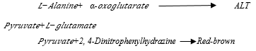
ALT catalyses the transfer of amino group from alanine to alpha oxoglutarate forming L-glutamate and pyruvate. The pyruvate reacts with 2, 4-dinitrophenyl hydrazine which in an alkaline medium gives a reddish brown colour which is measured spectrophotometrically at 546nm wavelength. The intensity of the colour is directly proportional to the enzyme activity.
Estimation of Aspartate Aminotrasferase (AST)
The AST activity in the sample was determined using the method described by Rietman and Frankel, (1957).
Principle
AST catalyses the transfer of an amino group from aspartate to alpha oxoglutarate forming L-glutamate and oxaloacetate which reacts with 2, 4-dinitrophenyl hydrazine which in an alkaline medium gives a reddish brown colour which is measured spectrophotometrically at 546nm wavelength. The intensity of the colour is directly proportional to the enzyme activity.

Estimation of Alkaline Phosphatase (ALP)
The ALP activity in the sample was determined using the Deutsche method described by Rec (1972).
Principle
ALP hydrolyses the substrate p-nitrophenol phosphate, liberating p-nitrophenol and inorganic phosphate. The reaction is stopped by the addition of sodium hydroxide (0.5N) and the absorbance of the coloured complex produced is read using a spectrophotometer at 405nm. The intensity of the colour is directly proportional to the activity of ALP in the sample.

Estimation of Gamma Glutamyl Transferase (GGT)
The GGT activity in the sample was determined using the method described by Szasz (1969).
Principle
The substrate L-Gamma-glutamyl-3-carboxyl-4-nitroanilide, in the presence of glycylglycine is converted by GGT in the sample to 5-amino-2-nitrobenzoate. The amount of 5-amino-2-nitrobenzoate formed is proportional to GGT activity and may be measured kinetically at 405 nm by the increasing intensity of the yellow colour formed.

Statistical Analysis
The results were presented using tables and figures. Data was presented as mean ± SD (standard deviation). Comparison of parameters between normal non-pregnant women (control) and pregnant women in different trimesters was done with student’s t-test (paired “t” test). A ‘p’ value less than0.05 was considered statistically significant. The data analysis was carried out using SPSS version 21.0.
Table 1 shows the comparison of serum LFTs (AST, ALT, ALP and GGT) levels in non-pregnant and pregnant women. The results obtained showed that the mean ALT in pregnant women (subjects) and non-pregnant women (control) was 2.27±1.75 and 12.17±2.47 (IU/L) respectively, while the mean AST was 3.44±1.88 and 14.76±2.34 (IU/L) respectively. Similarly, the mean ALP in pregnant women (subjects) and non-pregnant women (control) was 24.63±10.84 and 41.73±5.90 (IU/L) respectively, while the mean GGT was 3.60±1.88 and 11.01±2.19 (IU/L) respectively. There was no significant difference (p greater than0.05) in the mean ALT, AST and GGT of pregnant women (subjects) compared to non-pregnant women (control). However, there was significant difference (p less than0.05) in the ALP of pregnant women compared to non-pregnant women (control).
Figure 1 showed the result of the serum LFTs (AST, ALT, ALP and GGT) in the different trimesters of pregnancy. The mean ALT of pregnant women in the 1st, 2nd and 3rd trimesters were 14.56, 11.46 and 9.33 (IU/L) respectively. Similarly, the mean AST of pregnant women in the 1st, 2nd and 3rd trimesters were 12.60, 10.20, and 8.67 (IU/L) respectively. Furthermore, the mean GGT of pregnant women in the 1st, 2nd and 3rd trimesters was 15.14, 13.22, and 10.35 (IU/L) respectively. Figure 2 showed that the mean ALP of pregnant women in the 1st, 2nd and 3rd trimesters was 58.73, 69.55 and 82.31 (IU/L) respectively.
In table 2, the mean values ± SD of ALT showed an increasing trend in 1st, 2nd and 3rd trimester of pregnancies. The p-value is insignificant (p greater than0.05) in 1st, 2nd and 3rd trimesters respectively. The mean values ± SD of AST did not show much difference when compared with the control (non pregnant) group. The p-value was insignificant (p greater than0.05) all throughout the trimesters. The mean values ± SD of GGT also did not show much difference in pregnant women throughout different trimesters when compared with non-pregnant women (control), as p-values was greater than0.05 in all the trimesters. The mean values ± SD of ALP showed a significant increase (p less than 0.05) in 3rd trimesters as compared with non-pregnant women (control).

Table 1: Comparison of serum LFTs levels in non-pregnant and pregnant women
*Values of p greater than0.05 are significant
Keys:
ALT: AlanineAminotrasferase; ALP: Alkaline Phosphatase; AST: Aspartate Aminotrasferase; GGT: Gamma Glutamyl Transferase, SD: Standard Deviation; n: Number of Samples

Figure 1: Serum LFTs (AST, ALT and GGT) in the different trimesters of pregnancy

Figure 2: Serum ALP in the different trimesters of pregnancy

Table 2: Comparison of enzyme activities in pregnant women and control
*Values of p less than0.05 are significant
Keys: t-test: (T less than=t) two-tail (Paired Two Sample for Means); t Critical two-tail: 2.00; SD: Standard Deviation; CG: Control group, T1: 1st trimester, T2: 2nd trimester, T3: 3rd trimester, P less than0.05 = Significant; P greater than0.05 = Not Significant
Abnormal liver tests occur in 3–5% of pregnancies and show many different causes. Although alterations of liver enzymes could be a physiological phenomenon, it may also reflect potential severe liver injury, necessitating further assessment and accurate management. Some physiological changes occur in pregnant women in order to support fetal growth and development. Throughout pregnancy, levels of progesterone and estradiol gradually rise. Sex hormones had an impact on the metabolic, synthesis, and excretory activities of the liver. Serum liver function tests are crucial in monitoring hepatic disorders in adults who are not pregnant [21-22]. Numerous studies have demonstrated that the concentrations of certain trace elements change throughout pregnancy. Hence, this research was carried out to evaluate the liver serum enzyme activities (ALT, AST, ALP and GGT) in the different trimesters of pregnancy.
The result of this study showed that there was no significant difference (p greater than0.05) in serum AST and ALT between pregnant and non-pregnant women, but the value were slightly higher in pregnant women compared with non-pregnant women (table 1). The effects of pregnancy in serum ALT and AST activity levels are somewhat controversial. In agreement with this finding Mutua et al. [21] reported that a slight increase (p greater than0.05) in ALT and/or AST activity has been found during the third trimester compared with non-pregnant women. This finding is in contrast to the study by Bacq's [23], which stated that ALT and AST values remained below the normal upper limit during pregnancy. However, compared to non-pregnant women, serum ALT and AST activity levels were not altered during pregnancy or stay within the normal range in the majority of published research [24]. It is important to note that serum AST or ALT activity values over the normal upper limit during pregnancy should be regarded as pathogenic and trigger more research [25].
The AST activity as seen in table 2 showed no significant changes in all the trimesters (p greater than0.05). This supports majority of published studies that serum ALT and AST activities do not change during pregnancy or remain within normal limits established in adult population [25-26]. In the case of ALT, there is gradual increase in 1st, 2nd and 3rd trimester when compared with control (non pregnant) group. The increase was insignificant (p greater than0.05) in the 1st and 2nd trimesters compared to non-pregnant women (p greater than0.05). This is in agreement with other studies, which have shown a slight increase in ALT and or AST in the third trimester of pregnancy [26-28]. An increase in ALT or AST levels prior to labour might be due to contractions of the uterine muscles [29].
In the present study GGT level seems to decrease slightly in the 2nd and 3rd trimester but this decrease is not significant (p greater than0.05) as compared with non-pregnant women (control). Serum GGT activity has usually been considered to be normal during pregnancy. This study is agreement with the studies carried out by [30-32]. Lum & Gambino [30] reported a decrease in serum GGT level from 2nd to 3rd trimester in pregnant women, while Walker et al. [31] and Salgo and Pal [32] reported that serum GGT was within the normal range during pregnancy. These findings imply that sex hormones may impede the hepatic production of GGT, resulting in a decrease in serum GGT levels with gestational age.
Furthermore, the results showed that the serum ALP values of the test group (all three trimesters) increased significantly (p less than0.05) from 2nd trimester onwards compared with control. This study is also in agreement with the reports of [33-35]. Othman et al.[35] reported that serum alkaline phosphatase (ALP) was significantly higher (P˂0.001) during the third trimester (63-171) compared with the second trimester (33-137), and the first trimester (36-129), and with the control group (48-122) respectively. Bacq et al [23] also confirmed that serum ALP activity is significantly higher during the third trimester compared to non-pregnant women and during second trimester compared to first trimester.
The rise in ALP during the 3rd trimester of pregnancy is mostly caused by the creation of the placental isoenzyme by term, which may reach three times the usual adult upper reference value, rather than an increase in the hepatic isoenzyme. The growth of fetal bone also results in an increase in the synthesis of the bone isoenzymes with gestational age. Because of the lack of specificity, the measurement of serum ALP activity is a poor test for the diagnosis of cholestasis during the third trimester of pregnancy [36]. Alkaline phosphatase activity in the placenta is associated with preterm delivery and trophoblast alkaline phosphatase activity is altered in pre-eclampsia [37].
The study concludes that there was no significant difference (p greater than0.05) in the mean ALT, AST and GGT of pregnant women (subjects) in the different trimesters compared to non-pregnant women (control). However, there was significant difference (p less than0.05) in the ALP of pregnant women in the 3rd trimester compared to non-pregnant women (control). Liver function tests are crucial biological markers for adult-level alterations, and they should be routinely examined throughout pregnancy to identify any pathologic abnormalities. When interpreting LFT findings that might be affected by the typical changes of pregnancy, it's also crucial to take into account normal reference ranges particular to pregnancy.
Conflict of Interest
The authors declare no conflicts of interest. The authors alone are responsible for the content and the writing of the paper.
Funding
This research did not receive any grant from funding agencies in the public, commercial, or not-for-profit sectors.
Acknowledgements
The authors would like to thank Laboratory and Technical staff of the Department of Medical Laboratory Service, University Health Center, Bamidele Olumilua University of Education, Science and Technology, Ikere-Ekiti for their excellent assistance and Omon Emmanuel Research Institute (OMRI) for providing medical writing support/editorial support in accordance with Good Publication Practice (GPP3) guidelines.
Dear Editorial Team, Clinical Medical Reviews and Reports. My experience with the journal was highly positive. The peer-review process was rigorous, constructive, and completed in a timely manner. The reviewers provided valuable comments that helped improve the quality and clarity of our manuscript. The editorial office was professional, responsive, and supportive throughout all stages of the publication process. Communication was clear and efficient, and any questions were addressed promptly. Overall, I found the journal to maintain high scientific standards and an excellent publication workflow. I would be pleased to consider submitting future work to this journal. Best wishes from, Elena Popa.

It was my pleasure to submit my testimonial concerning the Reviewer Board of our Scientific Journal “Brain and Neurological Disorders”. The Reviewers focused on some modifications and their contribution was helpful. The ladies of our Editorial Office were also supported my efforts. It was my honor to have such a co-operation and I am looking forward for more collaboration.

Dear Grace Pierce, Editorial Coordinator of Journal of Clinical Research and Reports, Thank you for the speedy and efficient peer review process. I appreciate the fact that your peer reviewers do not take months to respond like with some other journals. I would also like to thank the editorial office for responding quickly to my questions. It is an excellent journal. I plan to submit more manuscripts in the future. Best wishes from, Robert W. McGee

Dear Grace Pierce, Editorial Coordinator of Journal of Clinical Research and Reports, Working with you and your team on our recent publication in JCRR has been a truly wonderful and enjoyable experience. The responses were prompt, and the reviewers were patient, constructive, and highly professional. One reviewer in particular gave me the feeling that a professor was carefully reading and commenting on my coursework, which was deeply touching. The entire process was straightforward and hassle‑free, with no tedious online forms to complete. I highly recommend this journal. Best wishes from, DR Aibing Rao, Head of R&D

I Appreciate the Opportunity to Share my Experience with the Journal of Clinical Research and Reports. The peer review process was timely and constructive, and the feedback provided helped improve the quality of our manuscript. The editorial office was professional, responsive, and supportive throughout the process, ensuring smooth communication and efficient handling of the submission. Overall, it was a positive experience collaborating with your team.

Dear Mercy Grace, Editorial Coordinator of Obstetrics Gynecology and Reproductive Sciences, We would like to express our gratitude for your help at all stages of publishing and editing the article. The editors of the magazine answer all the necessary questions and help at every stage. We will definitely continue to cooperate and publish other works in the Obstetrics Gynecology and Reproductive Sciences! Best wishes from, Alla Konstantinovna Politova,
