
Research Article | DOI: https://doi.org/10.31579/2641-0419/048
*Corresponding Author: Maciej Pawlikowski, AGH - Univ. of Science and Technology, Dept. Mineralogy, Petrography and Geochemistry, al. Mickiewicza 30, 30-059 Cracow, Poland.
Citation: Pawlikowski M. (2020) Artery Biomineralization and its Dissolution. J Clinical Cardiology and Cardiovascular Interventions, 3(4); DOI:10.31579/2641-0419/048
Copyright: © 2020 Maciej Pawlikowski, This is an open-access article distributed under the terms of The Creative Commons Attribution License, which permits unrestricted use, distribution, and reproduction in any medium, provided the original author and source are credited.
Received: 27 January 2020 | Accepted: 26 February 2020 | Published: 04 March 2020
Keywords: artery biomineralization; coronary artery; blood vessel
This publication presents the results of studies on biomineralization of “atherosclerotic plaque” forming in the coronary arteries of the heart. Obtained results were compared to studies of an artery unaffected by the process of atherosclerosis. Different types of artery biomineralization were marked, in terms of both mineral and chemical composition. Theories on the formation of crystallization centers where “atherosclerotic plaque” develops were also presented.
Results of preliminary in vitro studies on dissolving cholesterol and phosphate mineralization in arteries were presented. The results were interpreted, and conclusions were offered.
The so-called atherosclerotic plaque is the cause of many health problems. Its presence in arteries often leads to death. In this work, we present results of studies on biomineralization in coronary arteries which were conducted using, among others, classical methods of mineralogy. Types and structures of the developing mineral clusters were identified, as well as their mineral and chemical composition. Details concerning the development of artery biomineralization were presented, along with preliminary results of studies on its dissolution in vitro.
Presented results may be the basis for further research on this issue, which is vitally important for human health and life.
Histological, mineralogical and chemical tests of the biomineralization of coronary artery walls in the heart were conducted. Results of attempts to dissolve artery biomineralization in vitro were presented. Samples used in this study were obtained from deceased people at the John Paul II Hospital in Cracow. Due to reduction of the article’s length for publication, results of studies on 5 samples were chosen (Photo 1).
List of the samples for mineralogical and chemical analysis:
The goal of the study was to recognize the method of biomineralization in the wall and intima of the coronary arteries, both in cross-section, longitudinal section along the wall, as well as changes forming on the inner wall – intima. Therefore, before starting the analysis, the obtained fragments of arteries were cut in a way that made it possible to find the places where biomineralization was the strongest.
The studies were conducted using a polarizing and biological microscope, as well as Geol 540 scanning microscope. EDS chemical analyses were also conducted, using an attachment for the scanning microscope. Special attention was paid to the chemical composition of the soft tissue of arterial walls and mineral concentrations in the walls. Observed phenomena were documented with photos and graphs.
The results were compared and ordered by the archive data numbers. They were discussed and interpreted in the summary. Due to reduction of this publication’s length, its content was limited to consist mainly of documentation regarding the observed phenomena.
Studies of arterial biomineralization
Analysis using digital binocular microscope
Initial observations allowed us to choose five samples for analysis from the available material (Photo 1). Sample A represents an artery without atherosclerotic process. Samples B, C, D and E are coronary arteries with varying levels of atherosclerotic plaque development, obtained from people of different age.

Studies of arterial walls using polarizing microscope
Observations of the areas in arteries chosen for analysis were carried out using a polarizing microscope equipped with a digital camera, in partially polarized light. The microscope was used to document biomineralization phenomena (in different magnifications) and to choose samples for further studies. Preliminary observations were conducted in low magnifications, which increased as the studies progressed (Photo 2, 3).
Forms of mineralization (both organic and inorganic) were observed and compared to the artery unaffected by those processes. Materials selected at this stage were further used for histological, scanning, chemical and other analyses.


Studies using biological microscope (histological)
Histological studies were conducted on preparations stained with hematoxylin. Below are the micrographs of selected preparations that offer the most information related to the studied issue. These studies allowed us to determine several main types of arterial wall biomineralization (Photo 4, 5, 6, 7, 8), which for obvious reasons were impossible to observe in the arteries themselves.


Studies of mineral clusters in arterial wall using scanning microscope with attachment for chemical analysis (EDS)
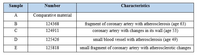
EDS chemical analyses of the types of biomineralization above proved very interesting. They showed elevated levels of calcium in spaces between cholesterol clusters, as well as in some clusters themselves (Figure. 1, Photo 4E5).


Studies of the intima surface using scanning microscope with an attachment for chemical analysis (EDS)
Organic concentrations
Organic concentrations forming on endothelium may be amorphous, in which case they take the form of infiltration (Photo 9A). They can also take crystalline forms. Cholesterol can form crystals. It builds aggregates or separate crystals. Such crystals develop in the spots where the inner wall of the artery has been damaged in some way (Photo 5). The crystals’ structure is inhomogeneous. Next to areas that are purely cholesterol we find spots with elevated calcium levels, as indicated by EDS chemical analyses (Photo 5, 4, 5B).

Crystallization of cholesterol
Cholesterol and cholesterol-fat concentrations develop in places where the atomic structures of the artery elements were damaged. That relates to both intima, the arterial wall, as well as its deeper parts (Photo 6).
Inorganic clusters
Those were also recognized in the walls and on the intima of arteries. The initial form of these accumulations – mostly calcium phosphates at different level of crystallinity – is a type of thin, almost invisible film on the intima. It is harmful as it doesn’t allow the hormones (prostacyclin, prostaglandins and thromboxanes) to be released from the endothelium to blood.
Different stages of endothelium mineralization, from early to well-developed, can be seen in a single artery (Photo 7E). Phosphates often coexist with cholesterol clusters, and sometimes also with fats.

Crystallization of phosphates
Just like crystallization of cholesterol, formation of phosphate deposits starts in the places where biological structures of the tissues are damaged (Photo 8). Whether it is cholesterol or phosphates that crystallize in any given spot depends on what is present in the biological fluids near the damage. If calcium and phosphorus ions and cholesterol are all present in the fluids, they are all included in developing mineral cluster.


The organic “solvents” were evaporated after passing them through the artery, resulting in crystallization of the substances that had been dissolved in them. The crystals obtained as a result of evaporation were studied. It was determined that they were cholesterol crystals (Photo 10). Ethyl alcohol in an appropriate dilution proved to be the best solvent for cholesterol among those tested.

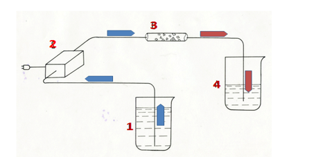
B. 2 Dissolving phosphate biomineralization (in vitro)
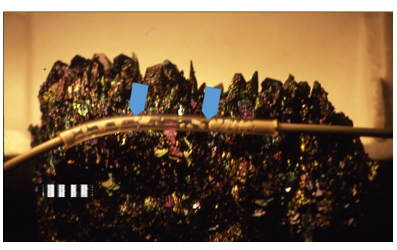
Photo 11: Tube with synthetic carbonate hydroxyapatite (arrows – material being dissolved).
Due to trouble with obtaining arteries with high concentrations of calcifications for experiments, carbonate hydroxyapatite with a microcrystalline structure was used in the experiments focused on dissolution of phosphate biomineralization. It had been synthetized in the laboratory of the Department of Mineralogy, Petrography and Geochemistry at the AGH – Univ. of Science and Technology. Before beginning the experiments, the synthetic carbonate hydroxyapatite was analyzed mineralogically to test its structure, chemical content and crystallinity. X-ray diffractometry and EDS analyzes performed during the observation of hydroxyapatite grains using SEM were used in the studies.
Obtained synthetic was crumbled and placed in a tube used in surgery for intravenous fluid administration. The speed of the flow of “solvents” was controlled by the peristaltic pump, matching the speed of the flow to the speed of blood flow in blood vessels (Photo 11, 12). Different kinds of inorganic fluids were used as solvents. The best solvent proved to be distilled water.
Studies of the surface of carbonate hydroxyapatite grains before and after dissolution were conducted, using scanning microscope.
The effect of solvents on hydroxyapatite leads to the formation of micro-holes in its grains, which increase as the solvent works. The dissolved material passes into the solvent in ionic form (Photo 13).

Studies show that biomineralization of arteries (atherosclerotic plaque) can be formed from cholesterol, phosphates or a mix of both, with varying proportions of both elements. The mixed, organic-inorganic type is the most typical.
Biomineralization (atherosclerotic plaque) forms and develops in places where the atomic structure of the artery was damaged – a place called the crystallization center. Electric field forms in the damaged spots and attracts electrically charged particles. After they attach, a deposit starts to form. The damage in the artery may occur in different places.
The reasons for the arterial damage and subsequent formation of crystallization centers vary. These could include: - genetic defects (damage),
- damage connected with physical effort and excessive mechanical “stress” on the circulatory system,
- damage caused by toxins resulting from the activity of microorganisms during an infection,
- chemical and physical factors (chemical compounds, mineral grains etc.) entering the circulatory system
- other.
The cause of damage to the arteries (in different places) and creation of crystallization centers may be one of the above factors, a few of them, or all of the above at the same time. It is worth noting that genetic defects favor repeating the atherosclerosis due to formation of biomineralization in specific places in arteries in subsequent generations.
Substances that cause the arterial biomineralization “wander" to the crystallization centers from the fluids present in the arterial wall and from the blood transported through the artery.
Complex chemical and mineral composition of analyzed deposits proves the existence of changes in the chemical composition of the fluids from which it comes.
In addition to different crystallization of biominerals in the arterial wall and its endothelium, several methods of biomineralization formation were found in the muscles of studied arteries. Its result are deposits of different structures.
For the “atherosclerotic plaque” and arterial biomineralization to form, elevated concentration of substances that crystallize in the blood and body fluids is necessary, as well as the formation of crystallization centers. Deposits will not develop without the centers present in arteries.
Therefore, fight against arterial biomineralization (atherosclerosis) should include not just lowering the levels of crystallizing substances (cholesterol, calcium etc.), but also blocking crystallization centers.
Obtained results suggest that dissolving the deposits in arteries should be attempted using mixed, organic-inorganic “solvents”. The proportion of both solvents used to fight biomineralization should be decided based on recognition of the type of biomineralization present. It should be based on mineralogical studies.
The author’s earlier research proves that described phenomena of biomineralization occur also in other arteries. However, due to the extraordinary role of the heart, their presence in the coronary arteries is particularly dangerous.
Studies on dissolving arterial biomineralization should be continued using in vivo experiments.
Dear Editorial Team, Clinical Medical Reviews and Reports. My experience with the journal was highly positive. The peer-review process was rigorous, constructive, and completed in a timely manner. The reviewers provided valuable comments that helped improve the quality and clarity of our manuscript. The editorial office was professional, responsive, and supportive throughout all stages of the publication process. Communication was clear and efficient, and any questions were addressed promptly. Overall, I found the journal to maintain high scientific standards and an excellent publication workflow. I would be pleased to consider submitting future work to this journal. Best wishes from, Elena Popa.

It was my pleasure to submit my testimonial concerning the Reviewer Board of our Scientific Journal “Brain and Neurological Disorders”. The Reviewers focused on some modifications and their contribution was helpful. The ladies of our Editorial Office were also supported my efforts. It was my honor to have such a co-operation and I am looking forward for more collaboration.

Dear Grace Pierce, Editorial Coordinator of Journal of Clinical Research and Reports, Thank you for the speedy and efficient peer review process. I appreciate the fact that your peer reviewers do not take months to respond like with some other journals. I would also like to thank the editorial office for responding quickly to my questions. It is an excellent journal. I plan to submit more manuscripts in the future. Best wishes from, Robert W. McGee

Dear Grace Pierce, Editorial Coordinator of Journal of Clinical Research and Reports, Working with you and your team on our recent publication in JCRR has been a truly wonderful and enjoyable experience. The responses were prompt, and the reviewers were patient, constructive, and highly professional. One reviewer in particular gave me the feeling that a professor was carefully reading and commenting on my coursework, which was deeply touching. The entire process was straightforward and hassle‑free, with no tedious online forms to complete. I highly recommend this journal. Best wishes from, DR Aibing Rao, Head of R&D

I Appreciate the Opportunity to Share my Experience with the Journal of Clinical Research and Reports. The peer review process was timely and constructive, and the feedback provided helped improve the quality of our manuscript. The editorial office was professional, responsive, and supportive throughout the process, ensuring smooth communication and efficient handling of the submission. Overall, it was a positive experience collaborating with your team.

Dear Mercy Grace, Editorial Coordinator of Obstetrics Gynecology and Reproductive Sciences, We would like to express our gratitude for your help at all stages of publishing and editing the article. The editors of the magazine answer all the necessary questions and help at every stage. We will definitely continue to cooperate and publish other works in the Obstetrics Gynecology and Reproductive Sciences! Best wishes from, Alla Konstantinovna Politova,
