

Research Article | DOI: https://doi.org/10.31579/2690-1919/643
Department of Cardiovascular Surgery, Fu Wai Hospital, Chinese Academy of Medical Sciences & Peking Union Medical College, Beijing, China.
*Corresponding Author: Xinjin Luo, MD. Department of Cardiovascular Surgery, Fu Wai Hospital, Chinese Academy of Medical Sciences & Peking Union Medical College No. 167, Beilishi Road, 100037, Beijing, China.
Citation: Yukun Wu, Shaoye Wang, Yang Yang, Juntao Qiu, Xinjin Luo, (2026), Application of a Dacron Prosthesis as a Subvalvular External Ring in Bicuspid Aortic Regurgitation Repair, J Clinical Research and Reports, 24(3); DOI:10.31579/2690-1919/643
Copyright: © 2026, Xinjin Luo. This is an open access article distributed under the Creative Commons Attribution License, which permits unrestricted use, distribution, and reproduction in any medium, provided the original work is properly cited.
Received: 13 July 2026 | Accepted: 27 July 2026 | Published: 30 July 2026
Keywords: bicuspid aortic valve; aortic regurgitation; repair
Background. Circumferential stabilization of the basal ring is a key step in improving the durability of aortic valve repair. Subvalvular external ring annuloplasty is an effective technique to achieve this goal.
Methods. From January to December 2025, 11 male patients with bicuspid aortic regurgitation underwent aortic valve repair using a Dacron prosthesis as a subvalvular external ring. The mean age was 36.1 ± 10.5 years (range, 20–47 years). Three patients had aortic root aneurysms and one had an ascending aortic aneurysm. All patients had severe aortic regurgitation except one patient with aortic root aneurysm who had moderate regurgitation. The Dacron ring diameter was selected based on body surface area.
Results. All procedures were completed without in-hospital mortality or complications. Valve function improved in all patients, with no more than mild residual regurgitation. The mean effective height was 9.0 ± 2.0 mm (range, 5.0–11.0 mm) and coaptation length was 5.7 ± 1.0 mm (range, 4.5–8.0 mm). At 6 months, aortic annular diameter decreased significantly (24.0 [23.5–24.5] mm vs. 26.0 [25.0–28.0] mm, p = 0.001), with no change in systolic transvalvular velocity (1.72 ± 0.41 m/s vs. 1.82 ± 0.19 m/s, p = 0.505).
Conclusions. Body surface area-based selection of Dacron ring size provides a practical and effective approach for subvalvular external ring annuloplasty in bicuspid aortic regurgitation repair.
Aortic annuloplasty, particularly at the basal ring plane of the aortic root, represents an important component of aortic regurgitation repair, providing annular stabilization and improving repair durability. The efficacy of aortic annuloplasty has been demonstrated in valve-sparing aortic root remodeling and bicuspid aortic valve (BAV) repair [1,2]. Several technical approaches to aortic annuloplasty have been described [3]. Among these, the subvalvular external ring annuloplasty proposed by Lansac et al. has shown consistent efficacy and has gained wide clinical acceptance [4].
Although Lansac et al. advocated selecting the size of the subvalvular external ring based on intraoperative measurement of the basal ring, variations in the materials used for the external ring across different periods led to notable changes in size selection criteria [1,4,5]. Using a Dacron prosthesis as the external ring, Basmadjian et al. proposed a sizing approach based on body surface area [6]. However, this method relies on a relatively simple stratification, with only two threshold values of 1.8 m2 and 2.2 m2.
This study hypothesizes that, in the repair of bicuspid aortic regurgitation, selecting the size of the subvalvular external ring based on body surface area can effectively guide treatment. Building on findings from previous anatomical studies7, more refined selection thresholds were proposed to better match patient body habitus. Through retrospective analysis of repair outcomes in patients with bicuspid aortic regurgitation treated under this strategy at our center, the study aimed to evaluate its effectiveness and to inform further standardization of key operative aspects of aortic valve repair.
Patients
This retrospective case series analyzed consecutive patients with bicuspid aortic regurgitation treated between January and December 2025. A total of 14 patients were admitted during this period. Two patients with concomitant aortic stenosis and one patient classified as type C according to the Brussels & Saarland classification [8] were excluded; these three patients underwent the Ozaki procedure.
The remaining 11 patients were included in the analysis. The mean age was 36.1 ± 10.5 years (range, 20–47 years), and all patients were male. Three patients had concomitant aortic root aneurysms, and one had an ascending aortic aneurysm. All patients had severe aortic regurgitation, except for one patient with an aortic root aneurysm (aortic sinus diameter of 52 mm) who had moderate aortic regurgitation on preoperative echocardiography. All clinical and follow-up data were collected with approval from the institutional ethics committee.
Technique
All procedures were performed under conventional cardiac anesthesia via median sternotomy, with perfusion through the ascending aorta or femoral artery, and supported by cardiopulmonary bypass.
After aortotomy, the configuration of the BAV was assessed, and the type and degree of cusp fusion were recorded. The quality of the leaflets was evaluated, and the geometric height of the non-fused cusp was measured. Repair was undertaken only when the geometric height exceeded 20 mm.
In patients with partial cusp fusion (n = 7), the fissure in the fused cusp was closed using interrupted 5-0 polypropylene sutures (PROLENE™, Ethicon LLC, Johnson & Johnson, New Brunswick, NJ, USA). The primary repair strategy involved achieving cusp alignment and correcting prolapse through central plication of the free margin using 6-0 polypropylene sutures (PROLENE™, Ethicon LLC, Johnson & Johnson, New Brunswick, NJ, USA). This was performed mainly by plicating the elongated fused cusp, guided by visual comparison of cusp height and free margin length relative to the non-fused cusp.
The aortic root was dissected along the lateral aortic wall to the subvalvular level of the basal ring. At the nadir of each sinus, at the level corresponding to the base of the interleaflet triangle, 5–6 stitches of 2-0 braided polyester sutures (Ethicon, 3/8 circle, 25-mm needle; Ethicon LLC, Johnson & Johnson, New Brunswick, NJ, USA) reinforced with pledgets were placed. These sutures were passed from the aortic lumen to the external aspect of the aortic wall and served as fixation sutures for the external ring. To avoid injury to the membranous septum, no sutures were placed at the interleaflet triangle of the right–noncoronary commissure. Instead, a 5-0 polypropylene suture with a pledget was placed on the roof of the right atrium, adjacent to the outer aortic wall, to serve as an additional external ring fixation point.
The external ring was fashioned from a polyester prosthesis by cutting standard Dacron tube grafts (Intergard, Marquet Cardiovascular LLC, Wayne, NJ, USA). For all patients, the target diameter of the Dacron external ring was selected according to body surface area, based on the criteria outlined in Table 1.
| Male | Female | ||
| BSA (m2) | External Ring Diameter (mm) | BSA (m2) | External Ring Diameter(mm) |
| BSA ≤ 1.5 | 24-25 | BSA ≤ 1.4 | 23-24 |
| 1.5 < BSA> | 26-27 | 1.4 < BSA> | 25-26 |
| 1.7 < BSA> | 27-28 | 1.6 < BSA> | 26-27 |
| 1.9 < BSA> | 28-29 | 1.9 < BSA> | 27-28 |
| 2.1 < BSA> | 29-30 | BSA > 2.4 | 28-29 |
| BSA > 2.5 | 30-31 | ||
BSA, body surface area.
Table 1: Dacron external ring diameters according to body surface area and sex.
Among patients who underwent isolated BAV repair (n = 8), a circular ring was fashioned from a Dacron graft with a diameter slightly larger than the target and a height of approximately 4 mm, corresponding to 3–4 graft folds. The ring was cut at one point to create a band, allowing it to pass beneath the ostia of the left and right coronary arteries. The two ends of the band were then sutured together using 5-0 polypropylene sutures. During suturing, the band length was adjusted to ensure that the final diameter of the reconstructed Dacron ring matched the target external ring diameter. The pre-placed fixation sutures were then passed around the Dacron ring and secured. Valve leaflet coaptation was assessed by direct visual inspection, and additional central plication of the free margins was performed as needed to optimize leaflet alignment.
One patient with aortic root dilatation (aortic sinus diameter of 46 mm) underwent the Florida sleeve procedure. A 32-mm Dacron graft was used as the sleeve, and keyholes were created at the corresponding locations of the coronary ostia. The aortic root was wrapped with the Dacron graft, allowing the left and right coronary arteries to exit through the prepared keyholes. The edges created at the lower margin of the sleeve during keyhole preparation were then sutured together using 5-0 polypropylene sutures. During this step, the spacing between sutures was adjusted to reduce the diameter of the sleeve base to the target value of 28 mm. The pre-placed fixation sutures were then secured to the appropriate positions along the lower edge of the sleeve. At the level of the sinotubular junction, the native aortic wall was imbricated and sutured to the sleeve using 5-0 polypropylene sutures. The ascending aorta was subsequently replaced with a separate 30-mm Dacron graft.
Two additional patients with aortic root aneurysms underwent root remodeling. The aortic sinus wall was trimmed, leaving a tissue margin of 3–5 mm from the fibrous annulus for suturing. One end of a 28-mm Dacron graft was divided into two equal segments using two longitudinal incisions, and each segment was shaped into a scalloped configuration. The height of each longitudinal incision, corresponding to the apex of the new commissural junction after reconstruction, was matched to the height of the native primary commissure. The two scalloped graft segments were anastomosed to the residual sinus wall margins using 5-0 polypropylene sutures. A 28-mm Dacron external ring was then positioned in the subvalvular location along the reconstructed graft. The pre-placed fixation sutures were passed around the Dacron ring and secured. Openings were created at appropriate positions in the graft using electrocautery, trimmed, and anastomosed to the left and right coronary ostia. Finally, the graft was anastomosed to the distal ascending aorta.
In the three patients who underwent aortic root remodeling or the Florida sleeve procedure, a temporary marking suture (Figure 1) was used intraoperatively to assess the effective height (eH) of the repaired valve and to correct any iatrogenic leaflet prolapse.
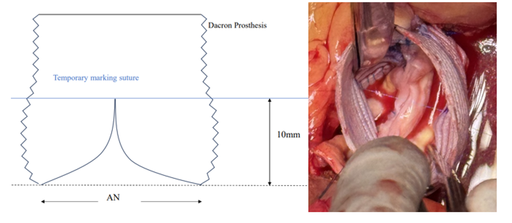
AN, aortic annulus.
Figure 1: Temporary marking suture. A Schematic diagram, B Intraoperative photo
A vertical measurement of 10 mm was taken from the lowest points of the two reconstructed aortic sinuses, and this level was marked on the Dacron graft. A 6-0 polypropylene suture was passed through the two marked points and tensioned externally to maintain a straight line, serving as a temporary marking suture for assessment of eH. Using two pairs of forceps, gradually spread out each of the two leaflets one by one to simulate the inflated state of the leaflet when the valve was closed, and evaluated whether the highest point of the free edge of the leaflets at this time could reach the level of the temporary marking suture. If this level was reached or exceeded, no further intervention was required. If not, central plication of the free margins of both leaflets was performed as needed, based on the discrepancy from the reference level, to ensure that the coaptation height reached the level of the temporary marking suture. Once satisfactory adjustment was achieved, the temporary suture was removed. This approach ensured that the postoperative eH measured by transesophageal echocardiography exceeded 9 mm.
The procedure was considered complete only when intraoperative transesophageal echocardiography confirmed no more than mild aortic regurgitation, or only trivial central regurgitation, after discontinuation of cardiopulmonary bypass. If this criterion was not met, cardiopulmonary bypass was resumed and further valve correction was performed. Echocardiography was used to assess aortic valve function and to measure both eH and leaflet coaptation length.
Statistics
Medical records were collected during hospitalization. Echocardiographic variables assessed preoperatively and at 6 months postoperatively included left ventricular end-diastolic diameter, left ventricular ejection fraction, aortic annular diameter, and transvalvular flow velocity. Continuous variables were tested for normality prior to analysis. Normally distributed data are presented as mean ± standard deviation and were compared using the paired t-test, whereas non-normally distributed data are presented as median (interquartile range) and were compared using the Wilcoxon signed-rank test. All tests were two-tailed, and p < 0>
All 11 patients were classified as type 1 (RL) BAV according to the Sievers classification. According to the Brussels & Saarland classification8, four patients were type B and seven were type A. The degree of cusp fusion and the geometric height of the non-fused cusp measured intraoperatively are presented in Table 2.
| No. | BSA (m2) | Preopeartive AR | Cusp fusion | gH (mm) | External ring diameter (mm) | eH (mm) | CL (mm) | Postopeartive AR |
| 1 | 1.98 | moderate | partial (2/3) | 20 | 28 | 9.5 | 6.0 | none |
| 2 | 1.84 | severe | complete | 21 | 28 | 9.5 | 5.5 | mild |
| 3 | 1.81 | severe | complete | 24 | 28 | 10 | 6.0 | trace |
| 4 | 1.93 | severe | complete | 20 | 29 | 9.0 | 5.0 | mild |
| 5 | 1.74 | severe | complete | 23 | 29 | 5.5 | 4.5 | trace |
| 6 | 1.77 | severe | partial (1/2) | 25 | 27 | 9.5 | 5.5 | none |
| 7 | 1.89 | severe | partial (1/3) | 23 | 27 | 11 | 8.0 | none |
| 8 | 1.77 | severe | partial (1/2) | 23 | 28 | 9.5 | 5.0 | trace |
| 9 | 2.33 | severe | partial (1/3) | 25 | 29 | 9.5 | 6.0 | mild |
| 10 | 2.39 | severe | partial (2/3) | 25 | 31 | 5.0 | 4.5 | trace |
| 11 | 2.05 | severe | partial (2/3) | 20 | 29 | 11 | 7.0 | none |
Preoperative refers to the last echocardiographic assessment performed before surgery, and postoperative refers to the echocardiographic assessment performed 6 months after surgery.
BSA, body surface area; gH, geometric height of the non-fused cusp; eH, effective height; CL, coaptation length; AR, aortic regurgitation
Table 2: Degree of aortic regurgitation and intraoperative parameters.
All patients underwent BAV insufficiency repair using a Dacron prosthesis as a subvalvular external ring. In nine cases, the Dacron ring diameter was determined solely based on body surface area. In the remaining two cases, intraoperative assessment showed that the native annulus was excessively large; therefore, Dacron rings with diameters 1–2 mm larger than the recommended size were selected.
No in-hospital deaths or complications occurred (e.g., bleeding, stroke, infection, renal failure). The mean postoperative hospital stay was 7.2 ± 1.6 days (range, 6–10 days). Two patients with aortic root aneurysms (root diameters of 52 mm and 50 mm) underwent modified root remodeling. One patient with a sinus of Valsalva dilated to 46 mm underwent the Florida sleeve procedure. One patient with an ascending aortic aneurysm underwent concomitant replacement of the ascending aorta using a 30-mm Dacron graft. The mean aortic cross-clamp time was 130.9 ± 39.5 minutes (range, 98–212 minutes), and the mean cardiopulmonary bypass time was 159.8 ± 46.4 minutes (range, 110–251 minutes).
Seven patients with incomplete cusp fusion underwent fissure closure using interrupted sutures. Five patients underwent central plication of the free margin of the fused cusp only, whereas in the remaining six patients, both leaflets underwent central plication. Aortic valve coaptation improved markedly in all patients after repair. Intraoperative transesophageal echocardiography showed a mean eH of 9.0 ± 2.0 mm and a coaptation length of 5.7 ± 1.0 mm (Table 2).
All patients returned for follow-up at six months after surgery. Transthoracic echocardiography showed no more than mild aortic regurgitation in all cases. Comparison of preoperative and postoperative results demonstrated a significant reduction in left ventricular end-diastolic diameter (51.1 ± 2.6 mm vs. 64.5 ± 6.5 mm, p < 0 xss=removed xss=removed xss=removed>
Discussion
With increasing understanding of aortic regurgitation, research has shown that achieving successful and durable aortic valve repair requires not only correction of leaflet pathology but also reconstruction and stabilization of the functional aortic annulus. Stabilization of the basal ring, a key component of the functional annulus, has become an essential step in improving repair durability [9]. Because earlier techniques such as subcommissural annular plication do not provide consistent stabilization, current approaches favor circumferential annuloplasty [10]. The same principles apply to BAV repair. Among the available techniques, the subvalvular external ring described by Lansac et al.4 and the suture annuloplasty technique described by Schäfers et al. [11] are widely used in clinical practice.
This study successfully applied a polyester Dacron ring as a subvalvular external ring in BAV repair and proposed a body surface area-based approach for selecting the ring diameter (Table 1), with favorable clinical outcomes.
Lansac et al. selected the size of the subvalvular external ring based on intraoperative measurement of the basal ring diameter. Initially, polyester Dacron prosthetic rings fashioned from standard Dacron tube grafts were used, and sizing recommendations for this material were reported in 20064. Subsequently, the group transitioned to a specially designed expansible extra-aortic ring (ExAo; Coroneo Inc, Montreal, QC, Canada), which has been shown to provide improved annular stabilization compared with polyester prostheses6. Based on the use of the extra-aortic ring, the recommended sizing criteria were substantially reduced4,5. However, limited commercial availability of the extra-aortic ring in many countries and regions, including China, restricts the ability of clinicians to adopt this approach in routine practice.
In addition, precise intraoperative measurement of the basal ring diameter can be challenging in some patients, as it requires careful handling to avoid damage to the preserved aortic valve leaflets. This limitation is particularly relevant in BAV anatomy, where the valve often maintains a “fish-mouth” configuration even when maximally opened. As a result, neither a Hegar dilator nor an appropriately sized valve sizer may pass smoothly through the valve orifice, limiting accurate annular measurement. This issue is especially pronounced in patients with markedly enlarged basal annuli.
Basmadjian et al. proposed determining the expected inner diameter of the aortic annulus based on body surface area, followed by selection of a Dacron ring approximately 5–6 mm larger than the desired annular diameter for use as a subvalvular external ring6. This approach was based on anatomical data reported by Capps et al., derived from analysis of 6,801 donor hearts across a wide age range, which established normative relationships between aortic annular diameter and body surface area7. However, to simplify clinical application, this approach used relatively few body surface area stratification thresholds. In the present study, anatomical data from Capps et al. were reanalyzed, grouping body surface areas with similar mean annular diameters, and a more refined recommendation table for Dacron external ring sizing was developed (Table 1), facilitating more practical and precise use in clinical settings. Although Capps' anatomical research data was derived from the American population, our study indicates that their research results can also be applied to the Chinese population in East Asia.
This study showed that reducing the basal ring with an appropriately sized external ring can increase the eH of the repaired bicuspid aortic valve. In two patients in this cohort (patients 5 and 10) who underwent isolated repair, eH values remained low (5.5 mm and 5.0 mm, respectively). Preoperative echocardiography showed relatively large aortic annular diameters (28 mm and 30 mm), whereas intraoperative assessment allowed passage of only 27 mm and 25 mm probes, limiting accurate evaluation of true annular size. Direct inspection suggested that the basal annulus was more dilated than indicated by echocardiography. According to body surface area-based recommendations, these patients would have been assigned Dacron external rings of 27–28 mm and 29–30 mm, respectively. However, given concerns about annular enlargement, larger rings (29 mm and 31 mm) were selected. This choice may explain why postoperative eH did not reach the target threshold. Nevertheless, both patients achieved a coaptation length of 4.5 mm, meeting expected values, and postoperative valve competence remained satisfactory.
Adequate eH is a key determinant of durable valve repair. Dedicated calipers have been described to guide intraoperative central plication of the leaflet free margins and achieve the target eH. In this cohort, such calipers were not used, yet most patients achieved satisfactory eH. In bicuspid cases undergoing root remodeling, the temporary marking suture technique described in this study was used to guide adjustment of leaflet free margin length. Because reconstruction creates two aortic sinuses, a single marking suture is sufficient, making the method simple and effective. In patients undergoing isolated BAV repair, the sinotubular junction is not intentionally reduced, and iatrogenic leaflet prolapse is uncommon. Following basal ring reduction with an appropriately sized external ring, eH typically meets the target threshold without the need for dedicated intraoperative measurement.
However, this study represents a single-center exploratory analysis with a small sample size and short follow-up duration. Validation in larger cohorts with longer-term follow-up is required.
In conclusion, this clinical experience suggests that selecting the size of a polyester Dacron ring based on body surface area and using it as a subvalvular external ring yields satisfactory outcomes in bicuspid aortic regurgitation repair.
Yukun Wu was a major contributor in writing the manuscript and conducted statistical analyses. Shaoye Wang took part in the operation and prepared the Tables and Figures. Yang Yang checked the manuscript and made correction. Juntao Qiu performed follow-up and analyzed the data; Xinjin Luo provided original ideals and conceived the study, performed the operations. All authors reviewed the manuscript.
Ethical approval and informed consent statements
This retrospective study did not obtain informed consent. The Ethics Committee of Fu Wai Hospital approved all clinical and follow-up data (approval number: 2025-3024).
Consent for publication
This manuscript does not contain data from identifiable individuals; therefore, consent for publication is not applicable
Data availability statement
The datasets used and/or analyzed during the current study are available from the corresponding author upon reasonable request. Anonymised data are available from the corresponding author upon reasonable request.
Declaration of conflicting interest
The authors declared no potential conflicts of interest with respect to the research, authorship, and/or publication of this article.
Dear Editorial Team, Clinical Medical Reviews and Reports. My experience with the journal was highly positive. The peer-review process was rigorous, constructive, and completed in a timely manner. The reviewers provided valuable comments that helped improve the quality and clarity of our manuscript. The editorial office was professional, responsive, and supportive throughout all stages of the publication process. Communication was clear and efficient, and any questions were addressed promptly. Overall, I found the journal to maintain high scientific standards and an excellent publication workflow. I would be pleased to consider submitting future work to this journal. Best wishes from, Elena Popa.

It was my pleasure to submit my testimonial concerning the Reviewer Board of our Scientific Journal “Brain and Neurological Disorders”. The Reviewers focused on some modifications and their contribution was helpful. The ladies of our Editorial Office were also supported my efforts. It was my honor to have such a co-operation and I am looking forward for more collaboration.

Dear Grace Pierce, Editorial Coordinator of Journal of Clinical Research and Reports, Thank you for the speedy and efficient peer review process. I appreciate the fact that your peer reviewers do not take months to respond like with some other journals. I would also like to thank the editorial office for responding quickly to my questions. It is an excellent journal. I plan to submit more manuscripts in the future. Best wishes from, Robert W. McGee

Dear Grace Pierce, Editorial Coordinator of Journal of Clinical Research and Reports, Working with you and your team on our recent publication in JCRR has been a truly wonderful and enjoyable experience. The responses were prompt, and the reviewers were patient, constructive, and highly professional. One reviewer in particular gave me the feeling that a professor was carefully reading and commenting on my coursework, which was deeply touching. The entire process was straightforward and hassle‑free, with no tedious online forms to complete. I highly recommend this journal. Best wishes from, DR Aibing Rao, Head of R&D

I Appreciate the Opportunity to Share my Experience with the Journal of Clinical Research and Reports. The peer review process was timely and constructive, and the feedback provided helped improve the quality of our manuscript. The editorial office was professional, responsive, and supportive throughout the process, ensuring smooth communication and efficient handling of the submission. Overall, it was a positive experience collaborating with your team.

Dear Mercy Grace, Editorial Coordinator of Obstetrics Gynecology and Reproductive Sciences, We would like to express our gratitude for your help at all stages of publishing and editing the article. The editors of the magazine answer all the necessary questions and help at every stage. We will definitely continue to cooperate and publish other works in the Obstetrics Gynecology and Reproductive Sciences! Best wishes from, Alla Konstantinovna Politova,
