
Case Report | DOI: https://doi.org/10.31579/2692-9422/084
1 Neurosurgery Service of the Maciel Hospital, Montevideo, Uruguay.
2 Department of Anatomy, Faculty of Medicine, University of the Republic, Montevideo,
*Corresponding Author: Alejandra Jaume, Neurosurgery Service of the Maciel Hospital, Montevideo, Uruguay.
Citation: Alejandra Jaume, Matilde Lissarrague, (2023), Anatomo-Imagenological Correlation of The Surcal Anatomy of The Lateral and Basal Aspects of The Temporal Lobe. 5(6): DOI:10.31579/2692-9422/084
Copyright: © 2023, Alejandra Jaume. This is an open-access article distributed under the terms of The Creative Commons Attribution License, which permits unrestricted use, distribution, and reproduction in any medium, provided the original author and source are credited.
Received: 07 November 2023 | Accepted: 16 November 2023 | Published: 05 December 2023
Keywords: temporal lobe; brain magnetic resonance; anatomical; imaging correlation ; epilepsy surgery
Introduction: within the anatomy of the temporal lobe, the mesial aspect of it has been studied extensively, due to its complexity and its relationship with the surgical treatment of epilepsy. The lateral and inferior aspects of the temporal lobe have simpler, but variable sulcal patterns. It is important to keep in mind that for surgery of the mesial temporal region, the lateral aspect is used as the approach, and therefore it is necessary to know the arrangement of the sulci in this region.
Materials and method: 6 temporal lobes (3 left and 3 right) obtained from adult cadavers fixed in formalin solution and without macroscopic pathology were used. The length, depth and direction of the grooves were measured in each of the preparations. After carrying out the measurements, the temporal lobes were cut in the 3 planes of space (two in the sagittal direction, 2 in the coronal direction and 2 in the horizontal plane) to perform a correlation with magnetic nuclear resonance (MRI) studies.
Results: on the lateral side, two constant grooves were found (superior and inferior temporal sulcus). The upper one measured between 72 and 99mm in length and between 5 and 13mm in depth. The inferior groove measured between 68 and 71mm in length and between 6 and 9mm in depth. On the lower surface, two constant grooves were found (temporo-occipital and collateral grooves). The temporo-occipital sulcus measured between 41 and 68mm in length and between 2 and 7mm in depth. The collateral sulcus measured between 54 and 69 mm in length and between 3 and 7 mm in depth.
Conclusions: the general direction of the temporal sulci points towards the temporal horn of the ventricle. Anatomical sections in the 3 planes of space provide excellent correlation with imaging studies.
The brain is composed of seven lobes: frontal lobe, parietal lobe, occipital lobe, temporal lobe, insular lobe, limbic lobe and paracentral lobe 5.
The temporal lobe is located below the sylvian fissure, which separates it from the frontal and parietal lobes; and, it is separated from the occipital lobe, by a line that joins the parieto-occipital fissure with the pre occipital recess. This lobe has four faces: superior, lateral, basal, and mesial2. The mesial surface is one of the most studied regions, due to its complexity and its relationship with the surgical treatment of Epilepsy3. However, the temporal lobe is the seat of multiple pathologies, which highlights the importance of its detailed anatomical knowledge. The lateral and basal aspects of the temporal lobe have simpler but variable sulcal patterns, and knowledge of them is essential for addressing different pathologies that occur in this region4. In turn, the lateral aspect is used as an approach for the mesial temporal region, therefore, it is necessary to know the arrangement of the sulci in said region5.
This work proposes to carry out a systematic study of the lateral and basal faces, and their imaging correlation, for a better microsurgical result.
Six temporal lobes (3 left and 3 right) obtained from adult cadavers fixed in formalin solution and without macroscopic pathology were used. Each hemisphere was studied, assessing its anatomy of grooves and turns, as well as different varieties according to each specimen. In each of the preparations, the length, depth and direction of the sulci on the lateral and basal surfaces of the temporal lobe were measured. After carrying out the measurements, the temporal lobes were cut in the 3 planes of space (two in the sagittal direction, 2 in the coronal direction and 2 in the horizontal plane) to perform a correlation with imaging studies. Subsequently, 20 MRI studies of patients without ostensible pathological findings at a temporal level were analyzed. A correlation was then made between the anatomical and radiological findings.

Figure 2: Vista inferior de cara basal. 1, giro temporal inferior; 2, surco temporo-occipital; 3, giro fusiforme; 4, polo temporal; 5, surco rinal; 6, surco colateral; 7, sector anterior del giro parahipocampal; 8, sector posterior del giro parahipocampal; 9, mesencéfalo.

Figure 3 and 4: Corte coronal en RNM, y en pieza anatómica. 1, fisura silviana; 2, giro temporal superior; 3, surco temporal superior; 4, giro temporal medio; 5, surco temporal inferior; 6, giro temporal inferior, 7, cuerno temporal.

Figure 5: Corte axial en pieza anatómica y en RNM. 1, fisura silviana; 2, giro de Heschl; 3, planum temporal; 4, atrío ventricular.

Figure 6: Corte sagital de RNM. 1, fisura silviana; 2, giro temporal superior; 3, surco temporal superior; 4, giro temporal medio; 5, surco temporal inferior; 6, giro temporal inferior; 7, giro supramarginalis; 8, giro angularis.
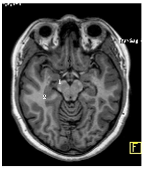
Figure 7: Corte axial de RNM. 1, cuerno temporal; 2, giro temporal medio.

Figure 8: Vista medial de cara mesial y basal. 1, segmento anterior del uncus; 2, área entorrinal; (1+2), lóbulo piriforme o sector anterior del giro parahipocampal; 3, surco rinal; 4, polo temporal; 5, sector posterior del giro parahipocampal; 6, surco colateral; 7, giro fusiforme; 8, surco hipocampal; 9, istmo del giro del cíngulo; 10, labio anterior de fisura calcarína; 11, giro lingual.

Figure 9: Corte sagital en RNM. 1, parte petrosa del hueso temporal piso de fosa media; 2, tienda del cerebelo; 3, atrío ventricular; 4, cuerno temporal.
The lateral aspect of the temporal lobe is divided into three gyri: superior, middle and inferior, by two sulci: superior and inferior. These grooves were constant in all specimens. The superior temporal sulcus measured between 72 and 99mm in length (mean: 85.5mm); and, between 5 and 13mm deep (average: 9mm). Its direction was transversal in all cases. The inferior sulcus measured between 68 and 71mm in length (mean: 69.5mm); and, between 6 and 9mm deep (average: 7.5mm). Its direction was slightly oblique
The general direction of the temporal sulci points towards the temporal horn of the ventricle. A correct anatomical-imaging correlation allows an approach minimally invasive, as well as preserving eloquent adjacent structures.
Dear Editorial Team, Clinical Medical Reviews and Reports. My experience with the journal was highly positive. The peer-review process was rigorous, constructive, and completed in a timely manner. The reviewers provided valuable comments that helped improve the quality and clarity of our manuscript. The editorial office was professional, responsive, and supportive throughout all stages of the publication process. Communication was clear and efficient, and any questions were addressed promptly. Overall, I found the journal to maintain high scientific standards and an excellent publication workflow. I would be pleased to consider submitting future work to this journal. Best wishes from, Elena Popa.

It was my pleasure to submit my testimonial concerning the Reviewer Board of our Scientific Journal “Brain and Neurological Disorders”. The Reviewers focused on some modifications and their contribution was helpful. The ladies of our Editorial Office were also supported my efforts. It was my honor to have such a co-operation and I am looking forward for more collaboration.

Dear Grace Pierce, Editorial Coordinator of Journal of Clinical Research and Reports, Thank you for the speedy and efficient peer review process. I appreciate the fact that your peer reviewers do not take months to respond like with some other journals. I would also like to thank the editorial office for responding quickly to my questions. It is an excellent journal. I plan to submit more manuscripts in the future. Best wishes from, Robert W. McGee

Dear Grace Pierce, Editorial Coordinator of Journal of Clinical Research and Reports, Working with you and your team on our recent publication in JCRR has been a truly wonderful and enjoyable experience. The responses were prompt, and the reviewers were patient, constructive, and highly professional. One reviewer in particular gave me the feeling that a professor was carefully reading and commenting on my coursework, which was deeply touching. The entire process was straightforward and hassle‑free, with no tedious online forms to complete. I highly recommend this journal. Best wishes from, DR Aibing Rao, Head of R&D

I Appreciate the Opportunity to Share my Experience with the Journal of Clinical Research and Reports. The peer review process was timely and constructive, and the feedback provided helped improve the quality of our manuscript. The editorial office was professional, responsive, and supportive throughout the process, ensuring smooth communication and efficient handling of the submission. Overall, it was a positive experience collaborating with your team.

Dear Mercy Grace, Editorial Coordinator of Obstetrics Gynecology and Reproductive Sciences, We would like to express our gratitude for your help at all stages of publishing and editing the article. The editors of the magazine answer all the necessary questions and help at every stage. We will definitely continue to cooperate and publish other works in the Obstetrics Gynecology and Reproductive Sciences! Best wishes from, Alla Konstantinovna Politova,
